Jump to content
In this section we have spot diagnoses posted on a daily basis since June 2010, now over 4000! You can review the archived cases and read the suggested diagnoses by users and the final comment by the contributors.
Case are uploaded each week day by 10 am UK time with the correct diagnosis will generally be posted at 8 pm UK time. Why not view the most recent spot diagnosis and proffer a diagnosis?

Case Number : Case 3031 - 17 February 2022 Posted By: Saleem Taibjee

Please read the clinical history and view the images by clicking on them before you proffer your diagnosis.
Submitted Date :
   (0 reviews)

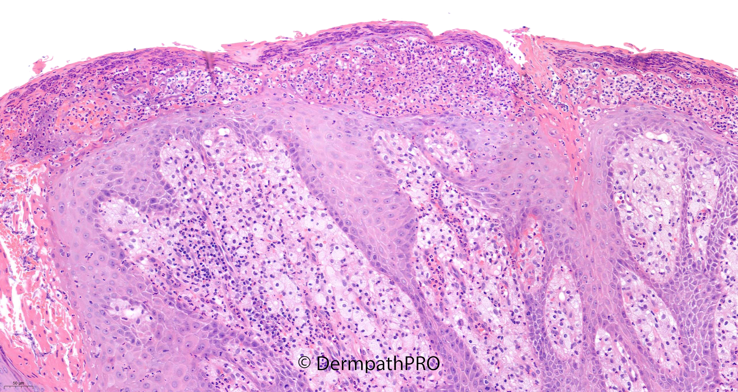
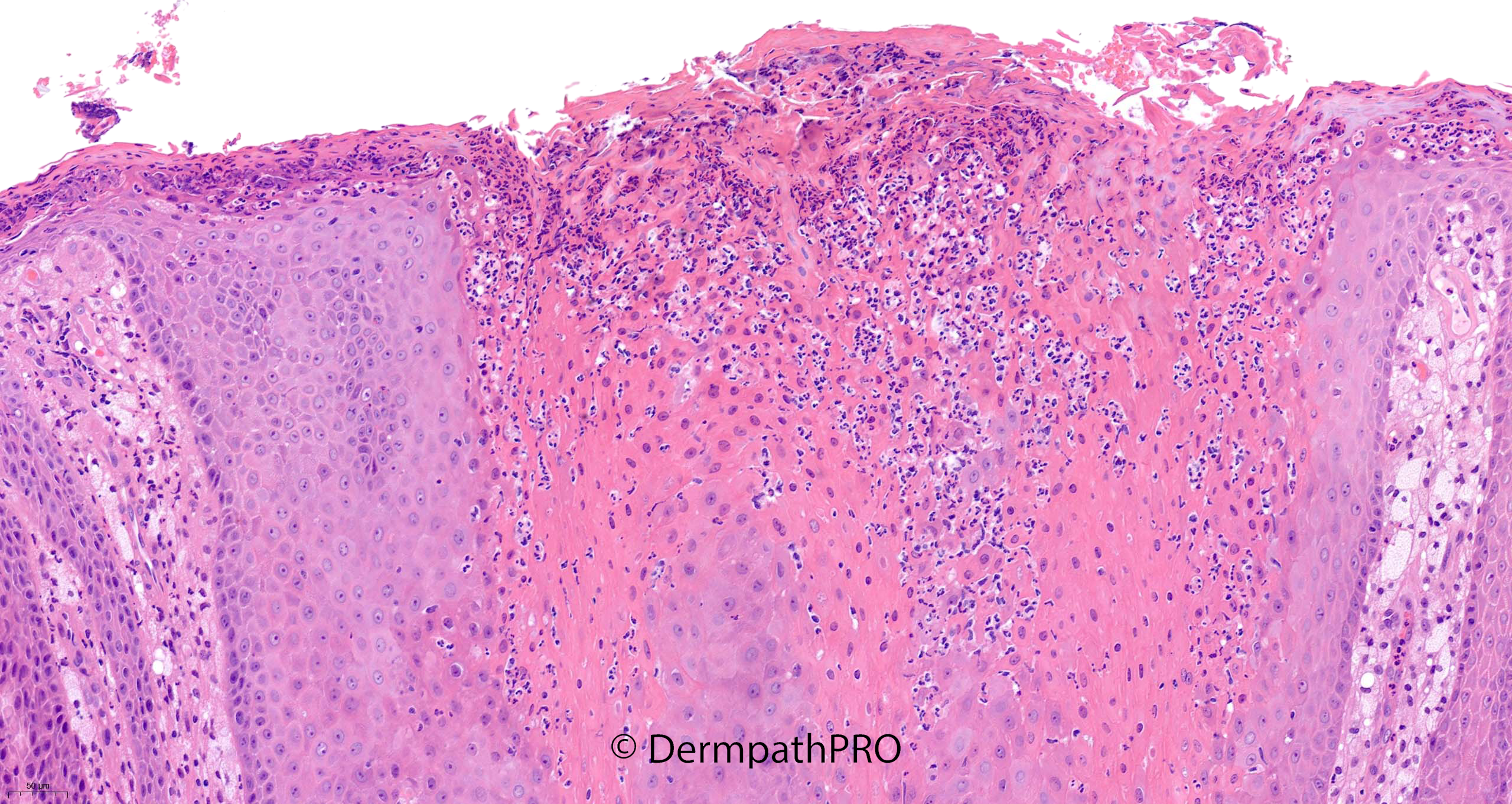
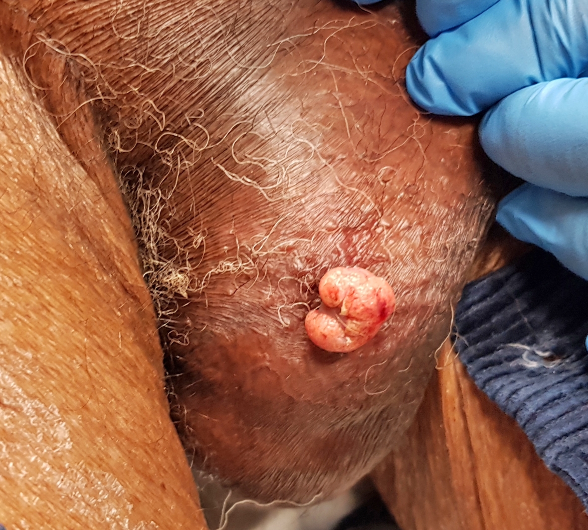
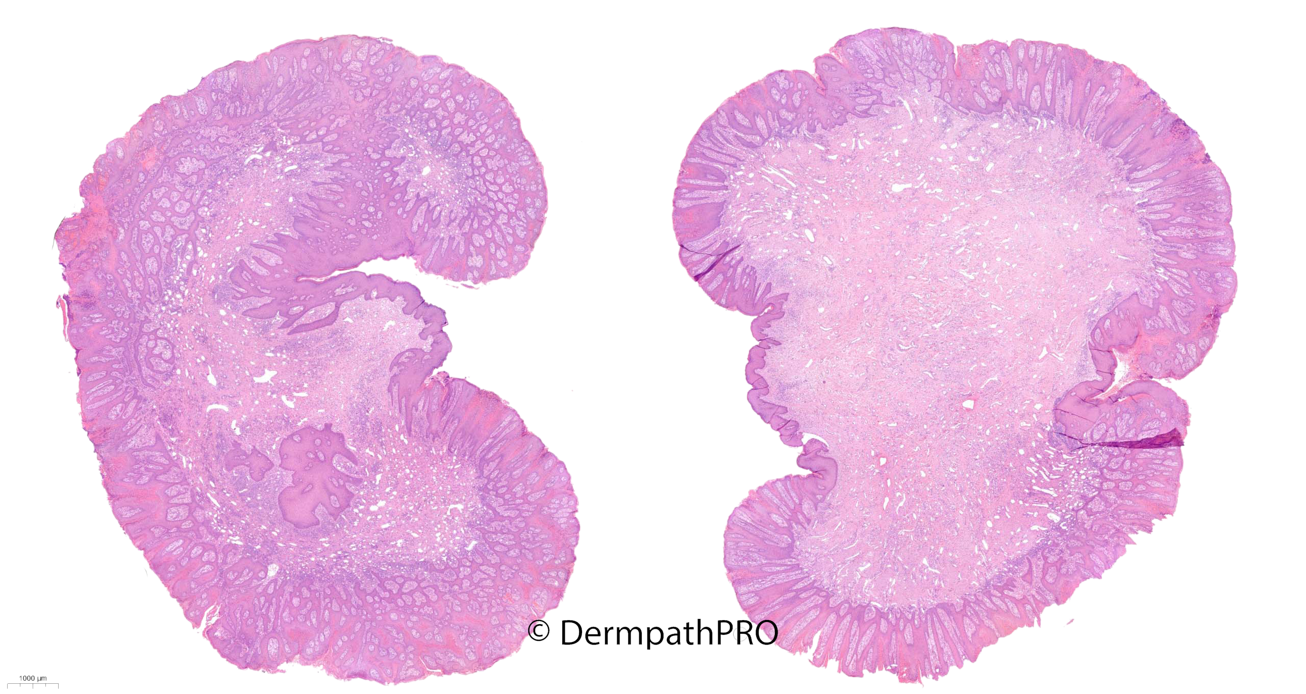
92M 2-year history of protuberant nodule right side of scrotum, recently bled ? traumatised papilloma

  • Like 1
  Report Record

User Feedback


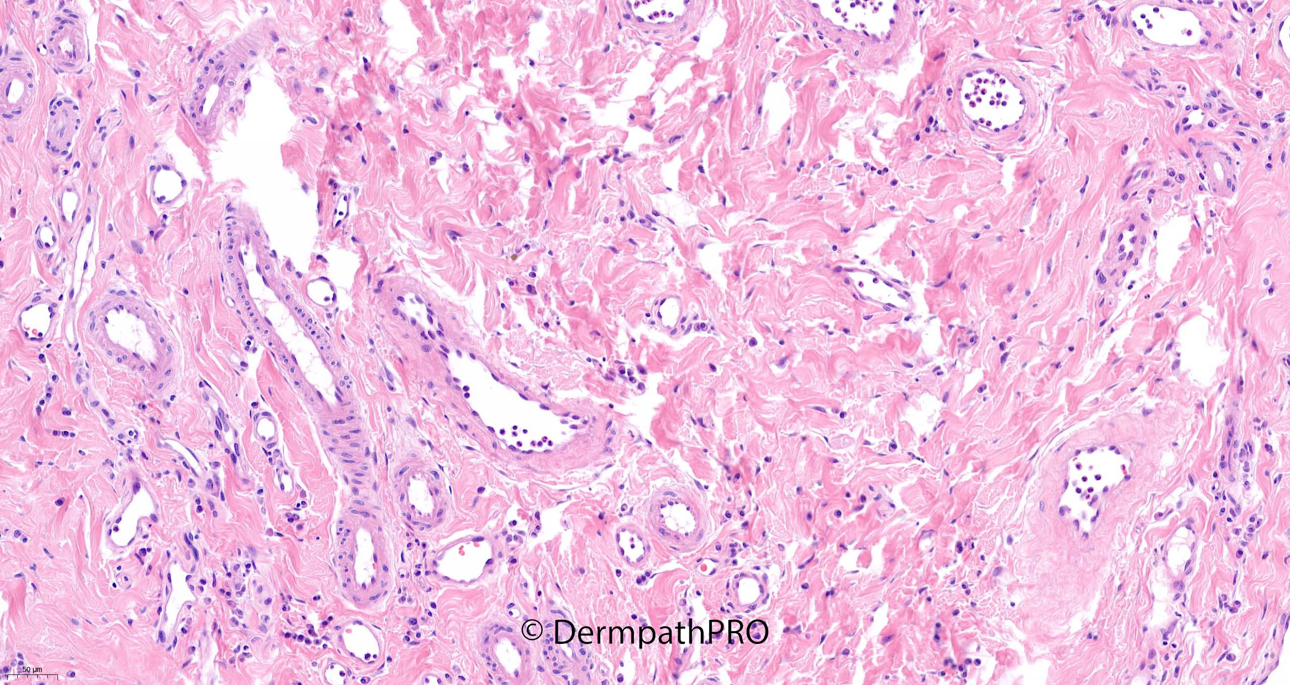
Verruciform xanthoma, the deeper vessels and plasma cell rich infiltrate are just reactive in nature?

Share this comment


Link to comment
share_externally

Richard Logan

Posted

There is no doubt that there are histological features of verruciform xanthoma present.  The clinical photo however, is very suggestive of a pyogenic granuloma and there is a prominent dermal vascularity which Saleem has highlighted in the last three slides.  There is a report (below) of a case where a verruciform xanthoma was associated with an arterio-venous malformation and I wonder if something similar is going on here.  Perhaps I am over-interpreting the dermal changes.

Ref; Kishimoto S, Takenaka H, Shibagaki R, Nagata M, Katoh N, Yasuno H. Verruciform xanthoma arising in an arteriovenous haemangioma. Br J Dermatol. 1998;139:546–548. doi: 10.1046/j.1365-2133.1998.02434.x. [PubMed] [CrossRef] [Google Scholar] [Ref list]

Share this comment


Link to comment
share_externally

6 hours ago, Richard Logan said:

There is no doubt that there are histological features of verruciform xanthoma present.  The clinical photo however, is very suggestive of a pyogenic granuloma and there is a prominent dermal vascularity which Saleem has highlighted in the last three slides.  There is a report (below) of a case where a verruciform xanthoma was associated with an arterio-venous malformation and I wonder if something similar is going on here.  Perhaps I am over-interpreting the dermal changes.

Ref; Kishimoto S, Takenaka H, Shibagaki R, Nagata M, Katoh N, Yasuno H. Verruciform xanthoma arising in an arteriovenous haemangioma. Br J Dermatol. 1998;139:546–548. doi: 10.1046/j.1365-2133.1998.02434.x. [PubMed] [CrossRef] [Google Scholar] [Ref list]

Agree. Traumatic scratching doesn’t fully explain the vascularized stroma. We pathologist like the single and exclusive diagnosis, but many times have to surrender to lesional association. 

Share this comment


Link to comment
share_externally

Saleem Taibjee

Posted

Hi. I must admit that I have just accepted this to be a nice example of verruciform xanthoma. It is either uncommon, or frequently mis-diagnosed as a squamous papilloma if one fails to notice the xanthomatised cells within the dermal papillae. I hadn't knowlingly seen a case for a while, and then funnily enough another case also came across my desk shortly after this one. With regards to the vascularised stroma which many of you have commented upon, I don't think it is very specific, and perhaps reflects the anatomical site. Even 'ordinary' fibroepithelial polyps/skin tags can sometimes show prominent vessels. So personally I do not think that there are 2 pathologies here.

BW, Saleem

Share this comment


Link to comment
share_externally


Join the conversation

You can post now and register later. If you have an account, sign in now to post with your account.

Guest
Add a comment...

×   Pasted as rich text.   Paste as plain text instead

  Only 75 emoji are allowed.

×   Your link has been automatically embedded.   Display as a link instead

×   Your previous content has been restored.   Clear editor

×   You cannot paste images directly. Upload or insert images from URL.

Loading...

×
×
  • Create New...