Jump to content
In this section we have spot diagnoses posted on a daily basis since June 2010, now over 4000! You can review the archived cases and read the suggested diagnoses by users and the final comment by the contributors.
Case are uploaded each week day by 10 am UK time with the correct diagnosis will generally be posted at 8 pm UK time. Why not view the most recent spot diagnosis and proffer a diagnosis?

Case Number : Case 3037 - 25 February 2022 Posted By: Dr. Richard Carr

Please read the clinical history and view the images by clicking on them before you proffer your diagnosis.
Submitted Date :
   (0 reviews)

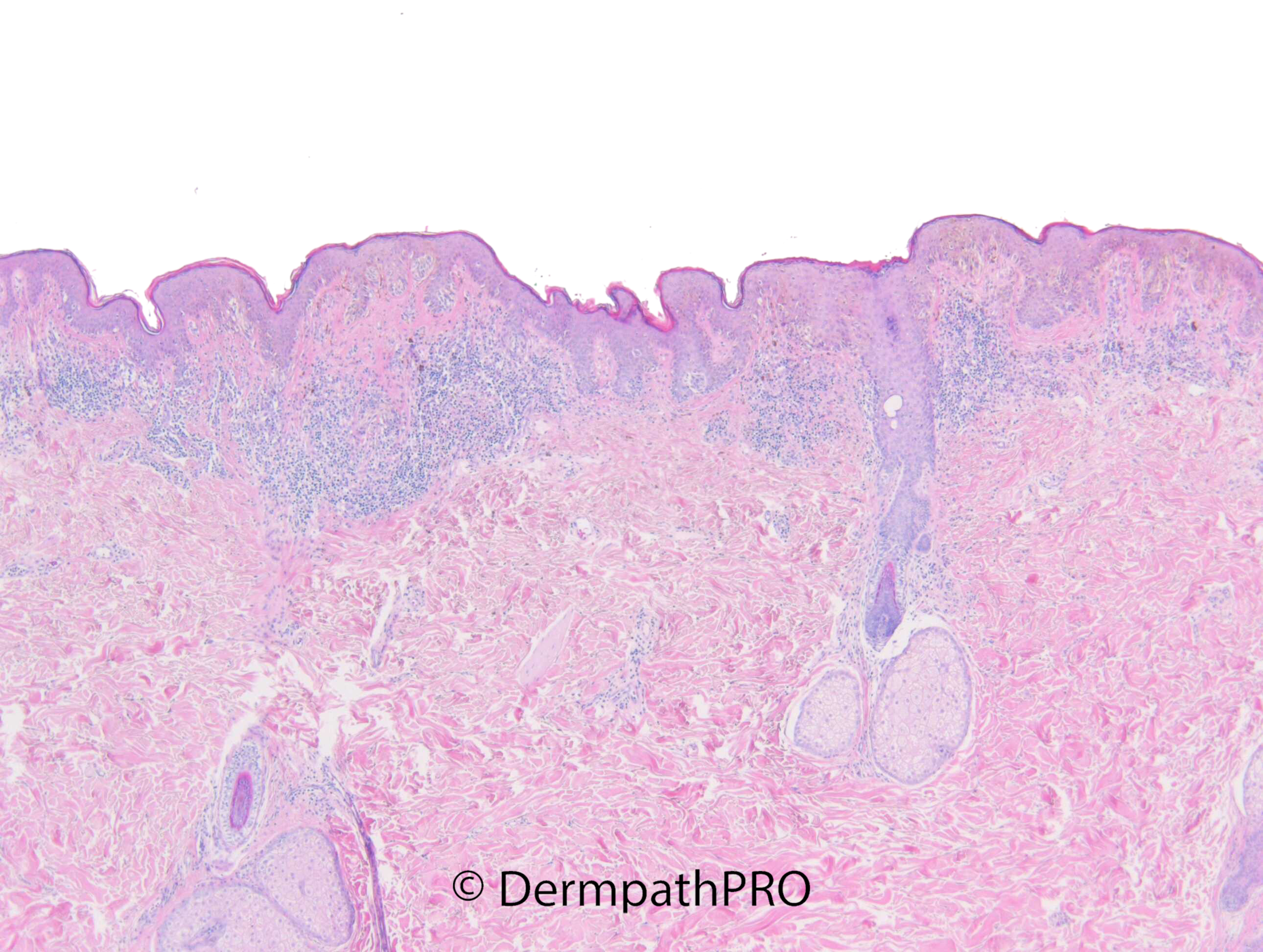
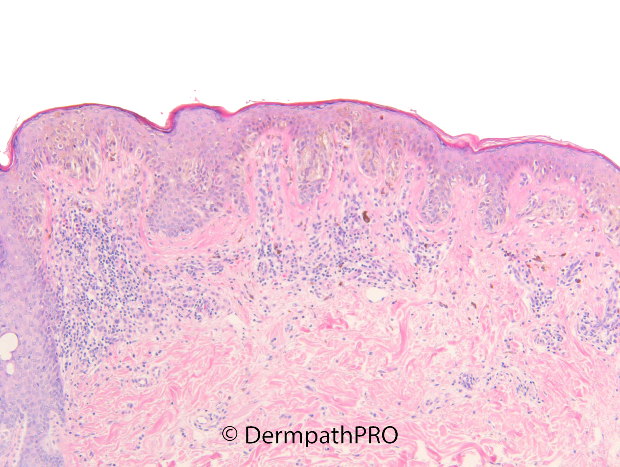
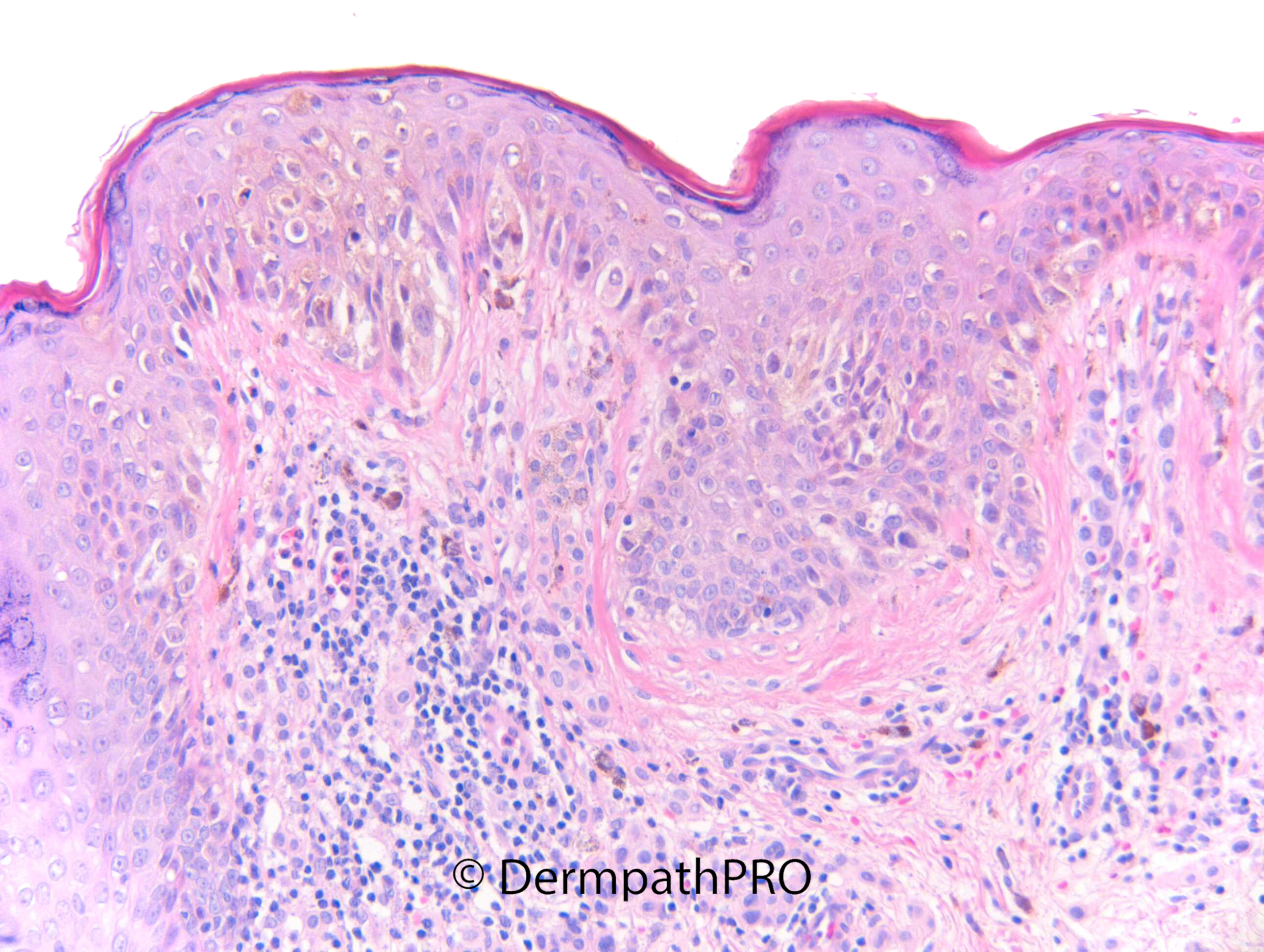
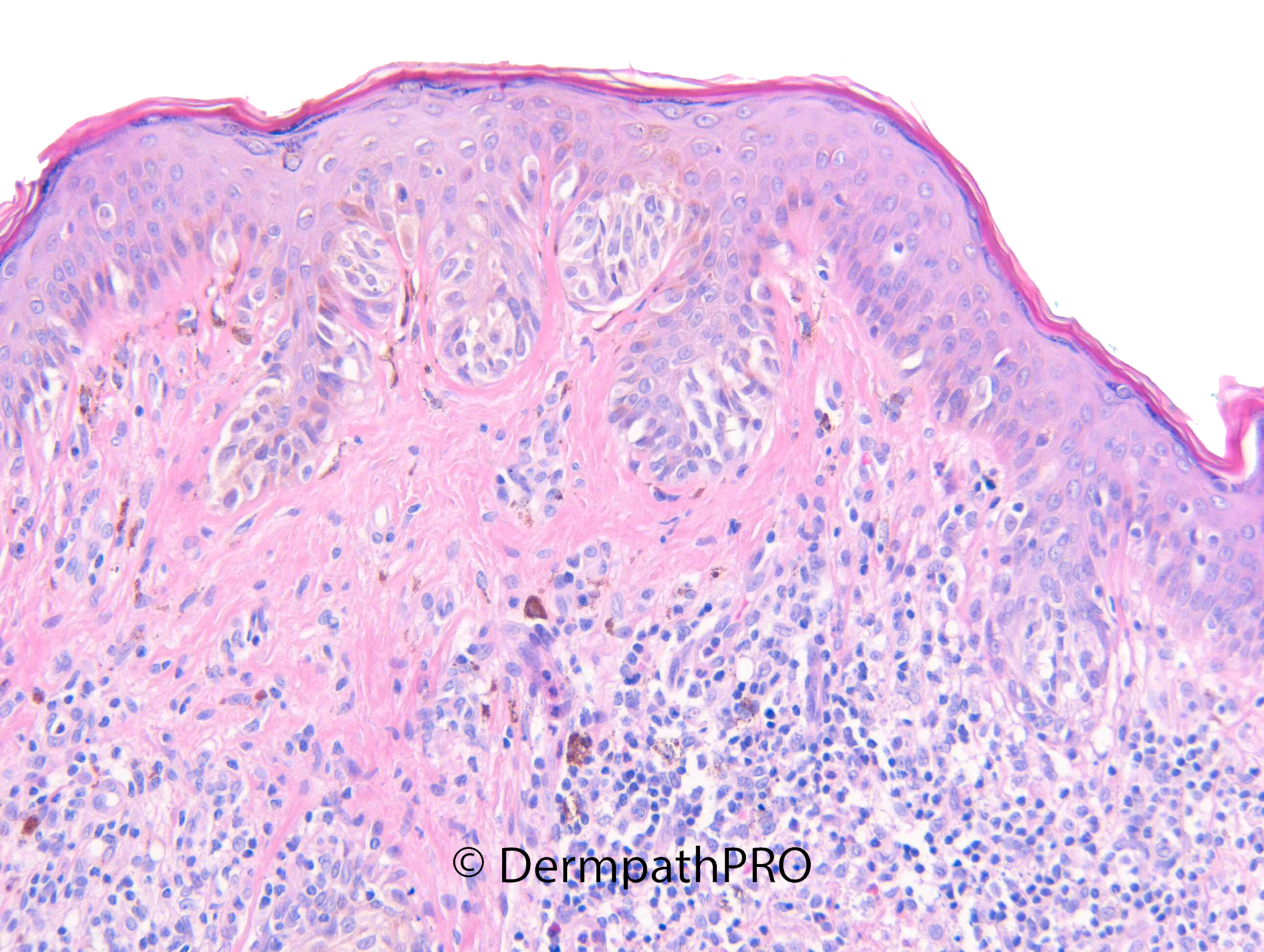
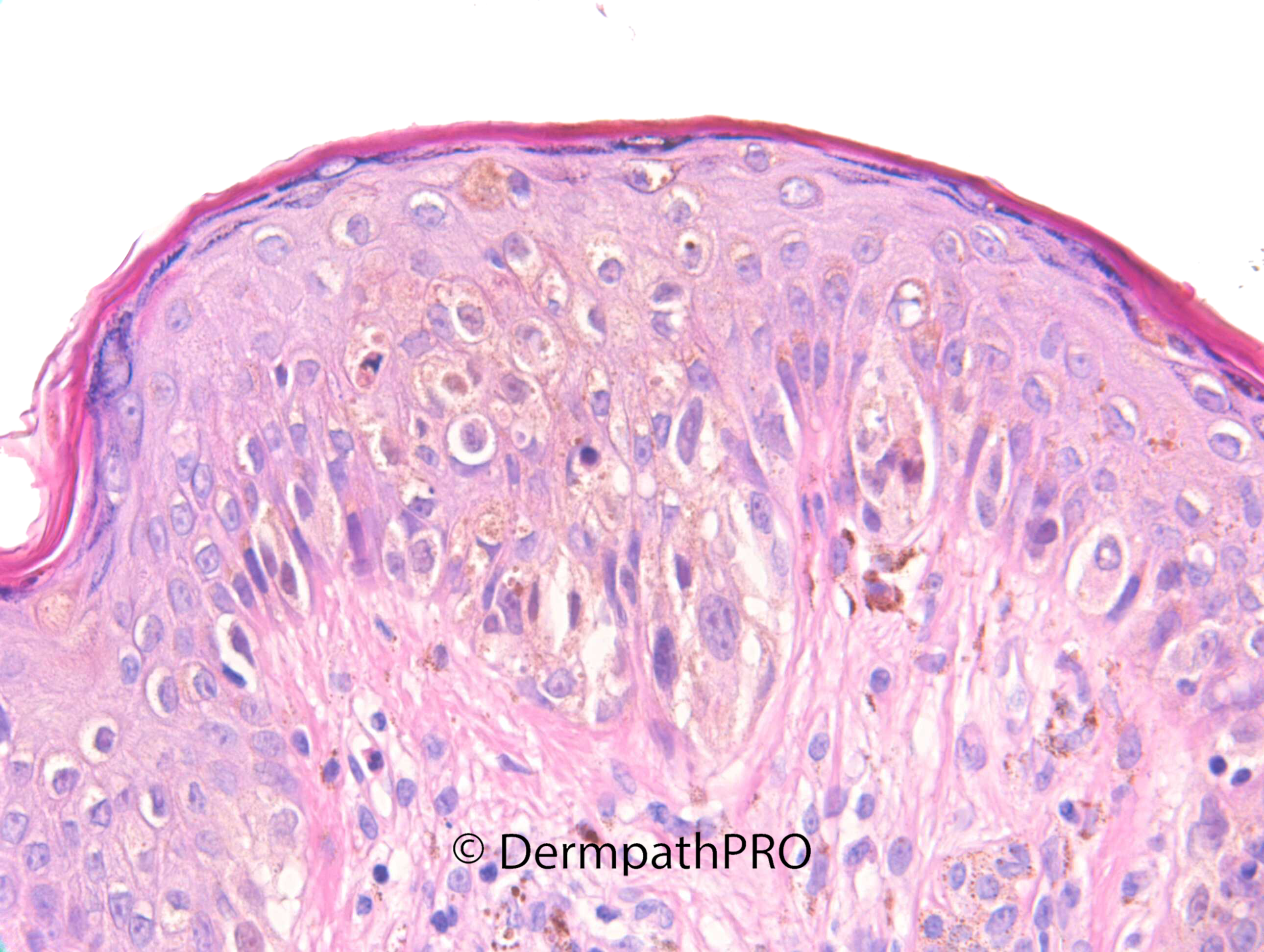
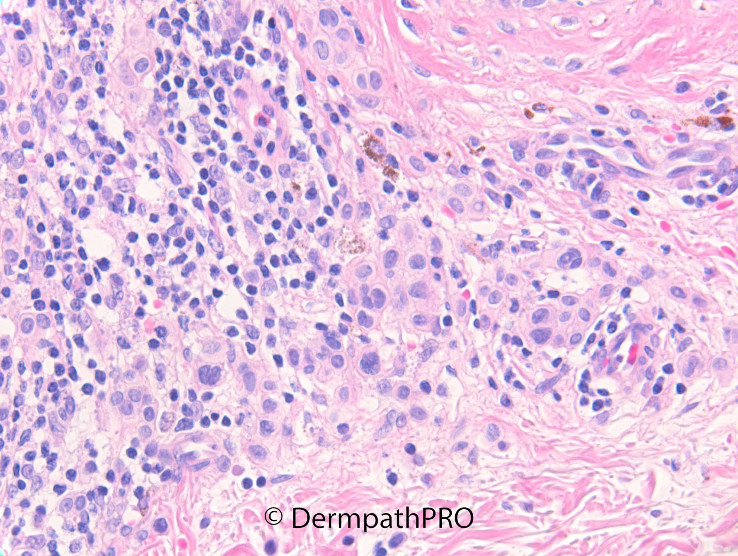
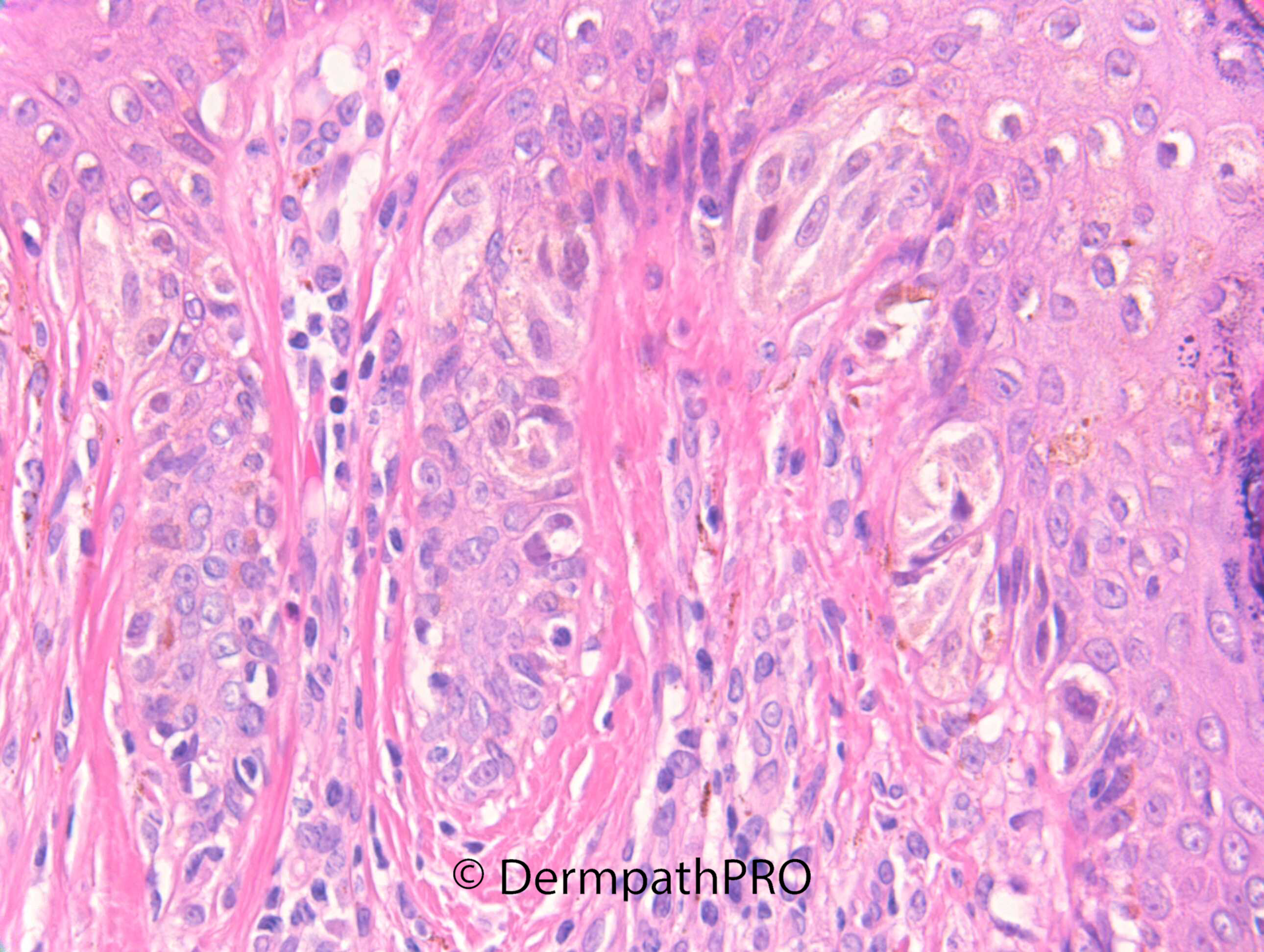
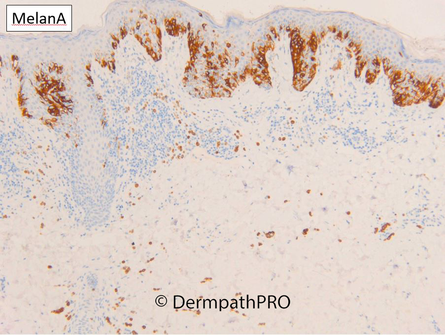
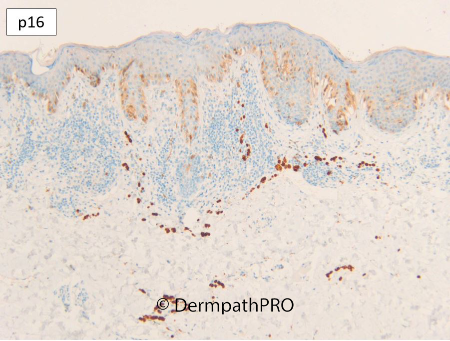
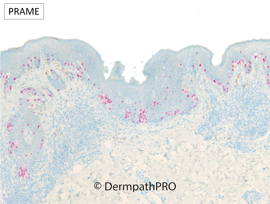
F25. Upper back. Mole. Approx. 1 year, change in size & border over last 6/12. Now irregular outline, 8mmd, prone to sunburn and has burnt in the past. No previous skin problems. ?Dysplastic naevus


  • Like 1
  Report Record

User Feedback


Two distinct population of melanocytes- junctional cells look Spitzoid, dermal cells are banal. Whether this is just a combined nevus or some kind of recurrence phenomenon in the context of previous sunburn... can't decide.


EDIT: IHC updated, loss of p16 and positive PRAME in the intraepidermal component seem to point to MIS and a dermal nevus

Share this comment


Link to comment
share_externally

I don’t know if it make sense calling this dysplastic nevus, but lamellar concentric fibrosis around rete ridge and a mild random atypia fit well the classic dysplastic nevus description. Anyway this is a nevus, for me. Am’I wrong?

Share this comment


Link to comment
share_externally

Meenakshi Batrani

Posted (edited)

On H&E alone was thinking more in terms of dysplastic nevus. Melan A does not show significant pagetoid spread, but there is loss of p16 and PRAME is expressed in epidermal component, although only on one  image difficult to say if it is above threshold of >75% cells. Tough one to choose between dysplastic nevus or early MIS.  

Edited by Meenakshi Batrani

Share this comment


Link to comment
share_externally

Dr. Richard Carr

Posted

It is interesting to see the quite different p16 in the bland dermal naevus and the junctional proliferation. Also agree I'd probably have signed out many lesions such as this as dysplastic naevus especially at this age. The PRAME is positive (4+, >75% of nuclei) so we favoured a melanoma in situ arising in a pre-existing naevus. Of course quite subjective how you approach these lesions but good to ensure complete excision with clear margins. 

Share this comment


Link to comment
share_externally


Join the conversation

You can post now and register later. If you have an account, sign in now to post with your account.

Guest
Add a comment...

×   Pasted as rich text.   Paste as plain text instead

  Only 75 emoji are allowed.

×   Your link has been automatically embedded.   Display as a link instead

×   Your previous content has been restored.   Clear editor

×   You cannot paste images directly. Upload or insert images from URL.

Loading...

×
×
  • Create New...