Jump to content
In this section we have spot diagnoses posted on a daily basis since June 2010, now over 4000! You can review the archived cases and read the suggested diagnoses by users and the final comment by the contributors.
Case are uploaded each week day by 10 am UK time with the correct diagnosis will generally be posted at 8 pm UK time. Why not view the most recent spot diagnosis and proffer a diagnosis?

Case Number : Case 3001 - 6 January 2022 Posted By: Saleem Taibjee

Please read the clinical history and view the images by clicking on them before you proffer your diagnosis.
Submitted Date :
   (0 reviews)

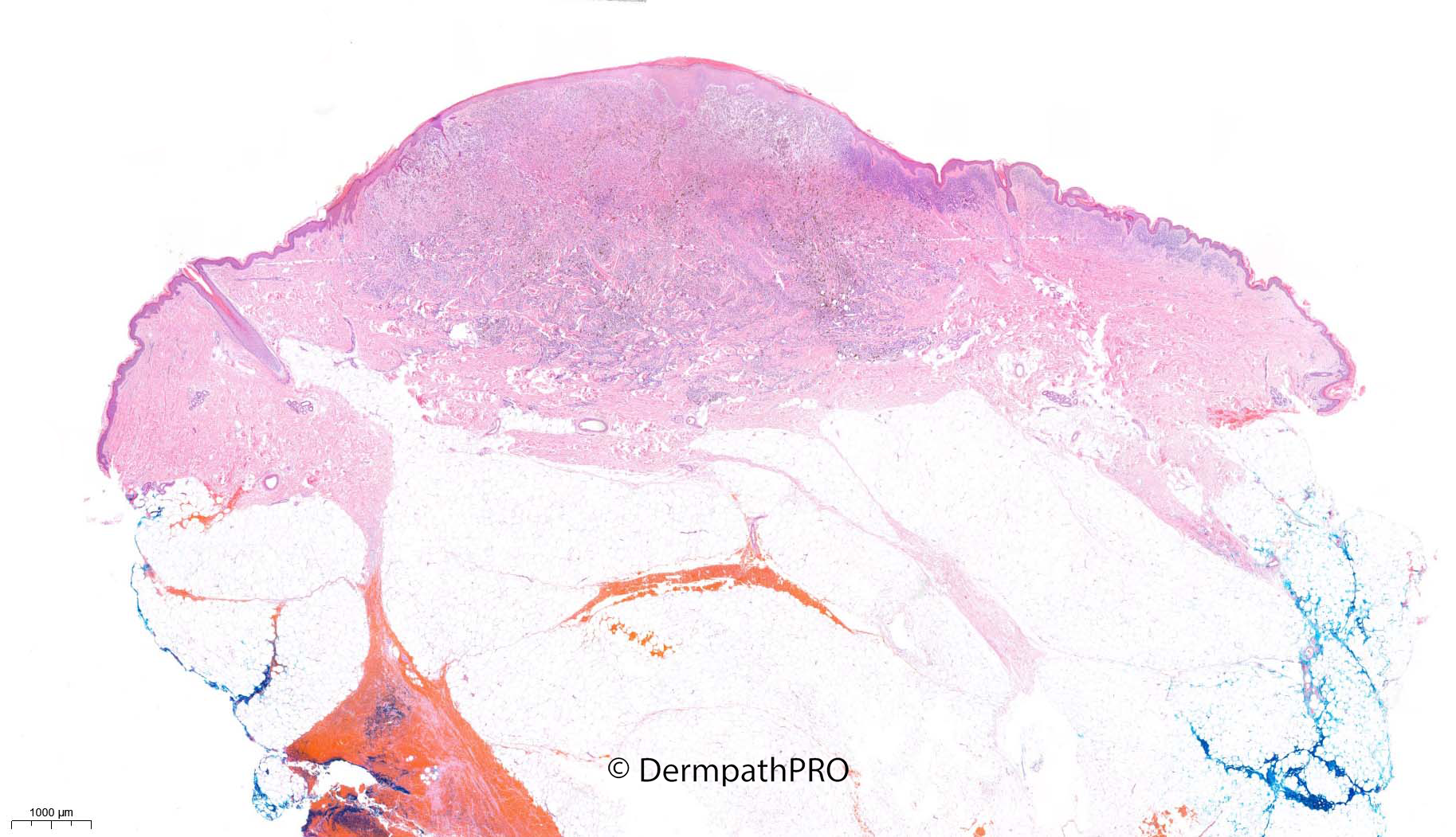
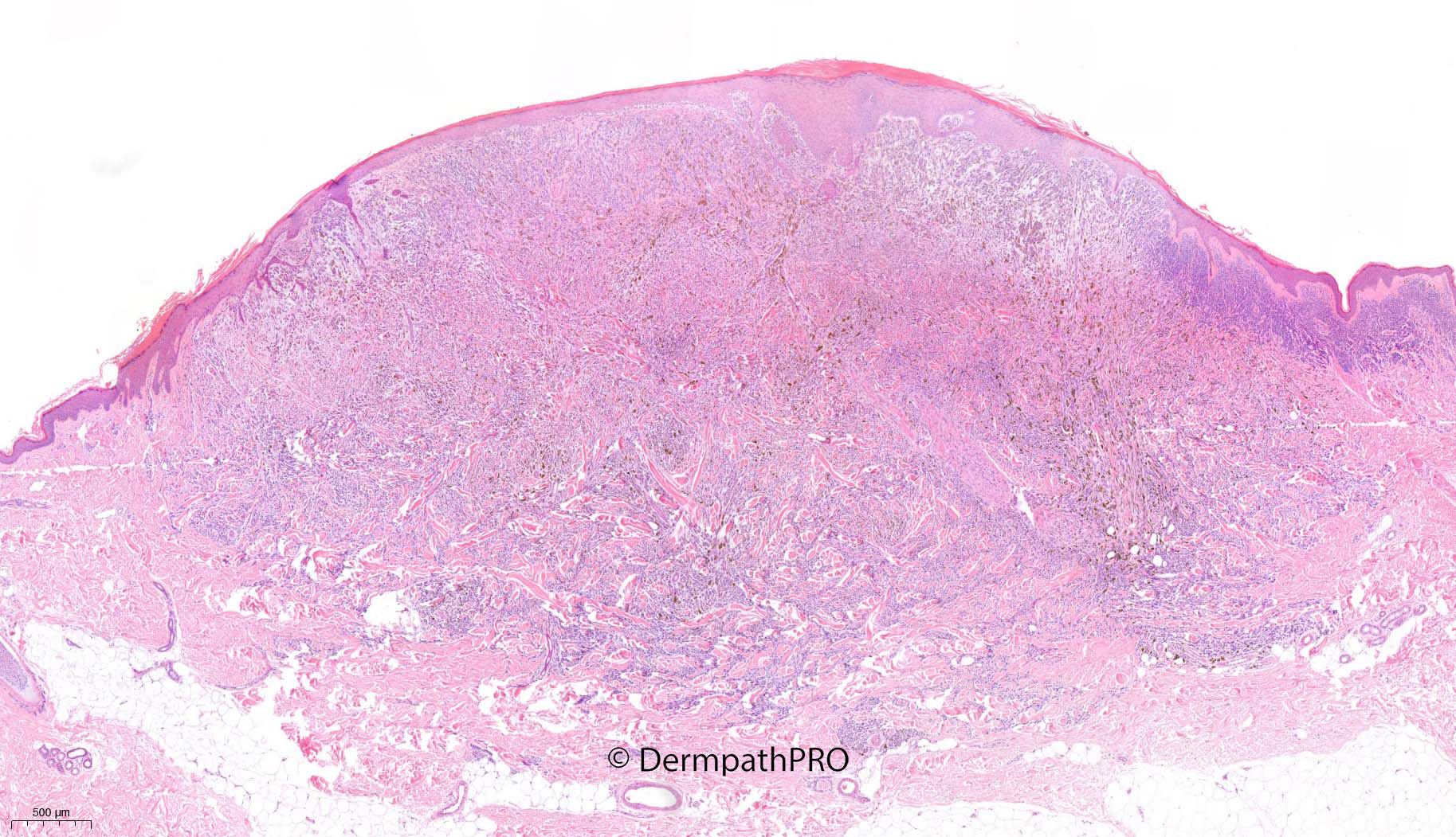
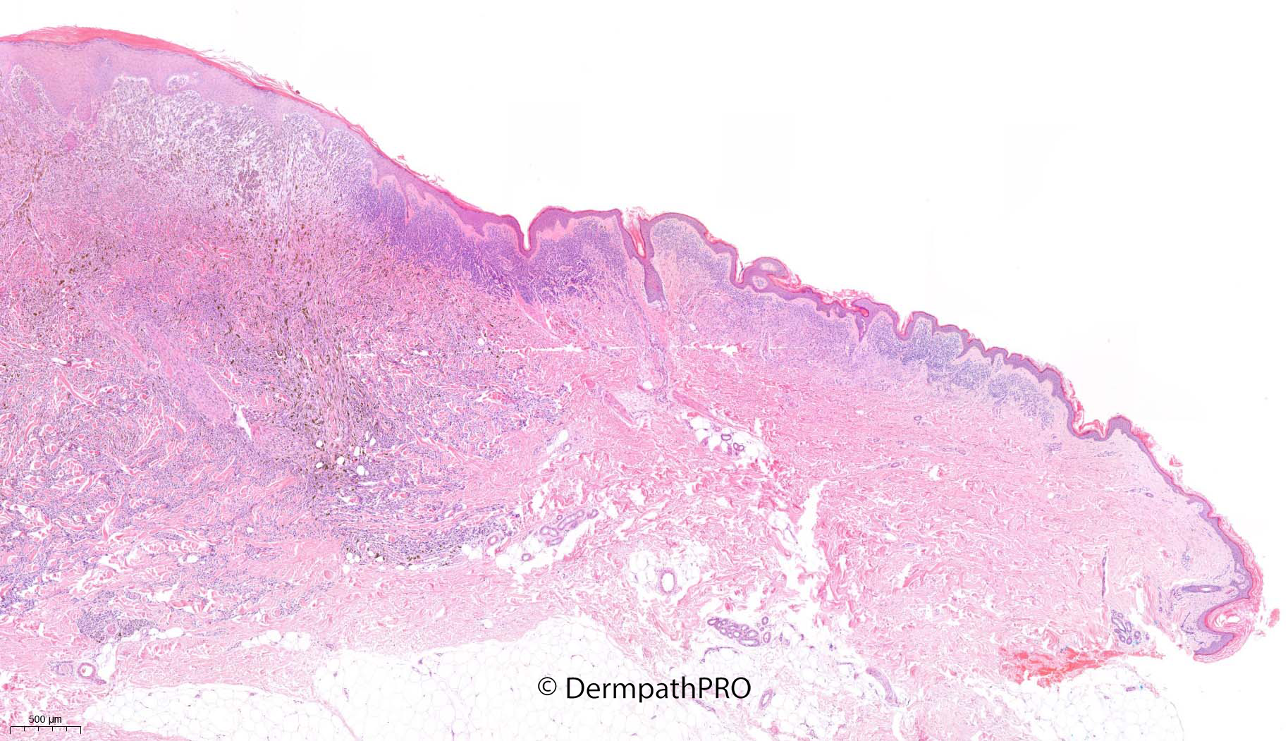
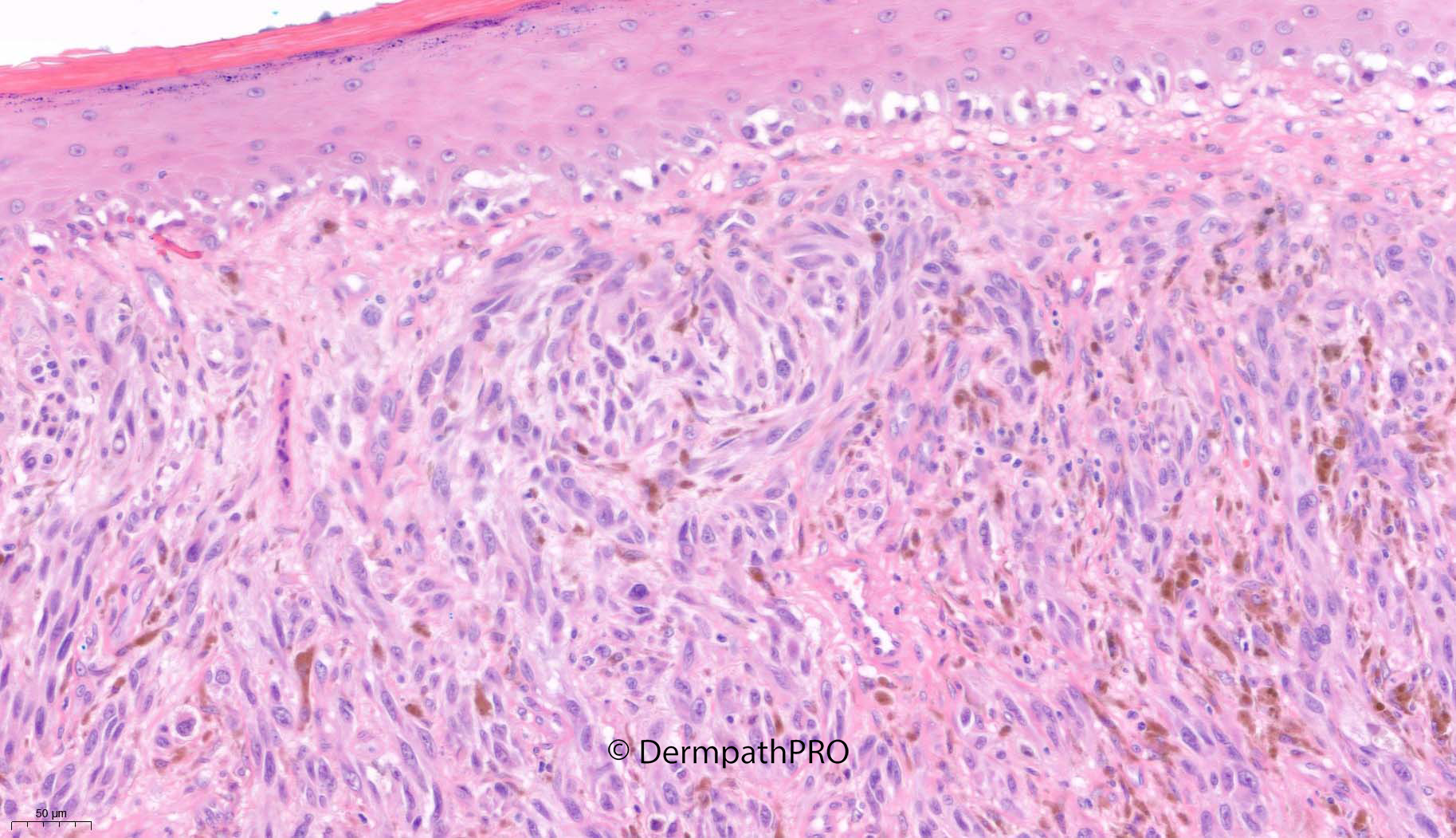
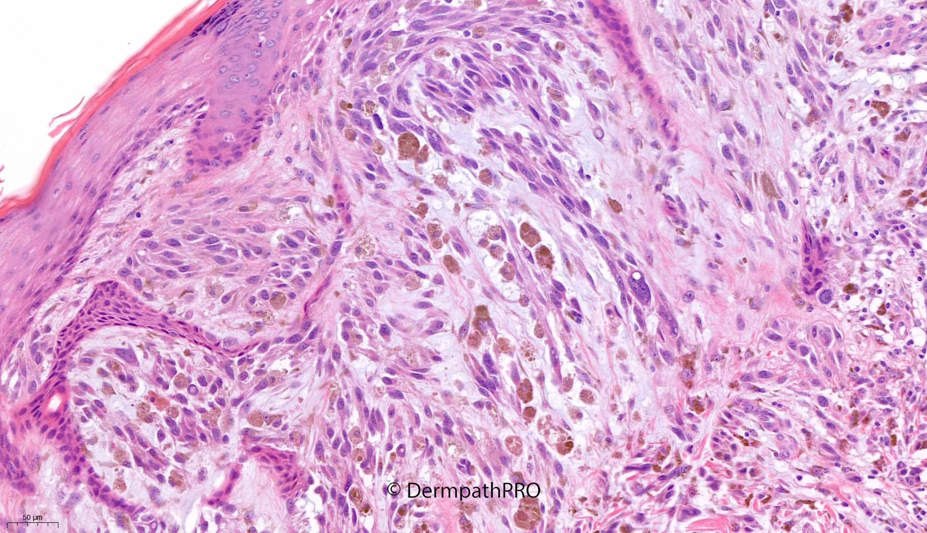
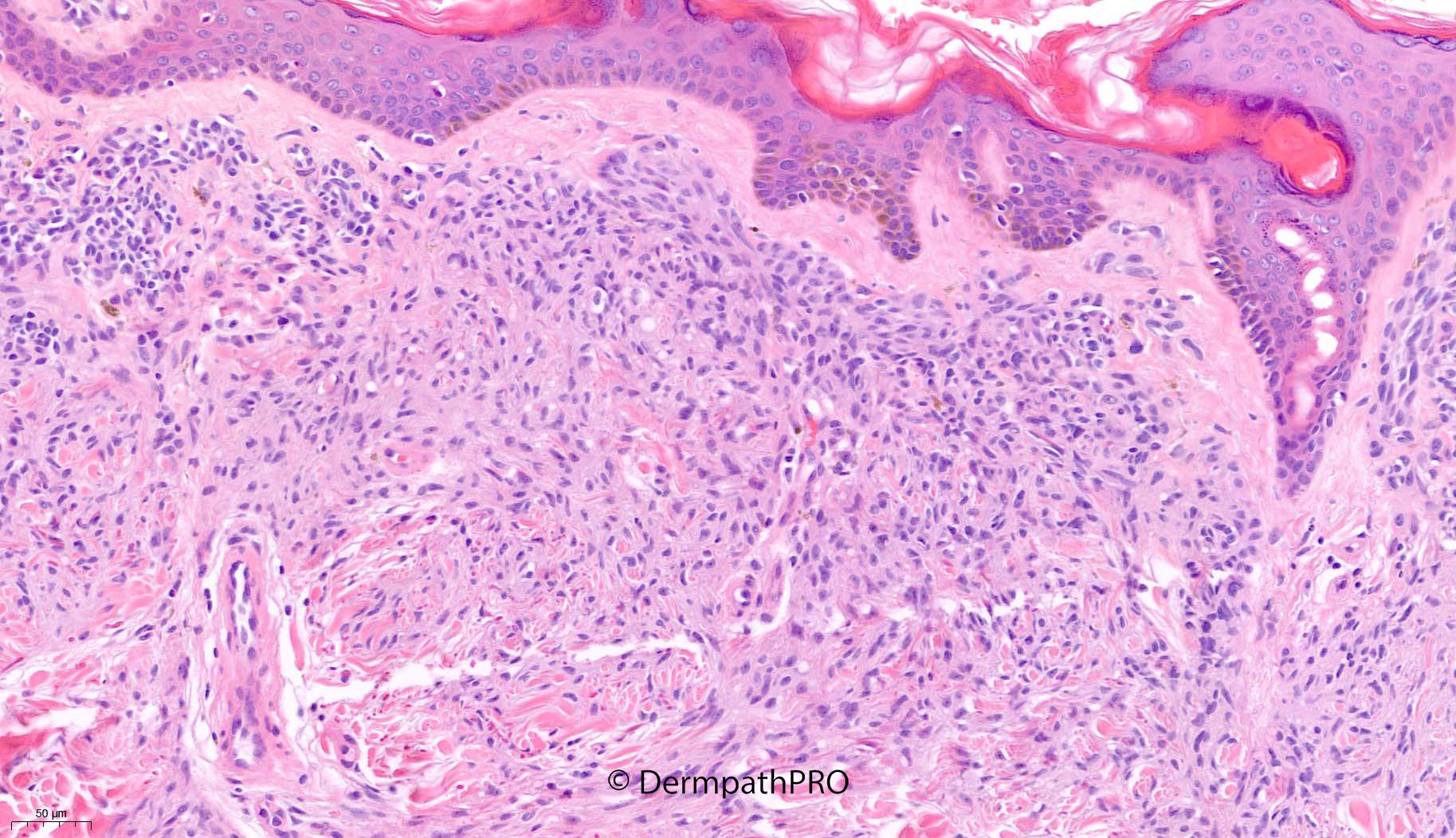
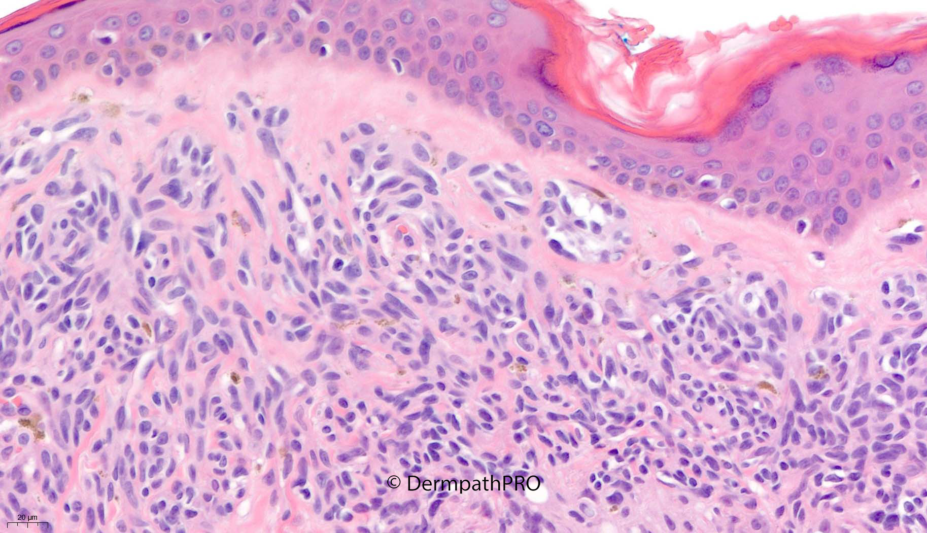
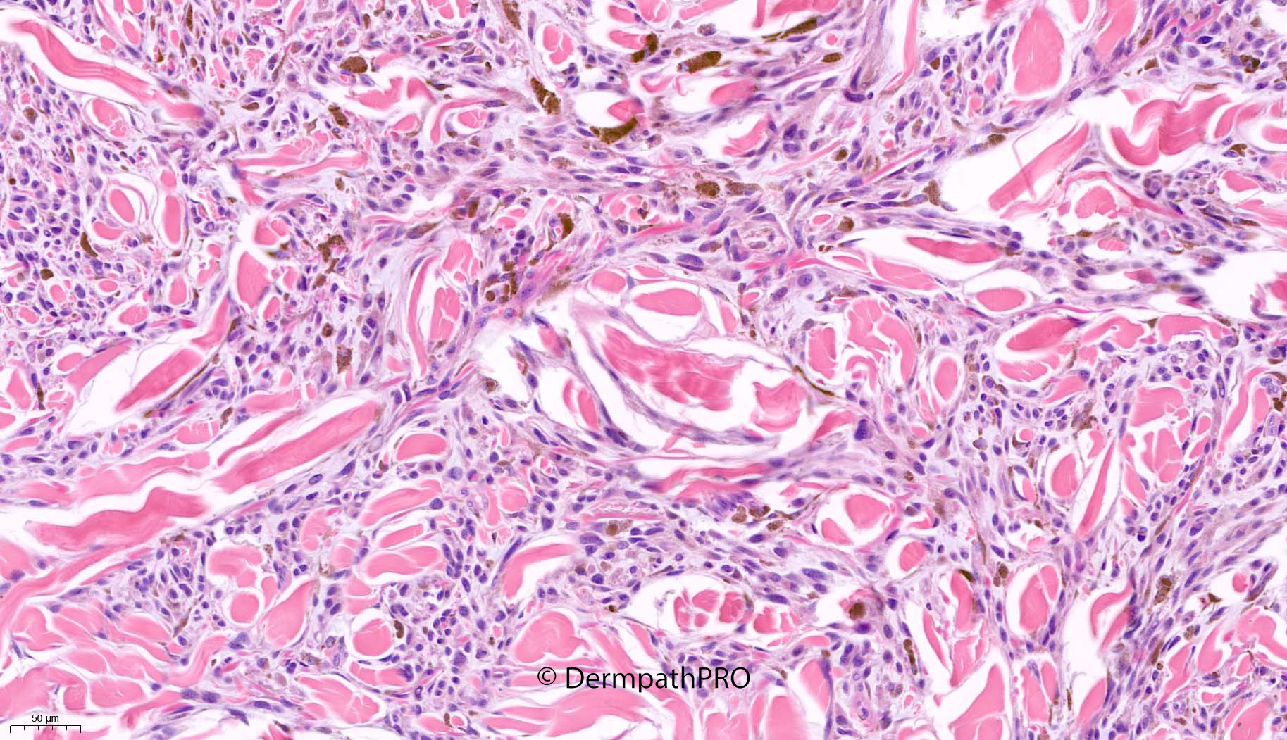
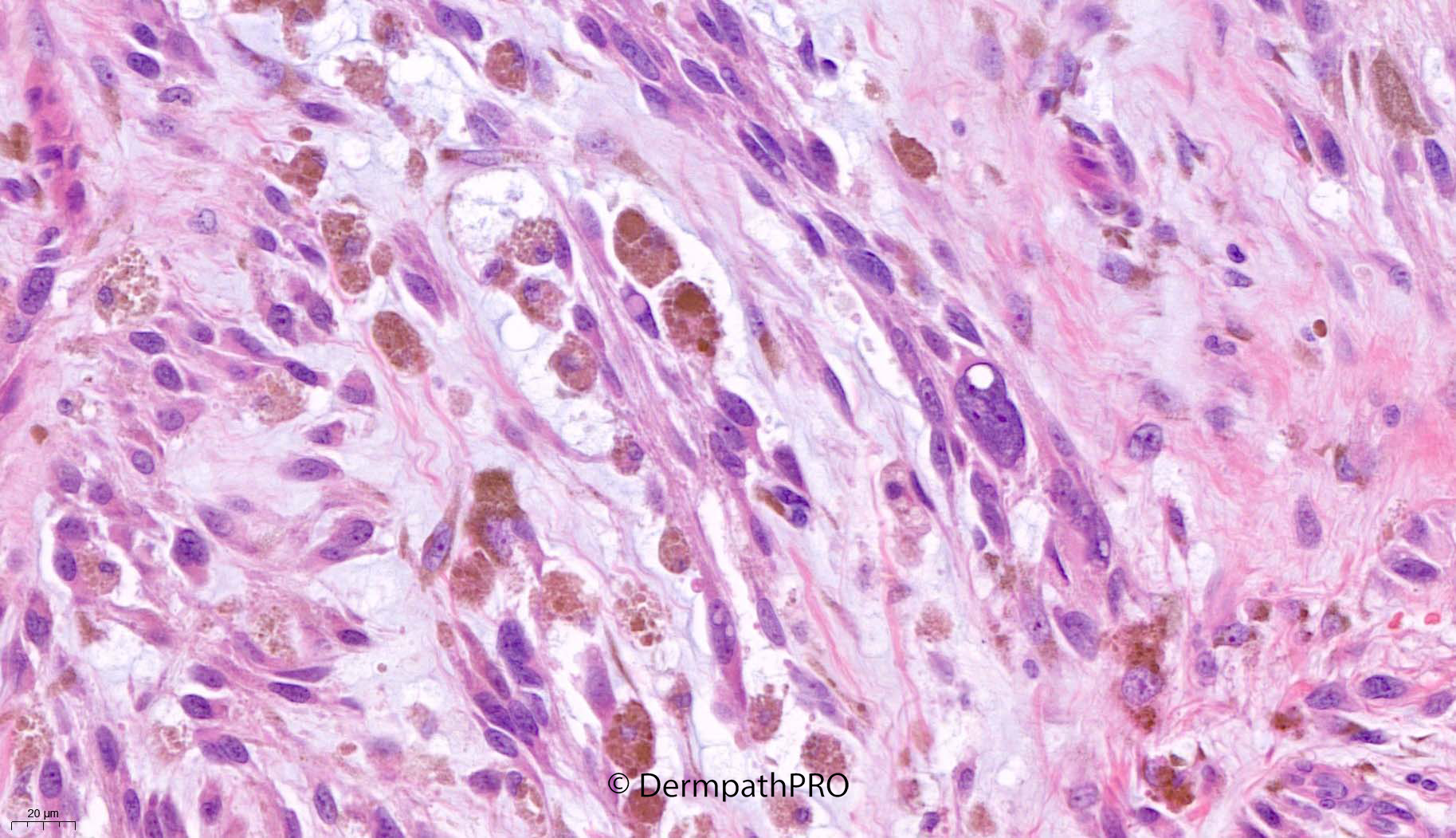
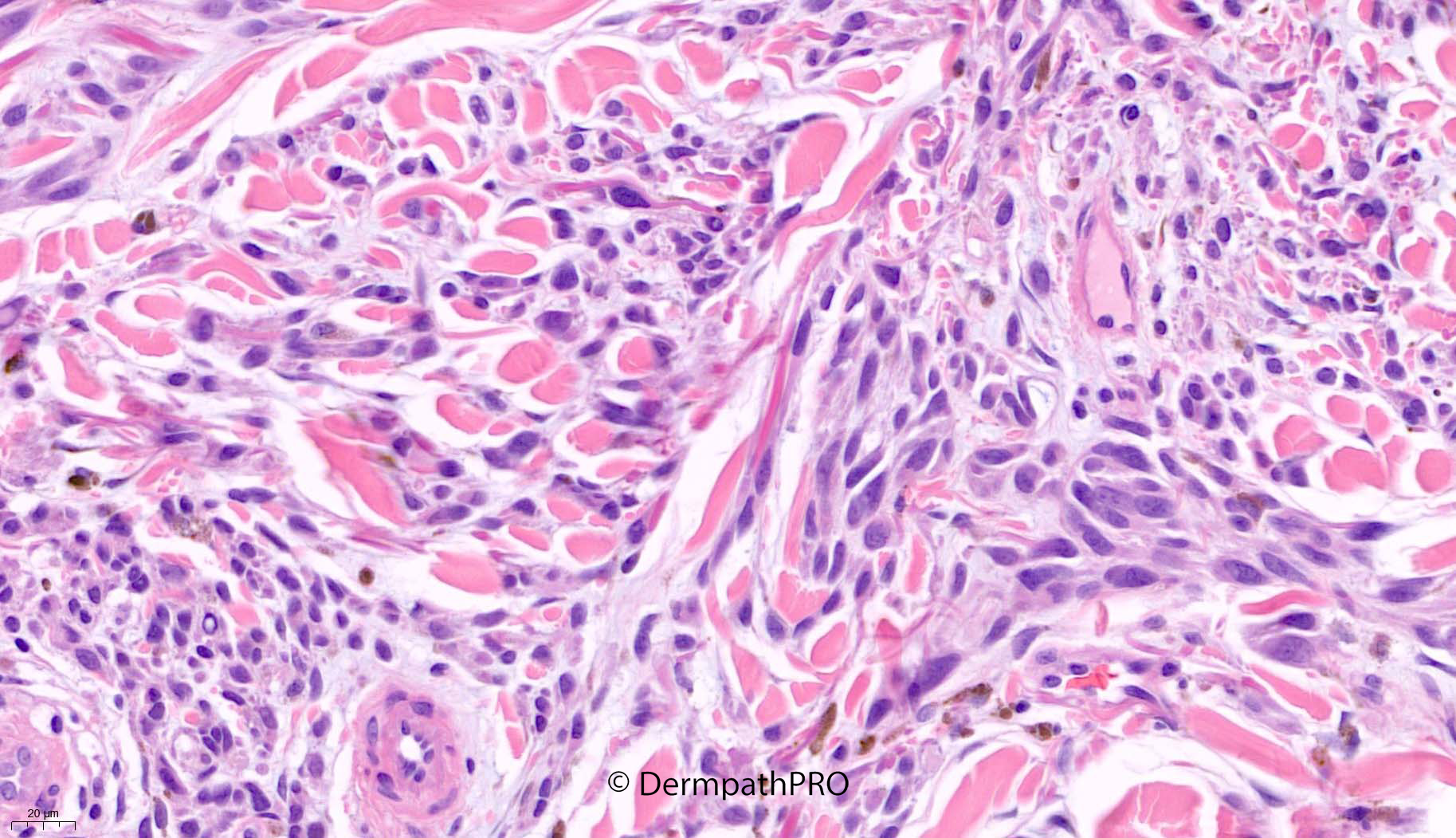
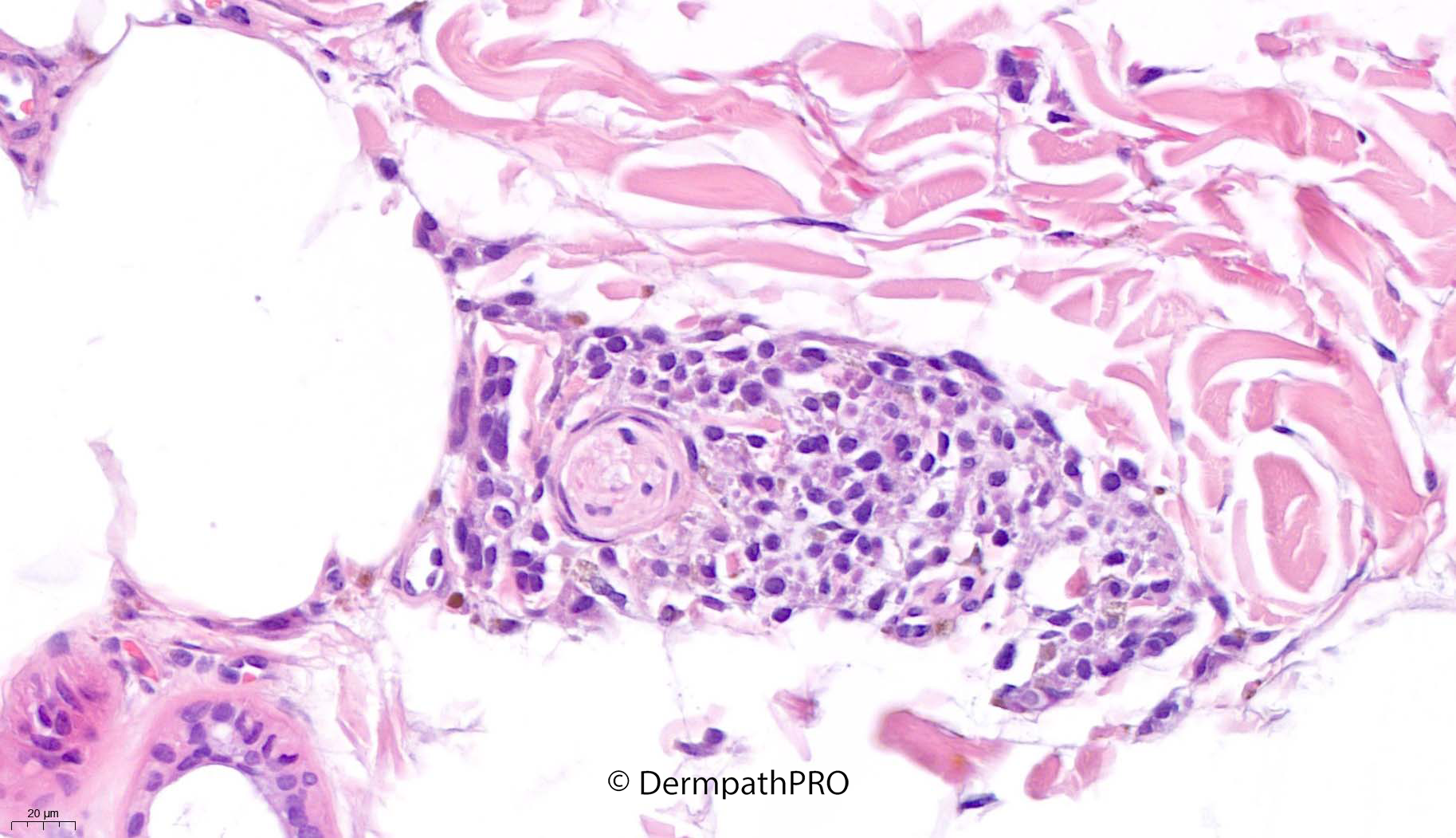
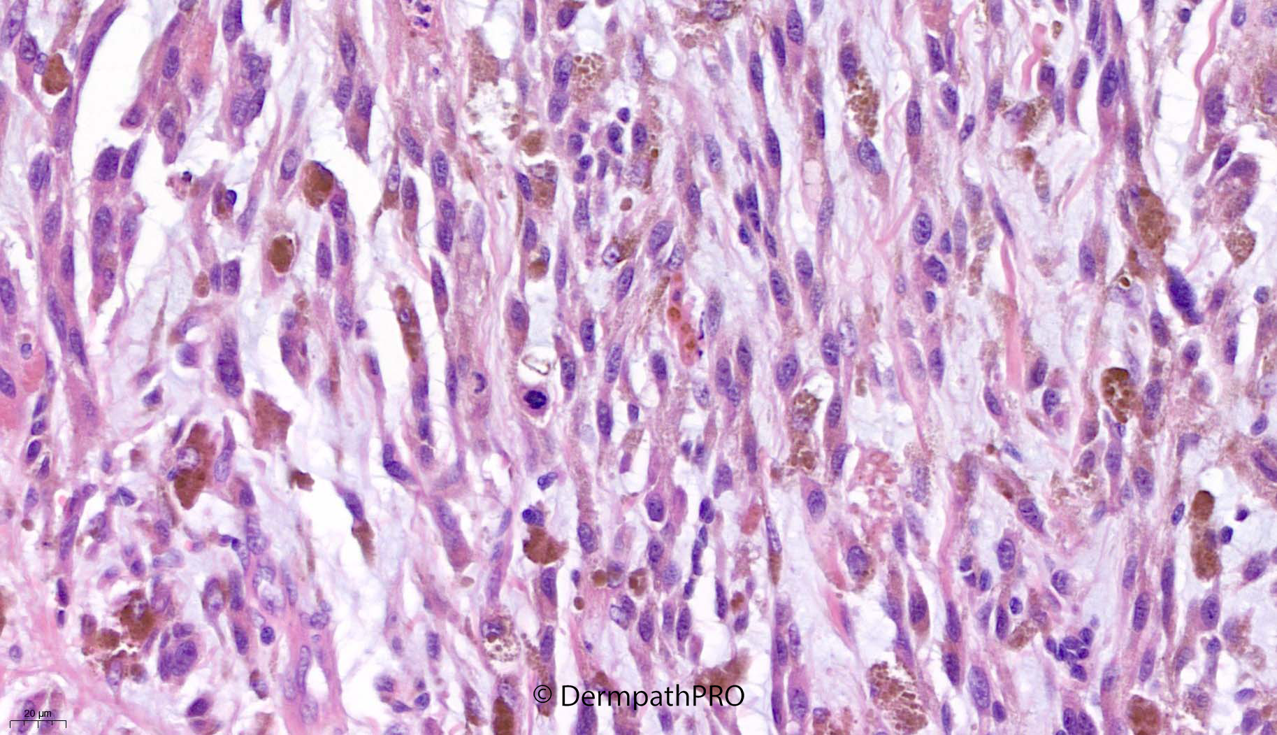
57M conspicuous pigmented lesion inner thigh


  Report Record

User Feedback


Cem Leblebici

Posted

Looks like melanoma arising in nevus. 

Share this comment


Link to comment
share_externally

Meenakshi Batrani

Posted

Challenging case. Overall silhouette makes me suspicious of DPN like borderline lesion. May be some markers and cytogenetic will help to delineate it further into benign or melanoma.

Share this comment


Link to comment
share_externally

On 08/01/2022 at 07:44, Meenakshi Batrani said:

Challenging case. Overall silhouette makes me suspicious of DPN like borderline lesion. May be some markers and cytogenetic will help to delineate it further into benign or melanoma.

Until proven otherwise, as TERT promoter or TP53, this should be a B-DPN ( borderline deep penetration nevus ) for me, based on typical epithelioid/spindle cells( half epithelioid half spindle the same cell ) marginated by parallel rows of melanophages. The lesion is combined with a conventional nevus, laterally juxtaposed. Beta-catenin Cyclin-D1 and LES would be supporting this diagnosis. 

Share this comment


Link to comment
share_externally

Saleem Taibjee

Posted

I'm guessing there is a bit of a lull whilst things pick up again after the New Year, judging from the relatively few responses on this recent very interesting and challenging melanocytic lesion.

But I can see that Meenakshi and Vincenzo had very similar thoughts to myself.

The architecture/silhouette and cytology of this lesion is on the spectrum of deep penetrating melanocytoma (I am now adopting this terminology to reflect the updated WHO classification and more recent medical literature). This particular lesion has a number of worrying features including definite cellular pleomorphism, phenotypic variability, scattered mitoses, focal perineural extension, and also the evidence that this lesion has originated from and now predominates over a much smaller conventional naevus towards one side.

The immunohistochemistry shows nuclear beta-catenin within the DPN-like component (but cytoplasmic only in the naevus), positive staining for PRAME and BRAFV600E (see below).

I deliberated over this case and shared with others, and although we elected not to label as clear-cut melanoma, I certainly emphasised the concerning features and potential metastatic risk (MELTUMP). As it happens, the patient has declined sentinel lymph node biopsy, but has opted for a further wide local excision given the narrow margins, and follow-up.

At the recent Anglo-French-Belgian Dermatopathology meeting there was some discussion about atypical DPN. There is a possible signal that those cases underpinned by an underlying NRAS mutation rather than BRAF mutation may be more likely to misbehave, but I still remain nervous about the current case.

BW
Saleem

DigitalSlide_B1M_4S_1_20.0x beta-catenin b labelled.jpg

DigitalSlide_B1M_6S_1_20.0x PRAME labelled.jpg

75203_10.0x BRAFV600E labelled.jpg

Share this comment


Link to comment
share_externally


Join the conversation

You can post now and register later. If you have an account, sign in now to post with your account.

Guest
Add a comment...

×   Pasted as rich text.   Paste as plain text instead

  Only 75 emoji are allowed.

×   Your link has been automatically embedded.   Display as a link instead

×   Your previous content has been restored.   Clear editor

×   You cannot paste images directly. Upload or insert images from URL.

Loading...

×
×
  • Create New...