Jump to content
In this section we have spot diagnoses posted on a daily basis since June 2010, now over 4000! You can review the archived cases and read the suggested diagnoses by users and the final comment by the contributors.
Case are uploaded each week day by 10 am UK time with the correct diagnosis will generally be posted at 8 pm UK time. Why not view the most recent spot diagnosis and proffer a diagnosis?

Case Number : Case 3007 - 14 January 2022 Posted By: Dr. Richard Carr

Please read the clinical history and view the images by clicking on them before you proffer your diagnosis.
Submitted Date :
   (0 reviews)

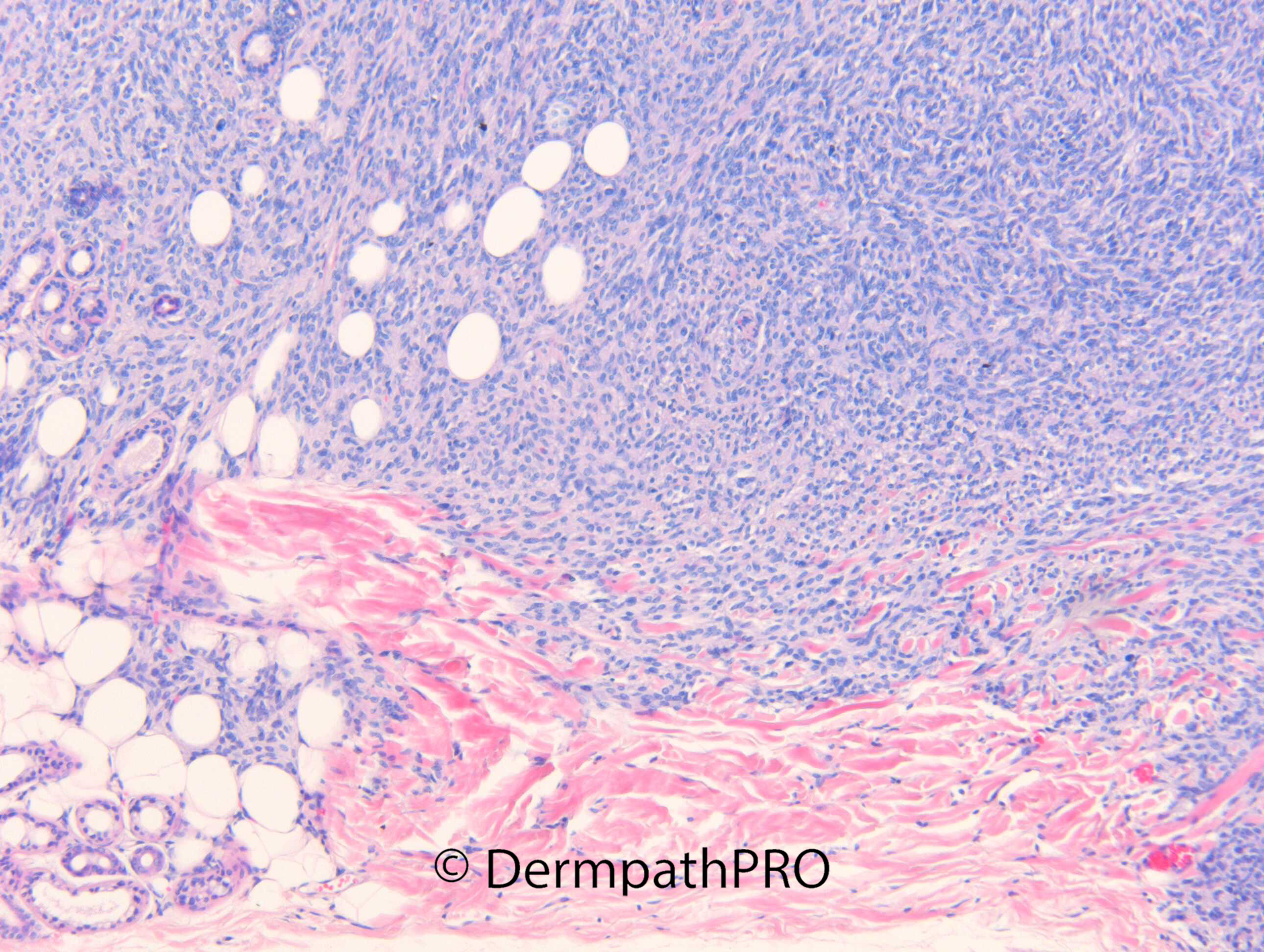
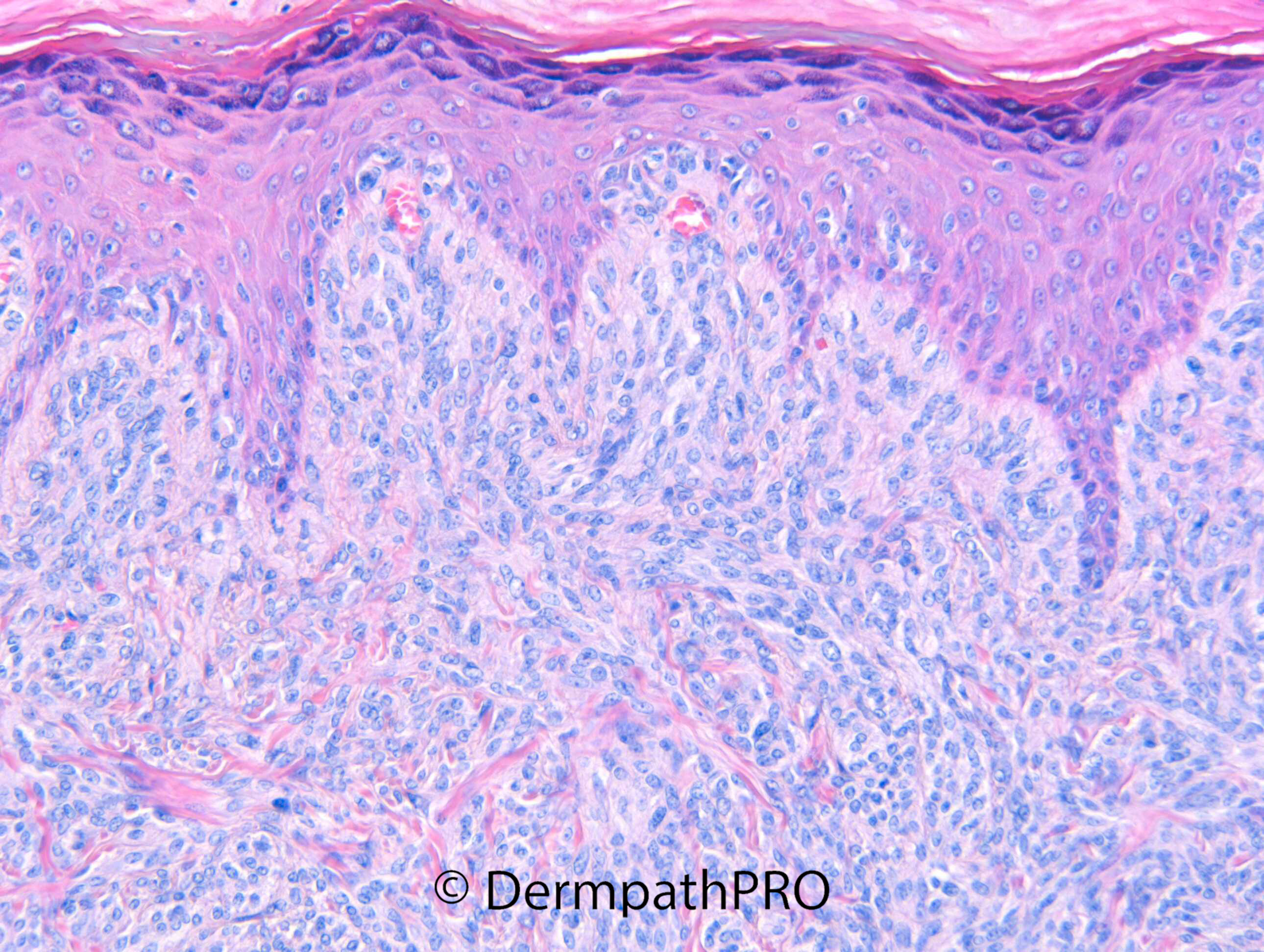
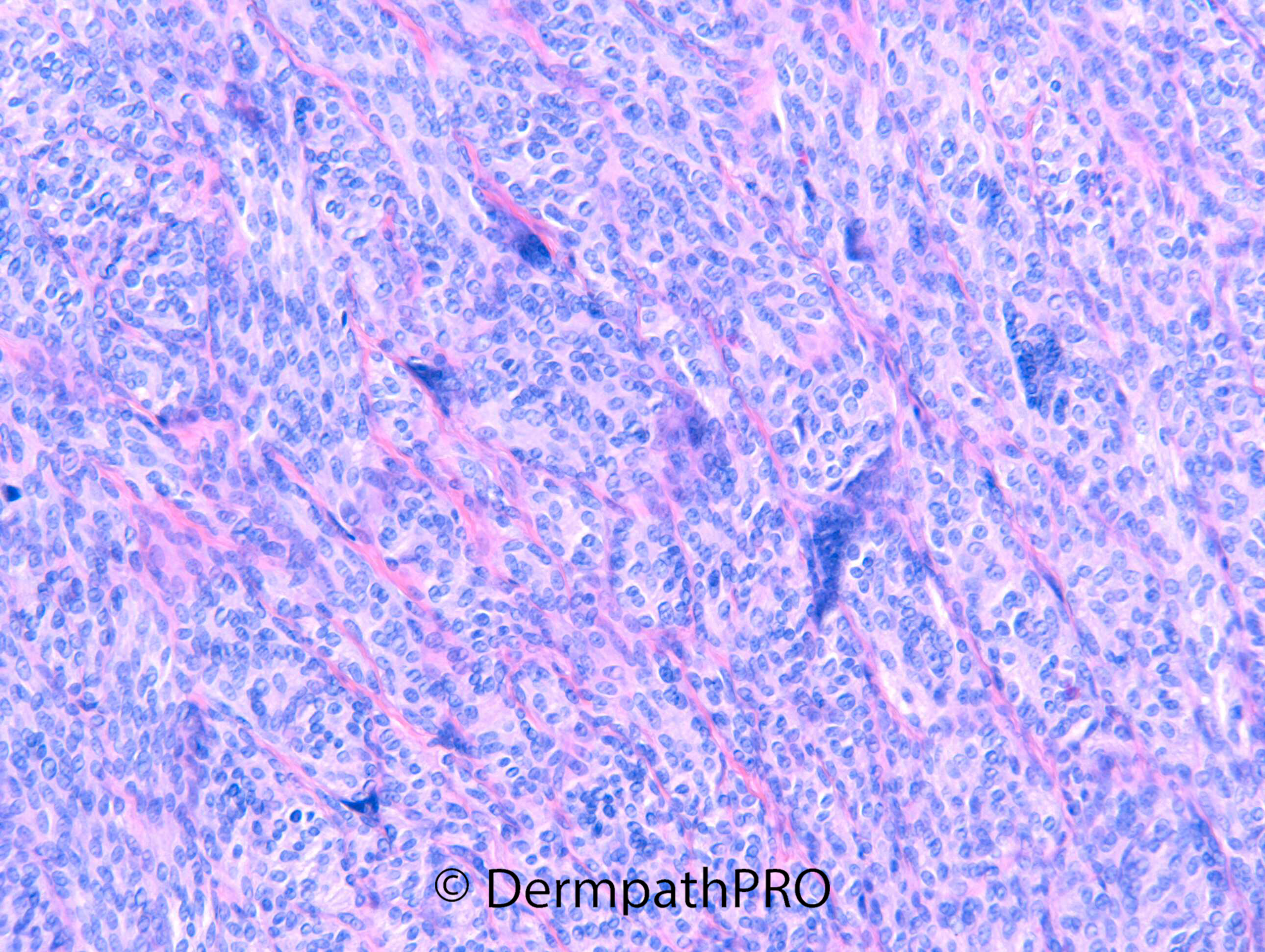
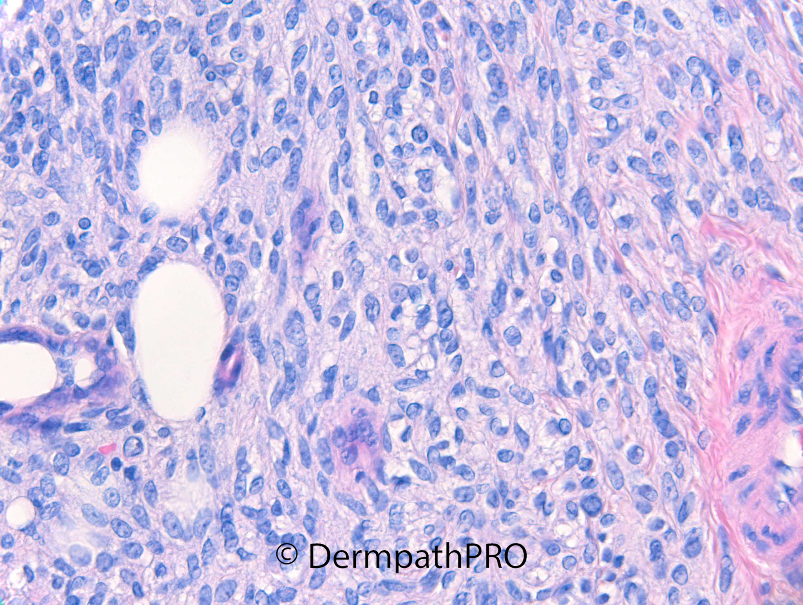
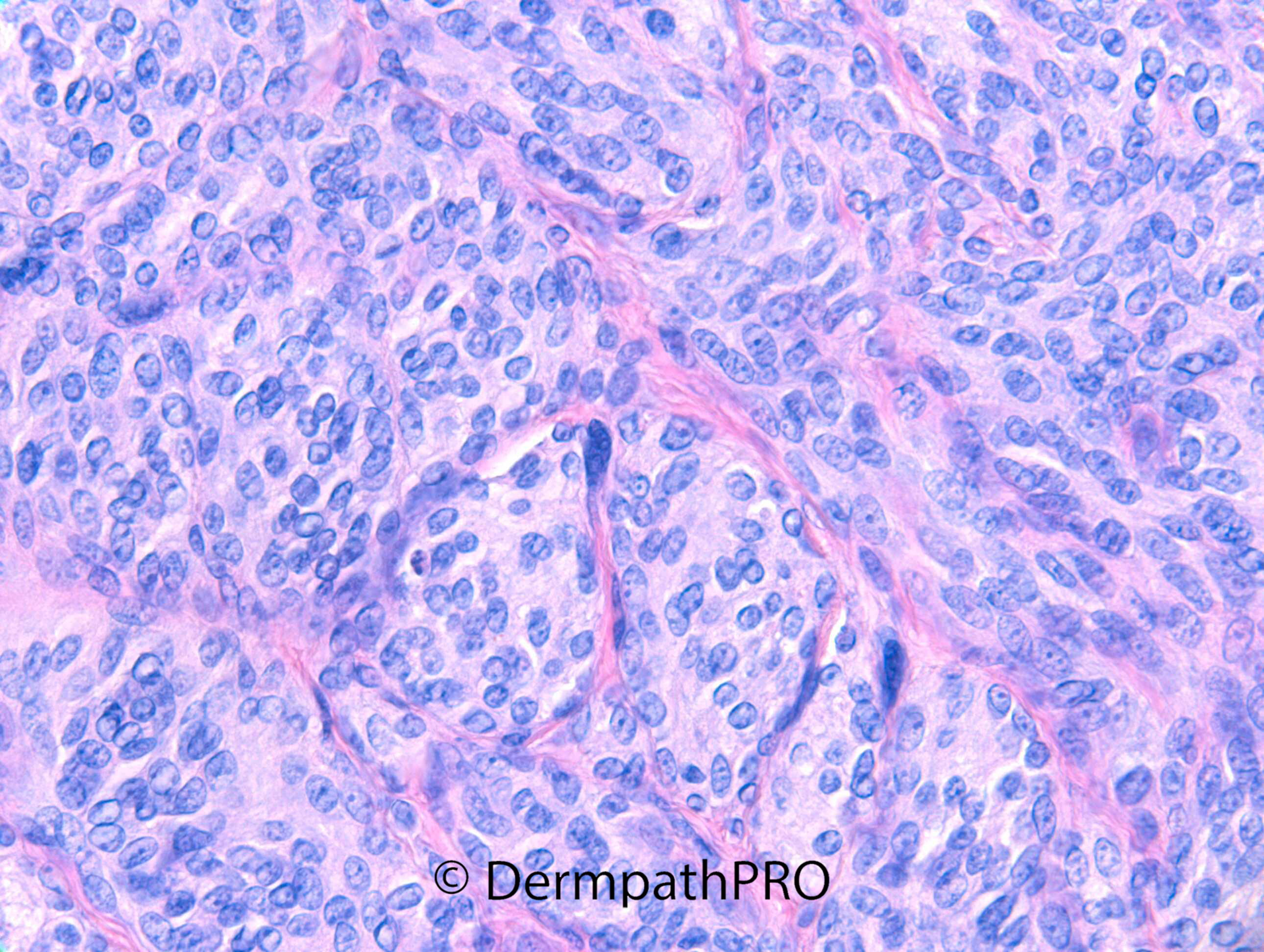
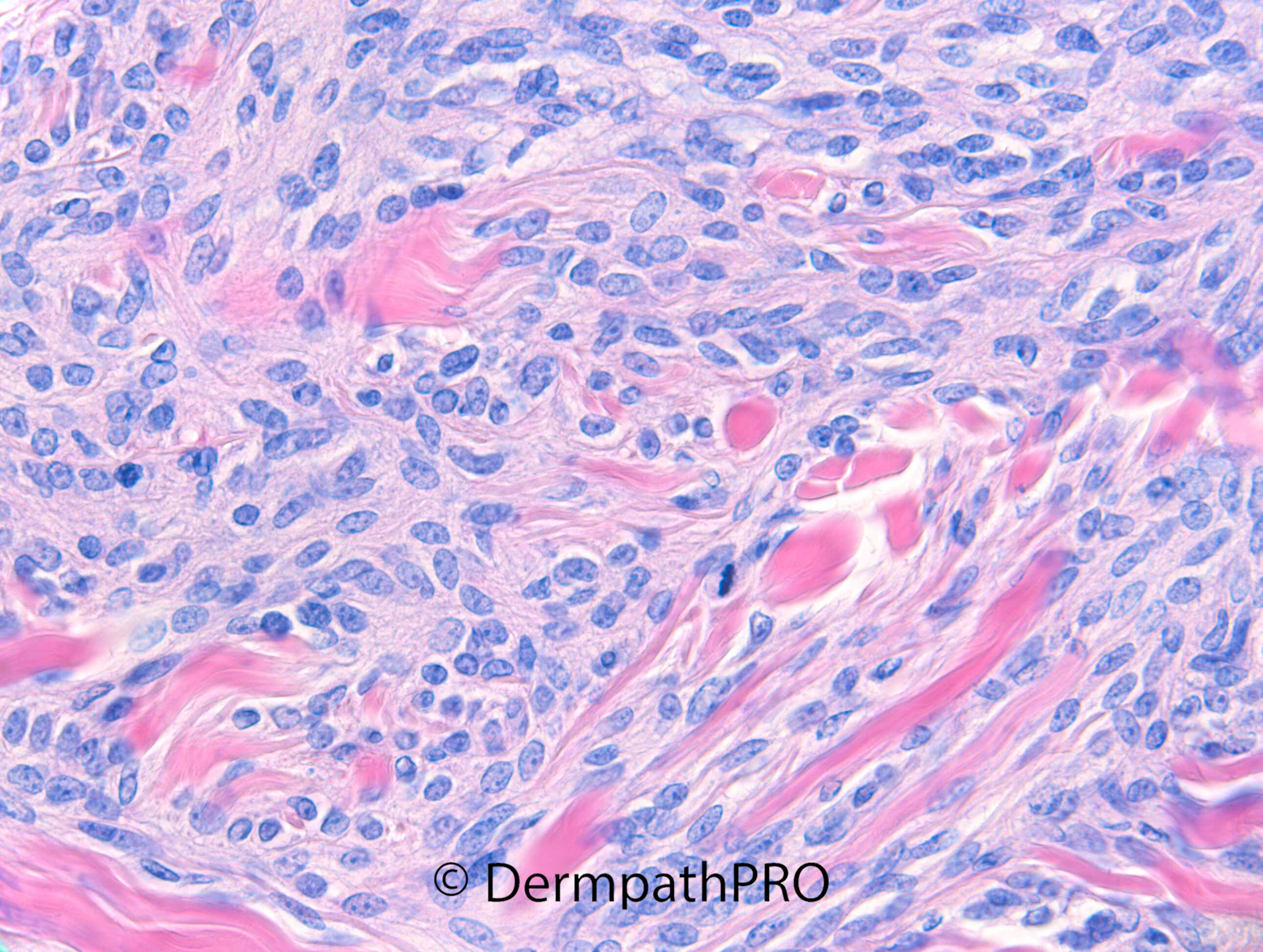
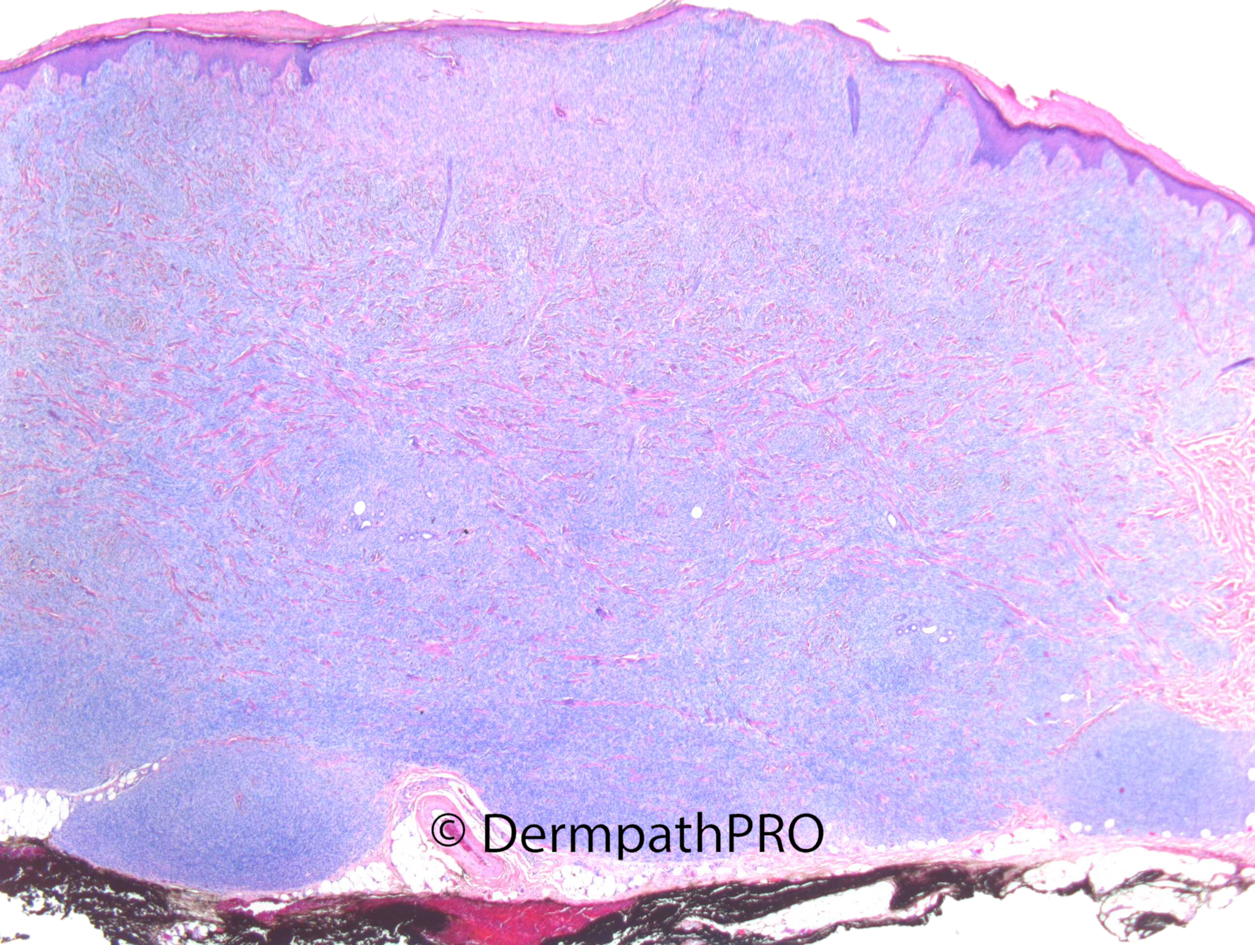
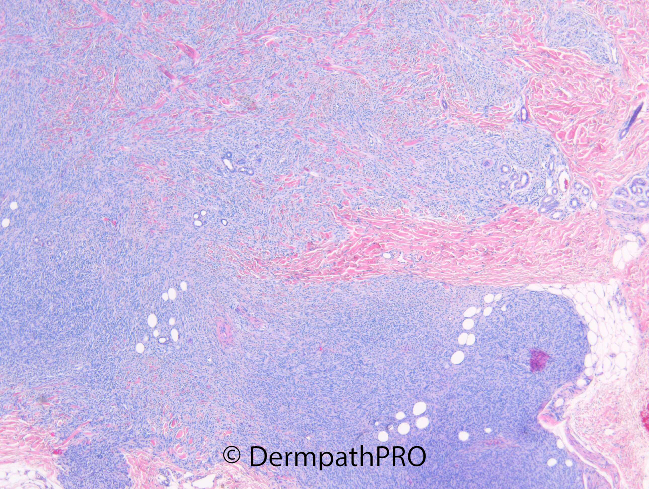
F15. Back of hand. Yellow firm dermal lump. 3-4 years. Growing for 2 years. Catching it on pocket and became ulcerated.

  Report Record

User Feedback


Dermal based tumor (not common for my hypothesis) but showing extension to subcutaneous fat, fat entrapment, adnexial sparing and the presence of osteoclast-like giant cells in the back of hand of a young female makes me think about Plexiform fibrohistiocytic tumor.

Share this comment


Link to comment
share_externally

A tricky case of Clear Cell Sarcoma of soft tissue?

Share this comment


Link to comment
share_externally

Richard Logan

Posted

I didn't know what this was.  However, I like Igor's suggestion of a plexiform fibrohistiocytic tumour.  There are only two previous examples of this entity on the website (Case 508 May 2012; On-line course case 82 , 21.2.12), which must reflect its rarity.

Share this comment


Link to comment
share_externally

Dr. Richard Carr

Posted

there should be some IHC I'll ask the dermpathpro team to post. I'm currently on hols in Barbados.

Share this comment


Link to comment
share_externally

Krishnakumar subramanian

Posted

dermal tumor some nesting pattern/plexiform pattern is seen

CD68 would be useful

Share this comment


Link to comment
share_externally

Dr. Richard Carr

Posted

Okay the IHC showed diffuse strong MelanA. Other markers including S100, epithelial, CD34 etc were negative. 

Share this comment


Link to comment
share_externally

Hmm. Unusual cellular blue nevus? Clear cell sarcoma?  What about other melanocyte markers like SOX-10, HMB-45, MiTF?

Share this comment


Link to comment
share_externally

Clear cell sarcoma of soft tissue. MelanA positivity is enough for me ( sic stantibus “morphologic” rebus ). 

Share this comment


Link to comment
share_externally

Meenakshi Batrani

Posted

11 hours ago, vincenzo said:

Clear cell sarcoma of soft tissue. MelanA positivity is enough for me ( sic stantibus “morphologic” rebus ). 

Agree with Clear cell carcinoma. There appears to be some junctional proliferation, although, very rarely, junctional component has been described in dermal clear cell sarcoma. 

Share this comment


Link to comment
share_externally

Krishnakumar subramanian

Posted

agree for clear cell sarcoma of soft tissue

Share this comment


Link to comment
share_externally

Dr. Richard Carr

Posted (edited)

While I considered clear cell sarcoma in the differential the signature diffuse strong melanA throughout and negative S100 is typical for a cellular blue naevus. The location and age are both typical for blue naevus but would also be commensurate with CCS. The bulbous downgrowths and preservation of adnexal structures together with the nested pattern and the giant cells are typical of cellular blue naevus although admittedly this is apigmented which is slightly unusual. I did not send the lesion for FISH but given the responses here will probably arrange this for completion as it's a recent case. Epidermal component has been recorded in CCS but also from personal communication in blue naevi. I'd have expected CCS to show focal melanA and S100 rather than the striking diffuse strong MelanA and S100 negative phenotype that in my experience is highly typical of blue naevi (>40 cases I've documented in my own unpublished spreadsheet and many more undocumented). 

Edited by Dr. Richard Carr

Share this comment


Link to comment
share_externally

You are right, Richard; and this is an amazing case. Thanks a lot. 

Share this comment


Link to comment
share_externally


Join the conversation

You can post now and register later. If you have an account, sign in now to post with your account.

Guest
Add a comment...

×   Pasted as rich text.   Paste as plain text instead

  Only 75 emoji are allowed.

×   Your link has been automatically embedded.   Display as a link instead

×   Your previous content has been restored.   Clear editor

×   You cannot paste images directly. Upload or insert images from URL.

Loading...

×
×
  • Create New...