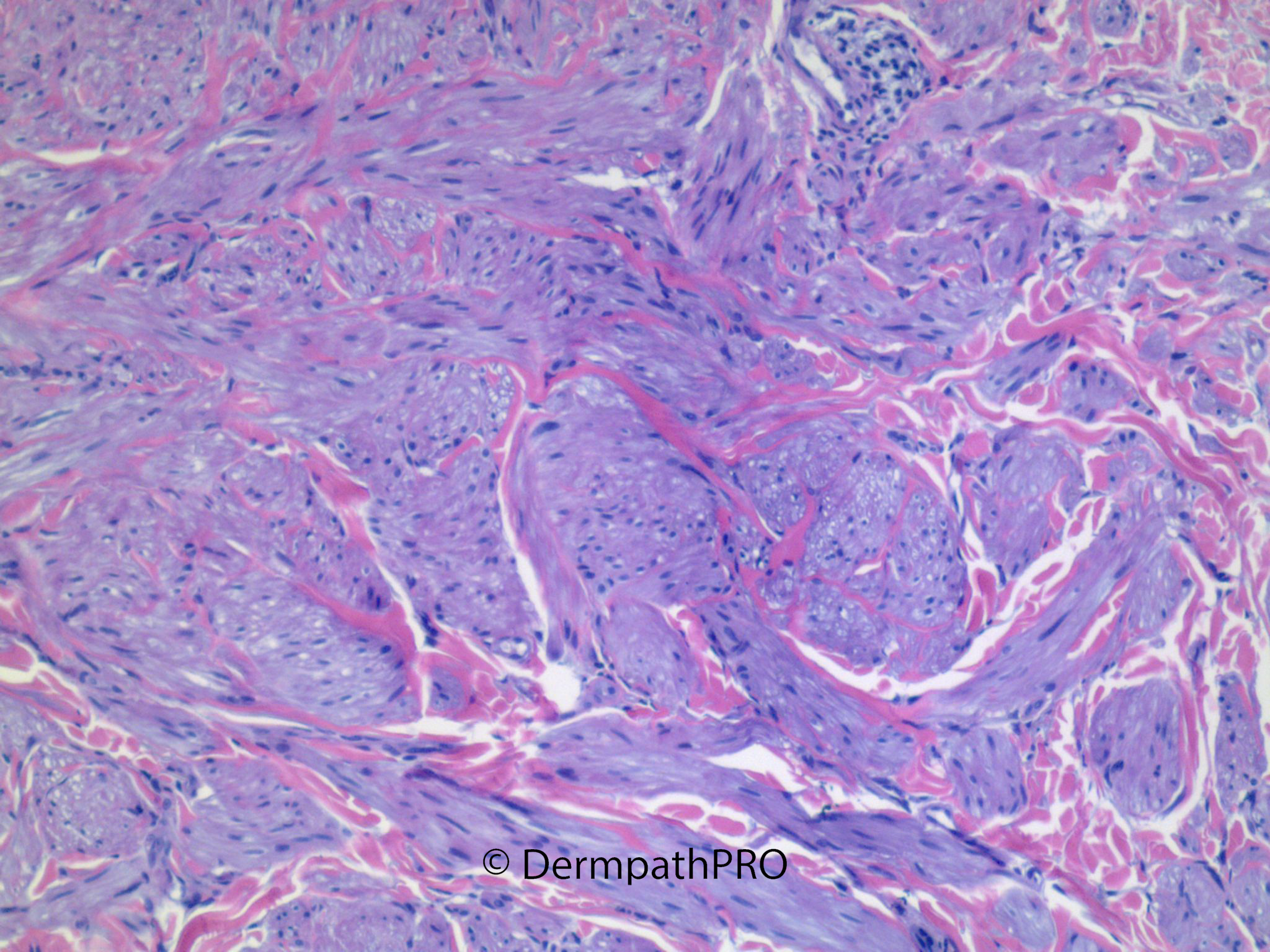
Jump to content
In this section we have spot diagnoses posted on a daily basis since June 2010, now over 4000! You can review the archived cases and read the suggested diagnoses by users and the final comment by the contributors.
Case are uploaded each week day by 10 am UK time with the correct diagnosis will generally be posted at 8 pm UK time. Why not view the most recent spot diagnosis and proffer a diagnosis?

Case Number : Case 3014 - 25 January 2022 Posted By: Uma Sundram

Please read the clinical history and view the images by clicking on them before you proffer your diagnosis.
Submitted Date :
   (0 reviews)

75 year old female with multiple lesions on the left leg.


  • Like 2
  Report Record

User Feedback


Pilar leiomyomas. Useful a HP-magnification to watch to nucleoli, so we can rule out Hereditary Leiomyomatosis( FH-mutation related ). I’m not able to see the prominent nucleoli with clear perinucleolar space, at these magnifications. Maybe negative. 

Share this comment


Link to comment
share_externally

Sorry. Not able to delete this comment. The above comment is my spot diagnosis. 

Share this comment


Link to comment
share_externally

Uma Sundram

Posted

Our concern also was hereditary leiomyomatosis syndrome, which we could not exclude based on morphology. A reminder that multiple leiomyomata can be syndromic, and should prompt us to alert the clinician.

Share this comment


Link to comment
share_externally


Join the conversation

You can post now and register later. If you have an account, sign in now to post with your account.

Guest
Add a comment...

×   Pasted as rich text.   Paste as plain text instead

  Only 75 emoji are allowed.

×   Your link has been automatically embedded.   Display as a link instead

×   Your previous content has been restored.   Clear editor

×   You cannot paste images directly. Upload or insert images from URL.

Loading...

×
×
  • Create New...