Jump to content
In this section we have spot diagnoses posted on a daily basis since June 2010, now over 4000! You can review the archived cases and read the suggested diagnoses by users and the final comment by the contributors.
Case are uploaded each week day by 10 am UK time with the correct diagnosis will generally be posted at 8 pm UK time. Why not view the most recent spot diagnosis and proffer a diagnosis?

Case Number : Case 4024 - 23 June 2022 Posted By: Saleem Taibjee

Please read the clinical history and view the images by clicking on them before you proffer your diagnosis.
Submitted Date :
   (0 reviews)

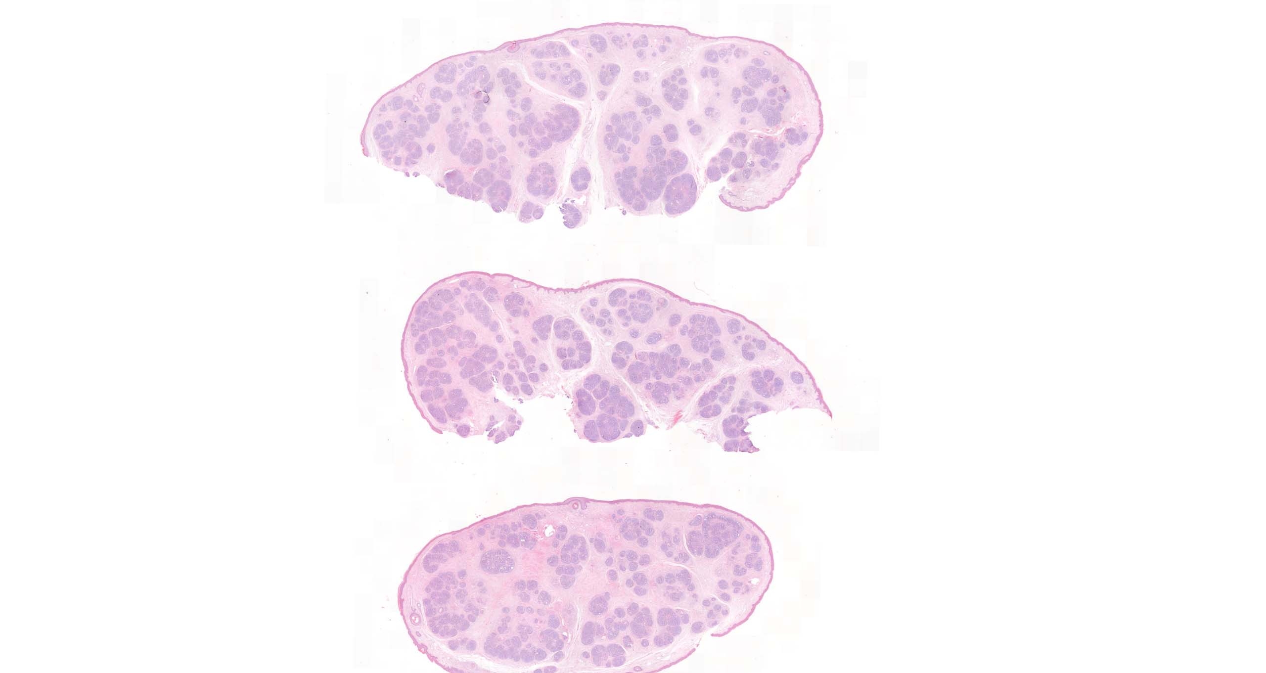
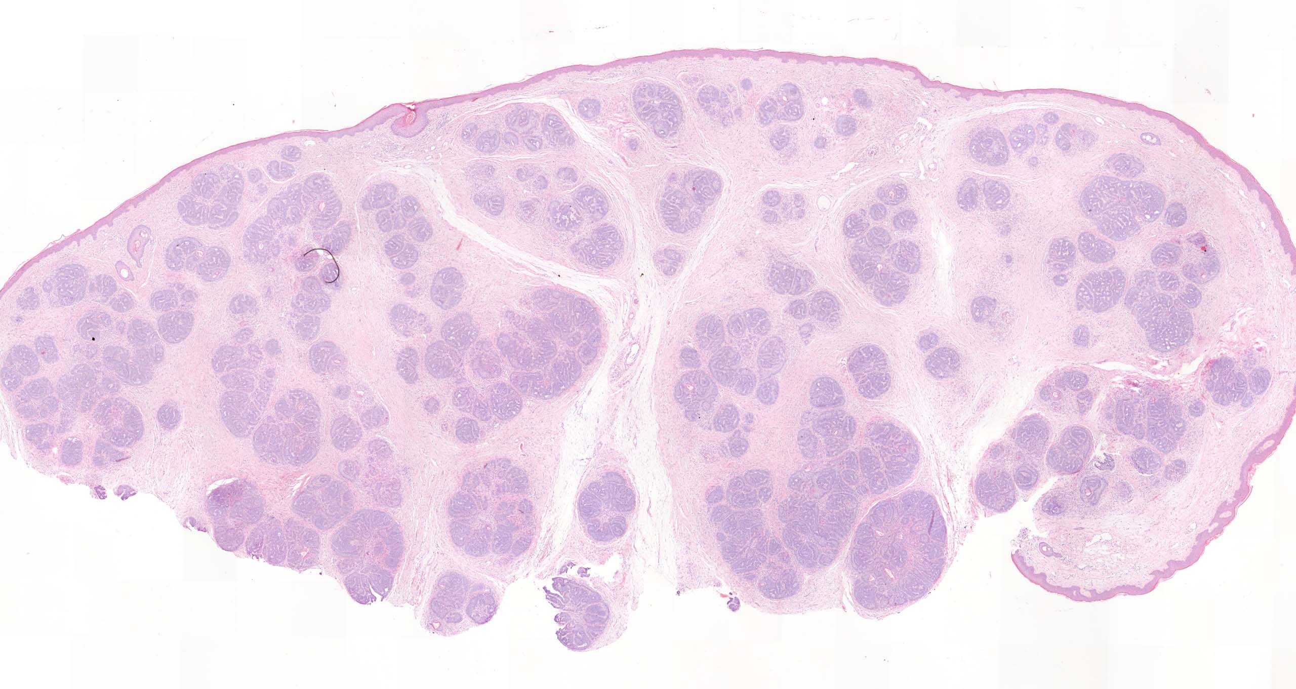
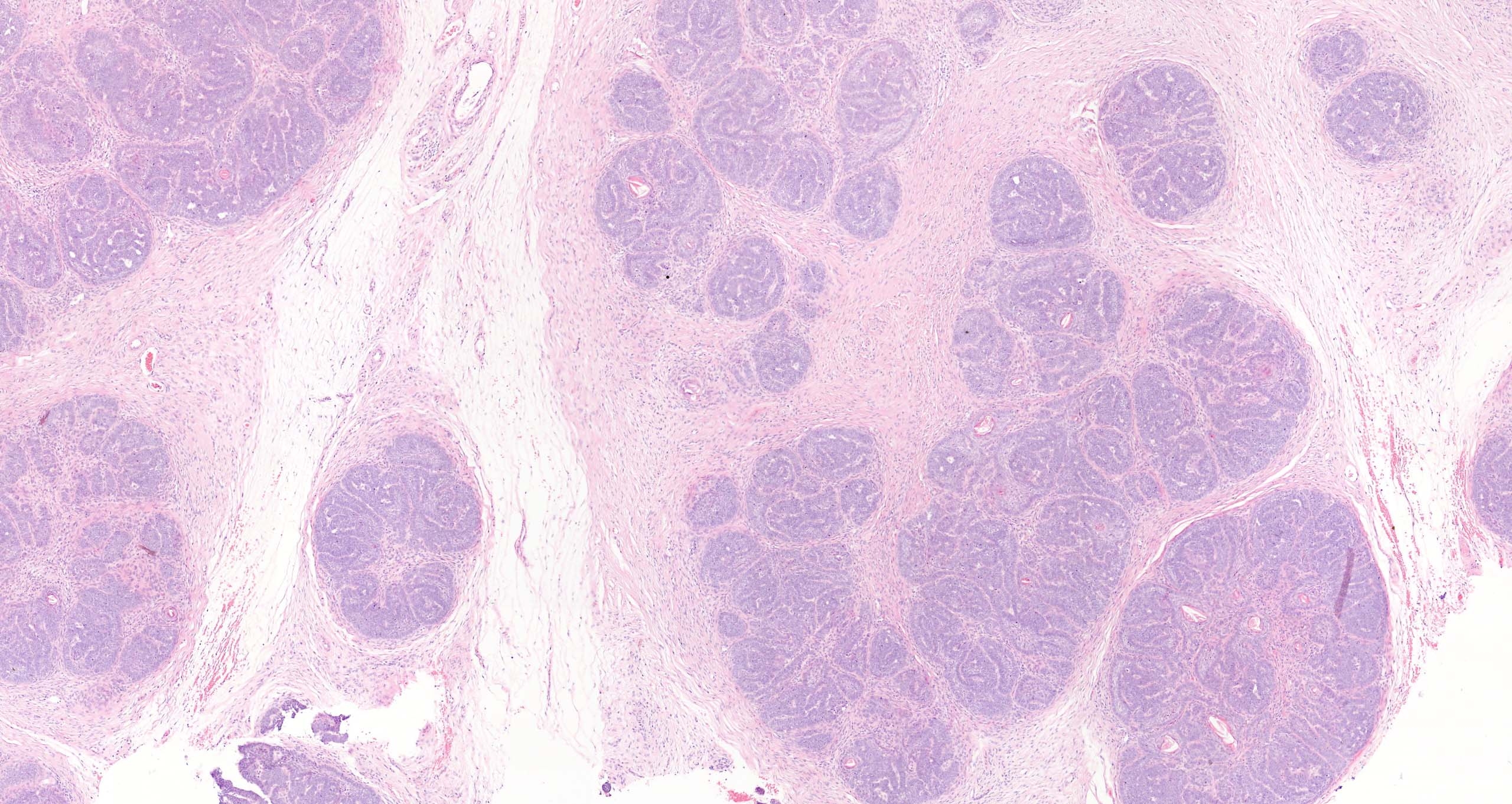
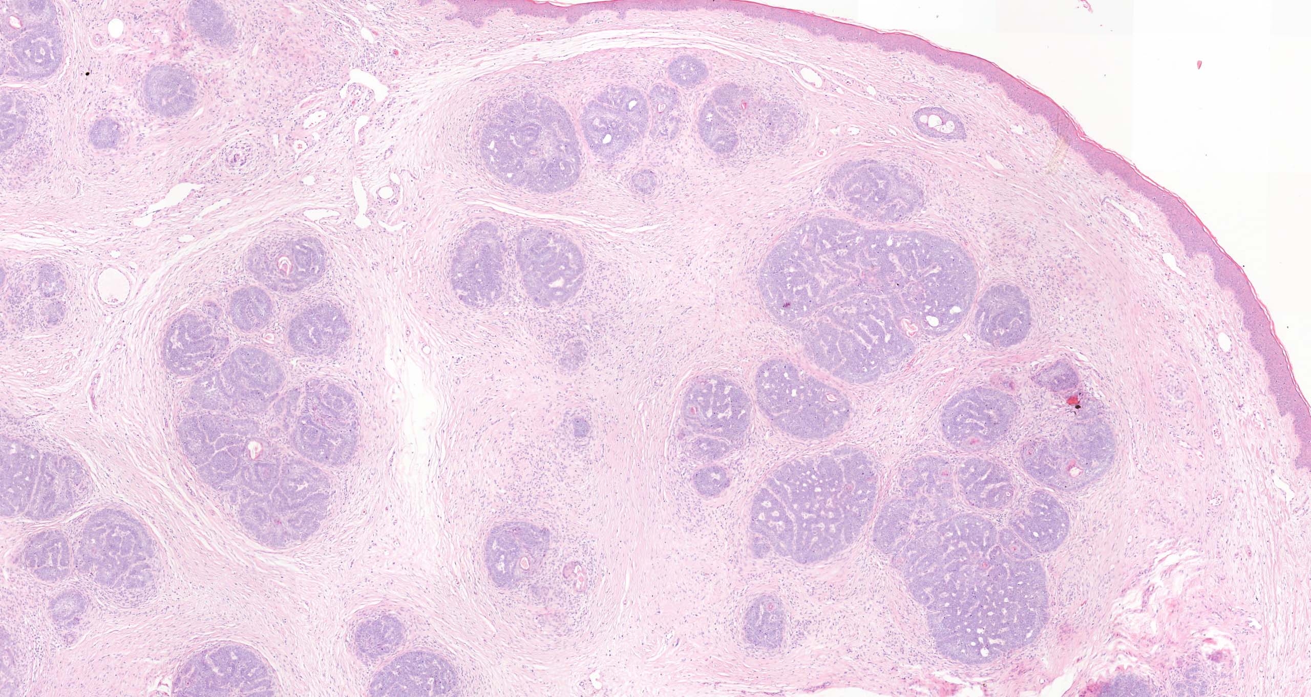
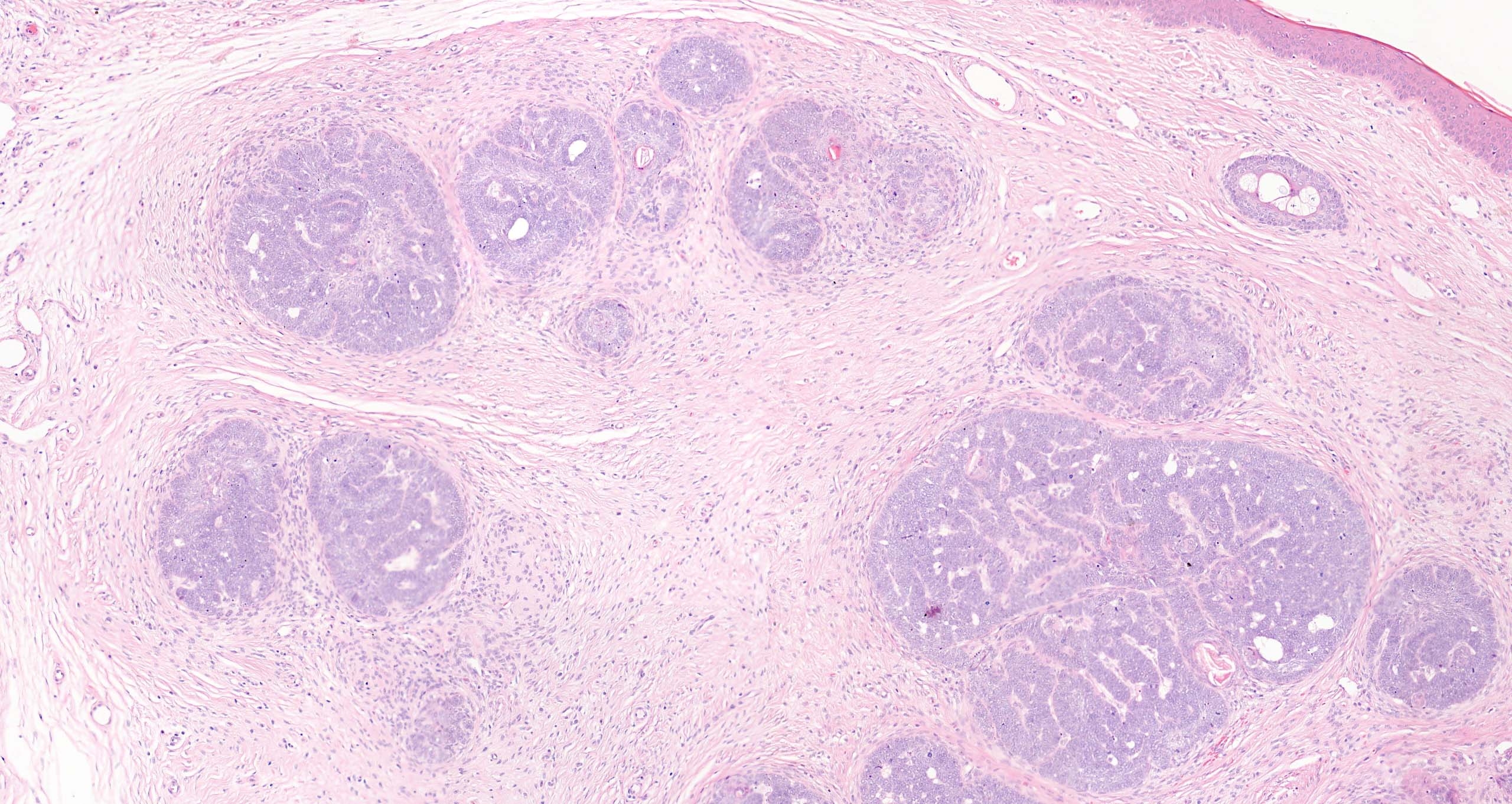
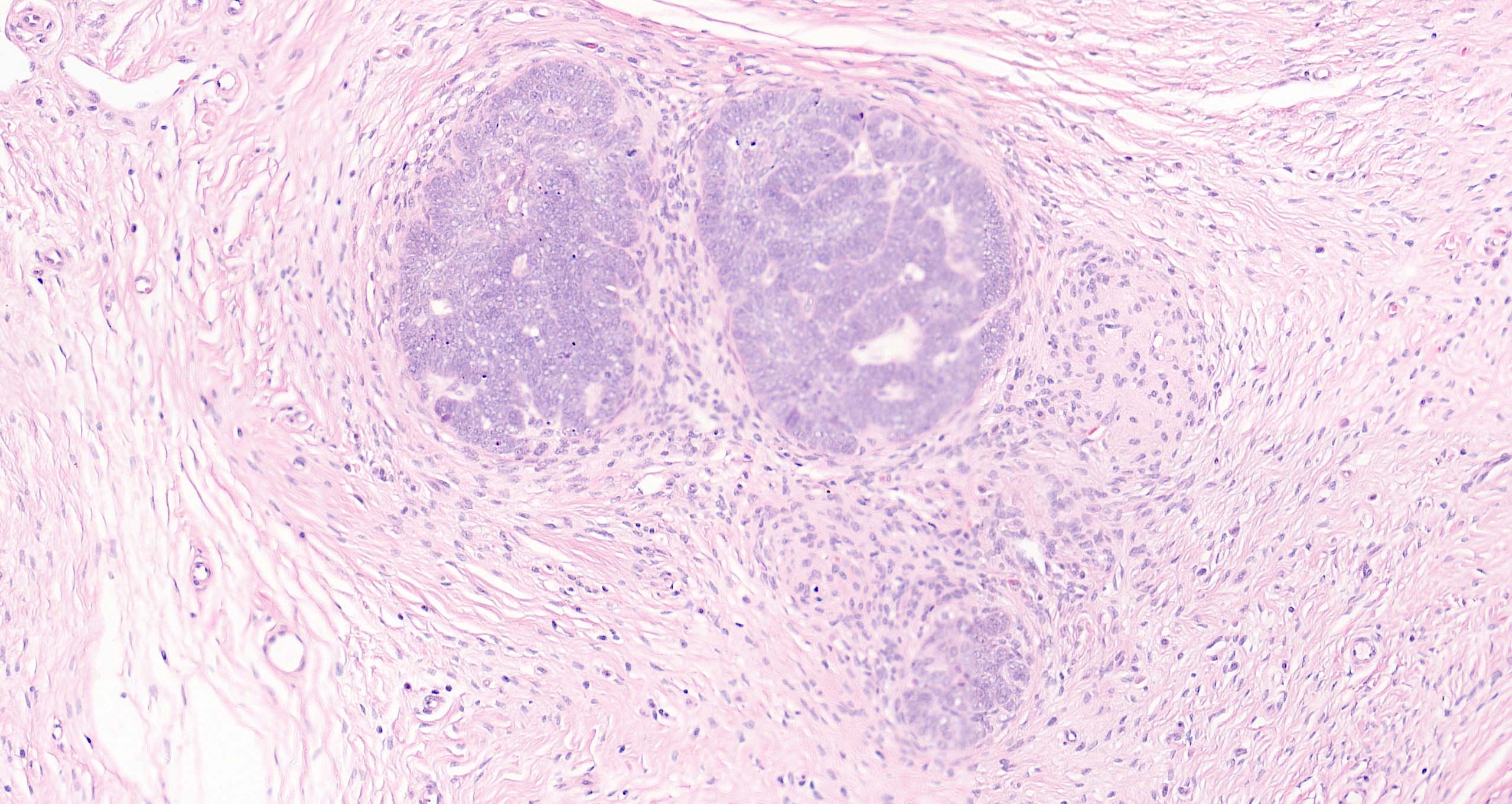
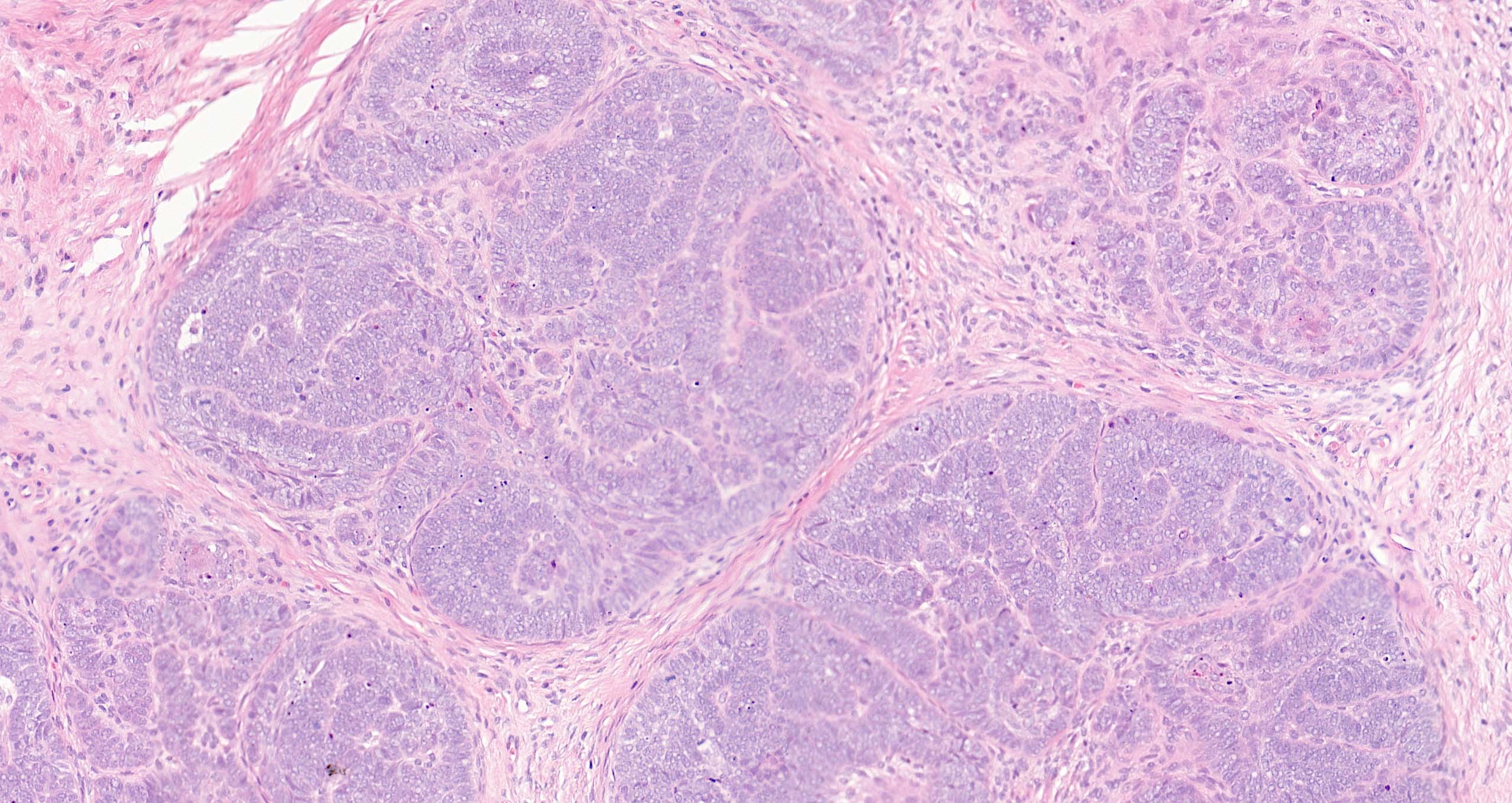
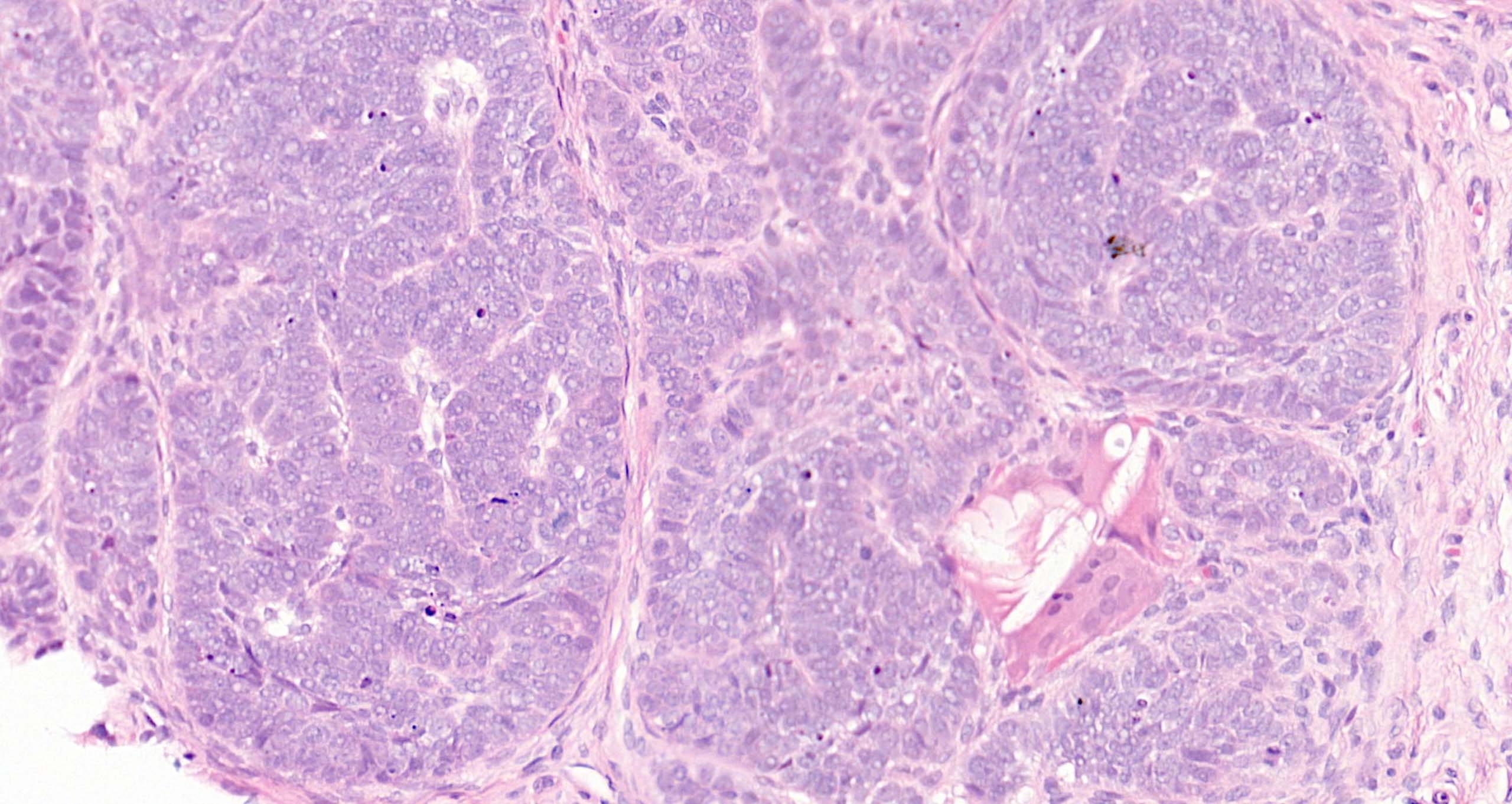
6-year-old boy, curettage right vertex of scalp ?fibroepithelial polyp


  • Thanks 1
  Report Record

User Feedback


Rippled pattern Trichoep/Trichoblastoma.

Share this comment


Link to comment
share_externally

Eman El-Nabarawy

Posted

Trichoepithelioma/Trichoblastoma.

Share this comment


Link to comment
share_externally

Trichoblastoma, stromal clefts, papillary-mesenchimal cells, scattered Lc

At that age not common, unless nevus sebaceous association

aslo CYLD gene mutation Brooke-spiegler syndrom association

Share this comment


Link to comment
share_externally

Saleem Taibjee

Posted

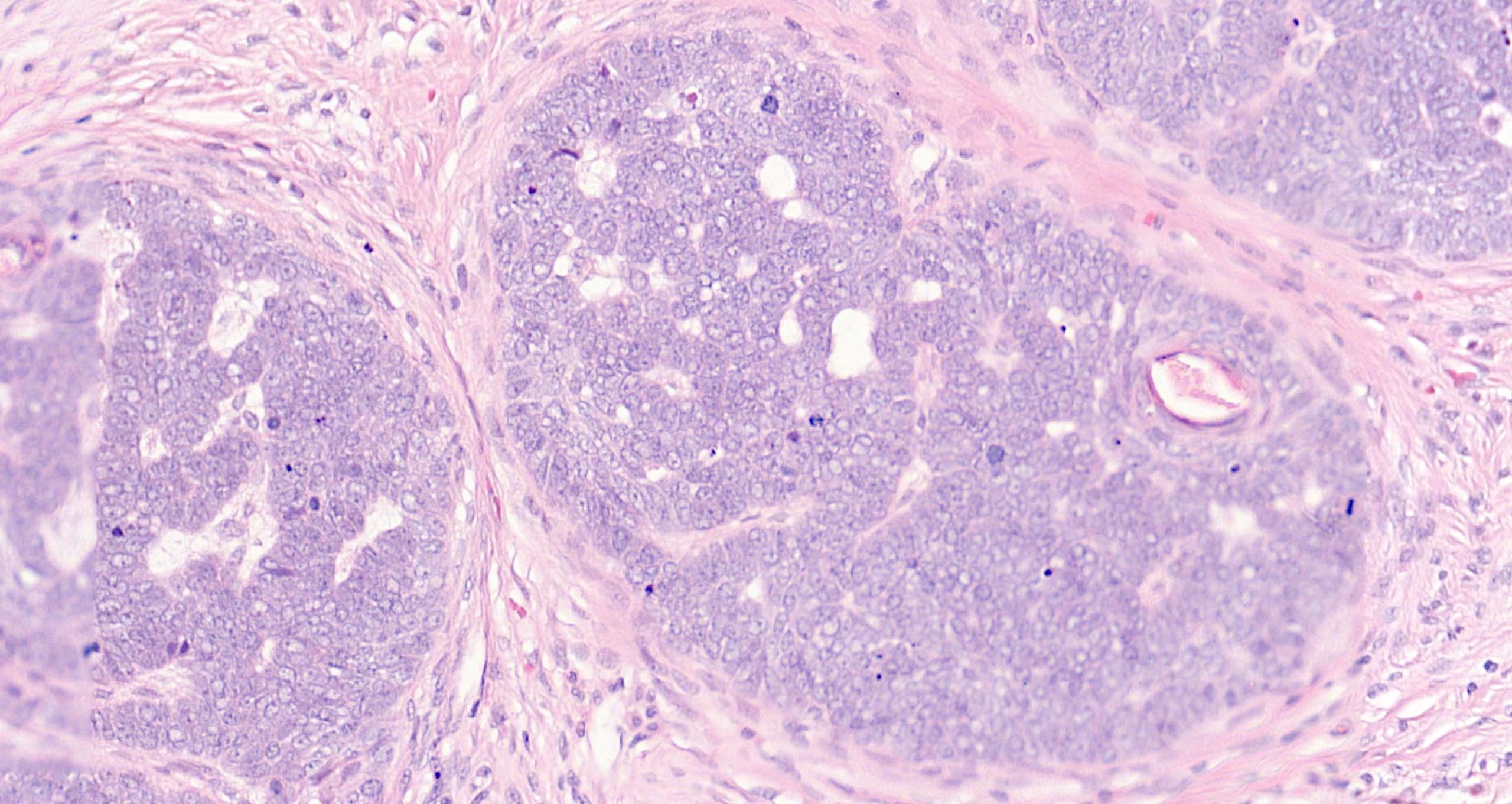
Does anyone have any comments/thoughts about the cytology (before I give my response)?

BW, Saleem

Share this comment


Link to comment
share_externally

The cytology looks like the blastic features well described in old books about tricho”blastoma”. Trichoepithelioma shows more mature, basaloid features. Am I wrong?

Used blastoid instead germinal, because , in this case, there might be confusion with the trichogerminoma variant ( squamoid morules ). 

Share this comment


Link to comment
share_externally

Saleem Taibjee

Posted

So I agree with trichoblastoma. But having seen regular examples of BCC with neuroendocrine differentiation, this case immediately struck me as showing a similar cytology with large relatively nuclei with dispersed chromatin.

The immunos are shown below. There is positive staining for neurofilament, and patchy staining for CD56, focal chromogranin (negative for synaptophysin). CK20 was negative, in fact not even showing entrapped Merkel cells which we might see in trichoblastoma. CD10 nicely highlighted the aggregates of papillary mesenchymal cells, but also showed some staining of the tumour epithelium.

I have trawled through the literature, and this seems to be a very rare occurrence. I found one reference to a trichilemmal carcinoma with neuroendocrine differentation, but not a whole lot else. Has anyone else come across this? But perhaps it is more common than we appreciate... We plan to write up the case.

BW. Saleem

image.jpeg

image.jpeg

image.jpegimage.jpeg

image.jpeg


image.jpeg

Share this comment


Link to comment
share_externally


Join the conversation

You can post now and register later. If you have an account, sign in now to post with your account.

Guest
Add a comment...

×   Pasted as rich text.   Paste as plain text instead

  Only 75 emoji are allowed.

×   Your link has been automatically embedded.   Display as a link instead

×   Your previous content has been restored.   Clear editor

×   You cannot paste images directly. Upload or insert images from URL.

Loading...

×
×
  • Create New...