Jump to content
In this section we have spot diagnoses posted on a daily basis since June 2010, now over 4000! You can review the archived cases and read the suggested diagnoses by users and the final comment by the contributors.
Case are uploaded each week day by 10 am UK time with the correct diagnosis will generally be posted at 8 pm UK time. Why not view the most recent spot diagnosis and proffer a diagnosis?

Case Number : Case 3041 - 03 March 2022 Posted By: Saleem Taibjee

Please read the clinical history and view the images by clicking on them before you proffer your diagnosis.
Submitted Date :
   (0 reviews)

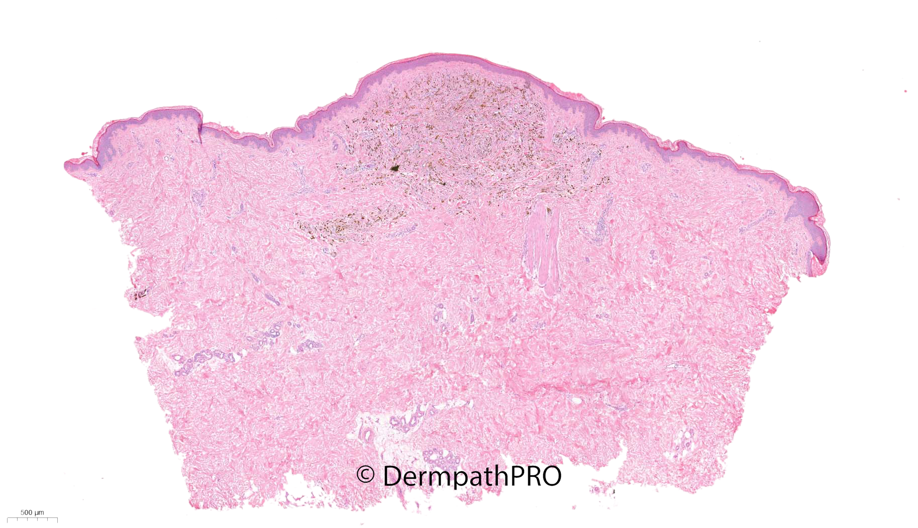
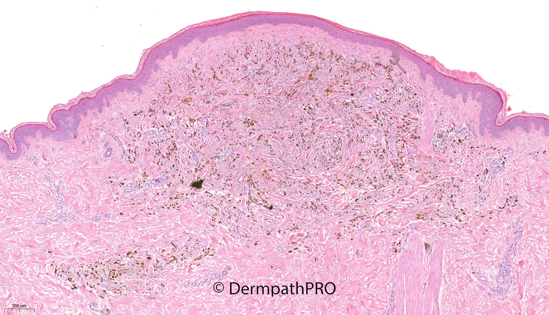
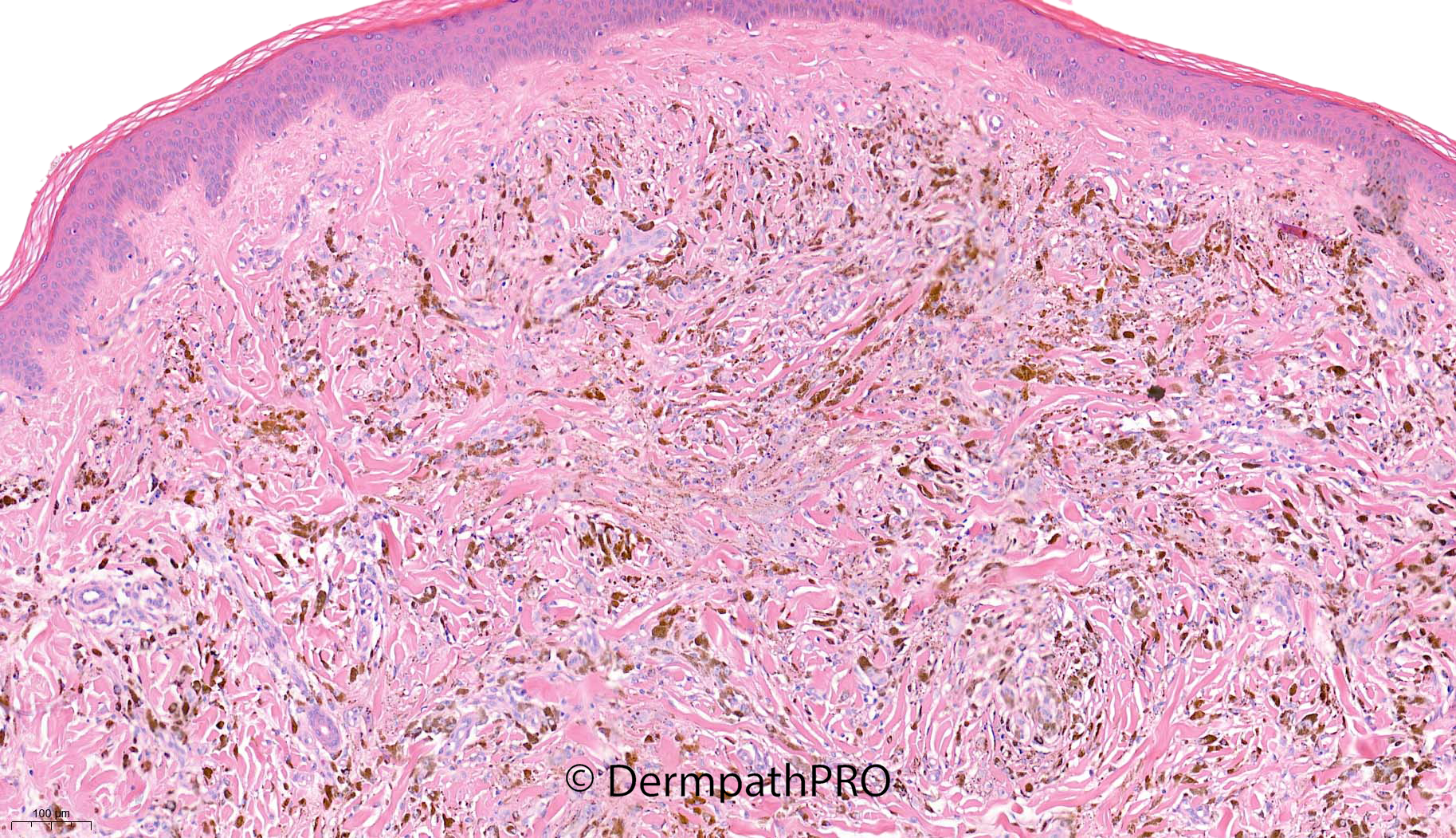
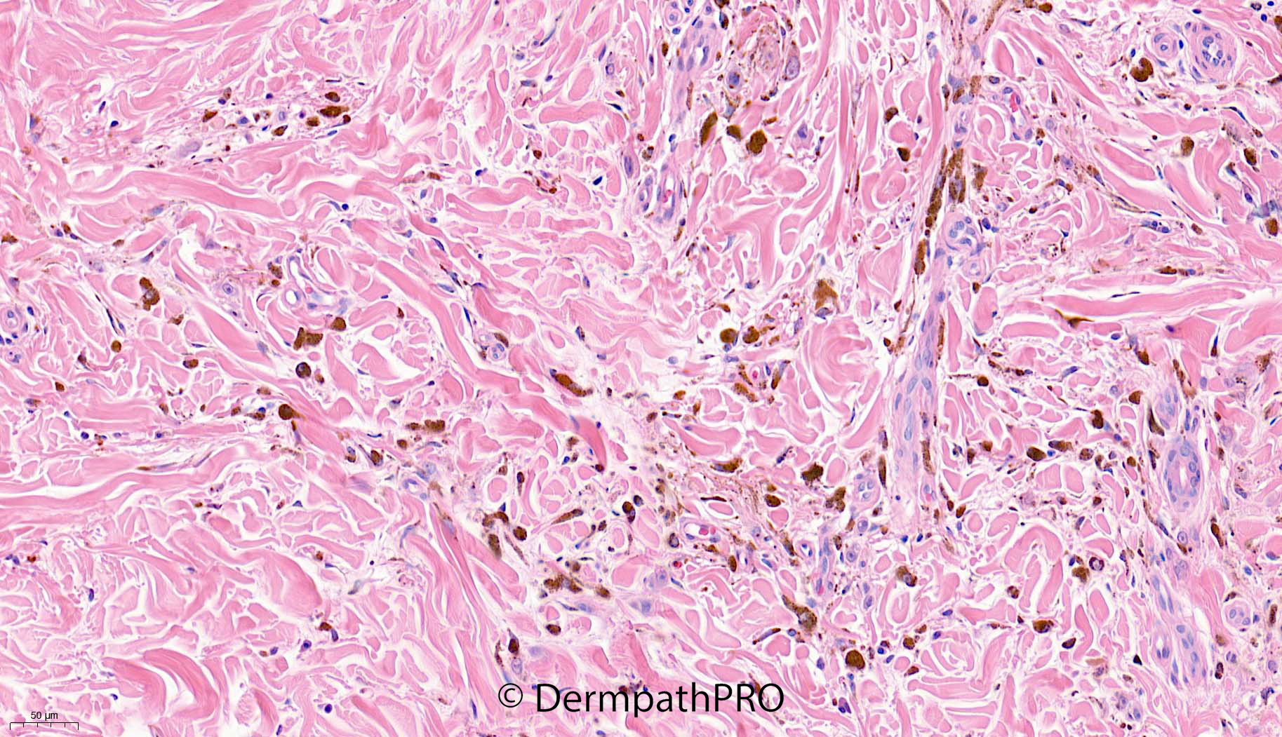
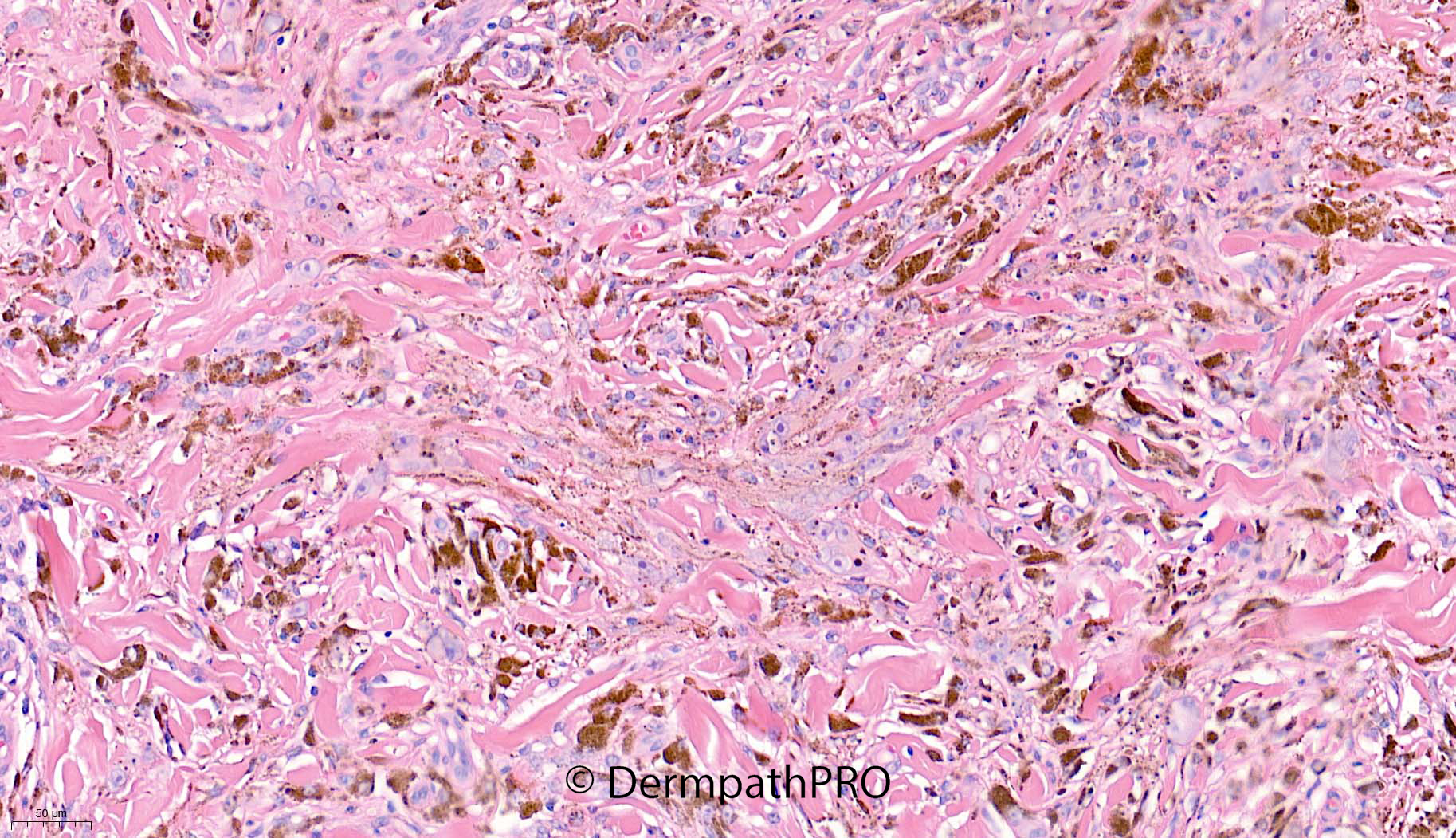
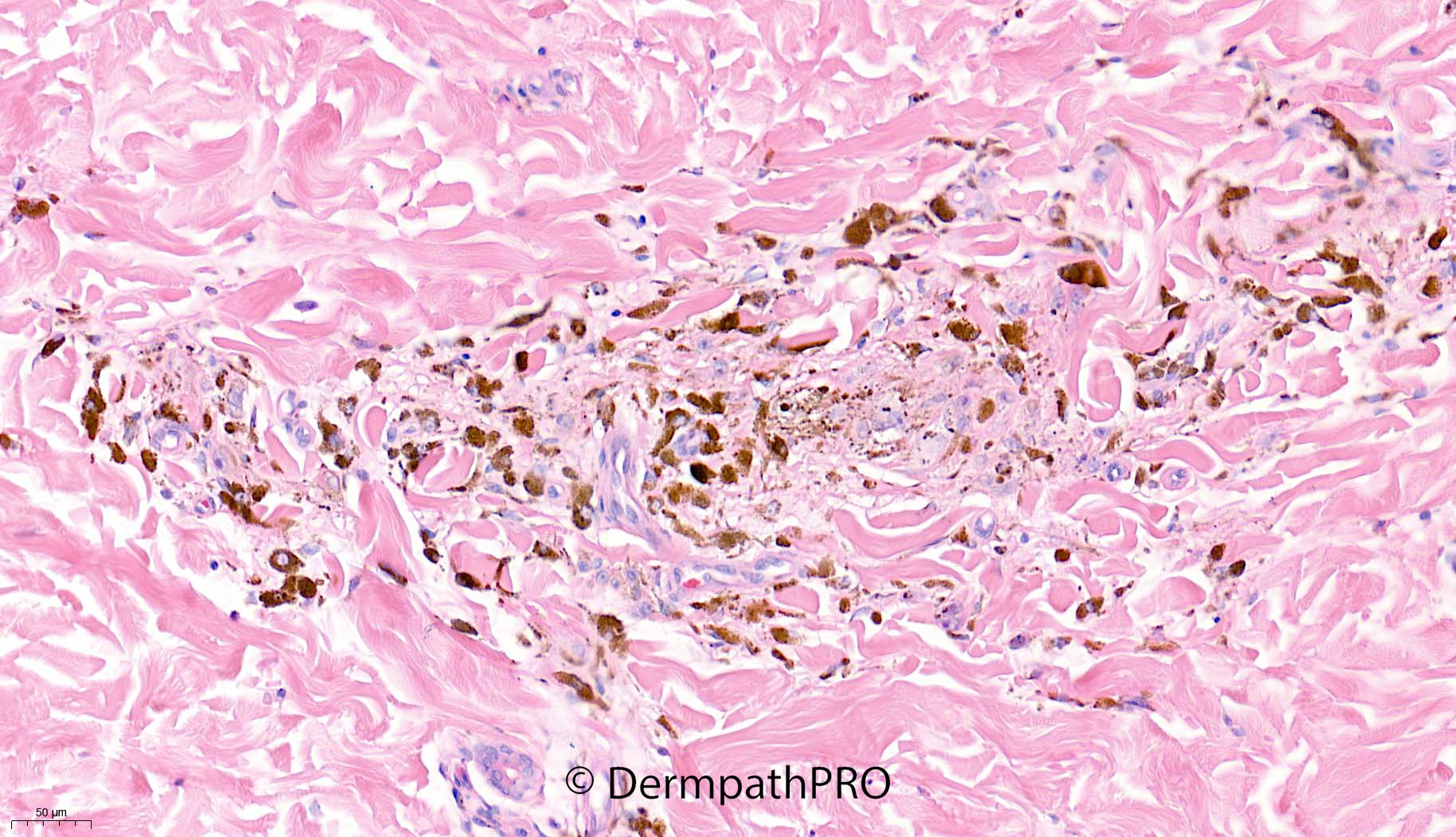
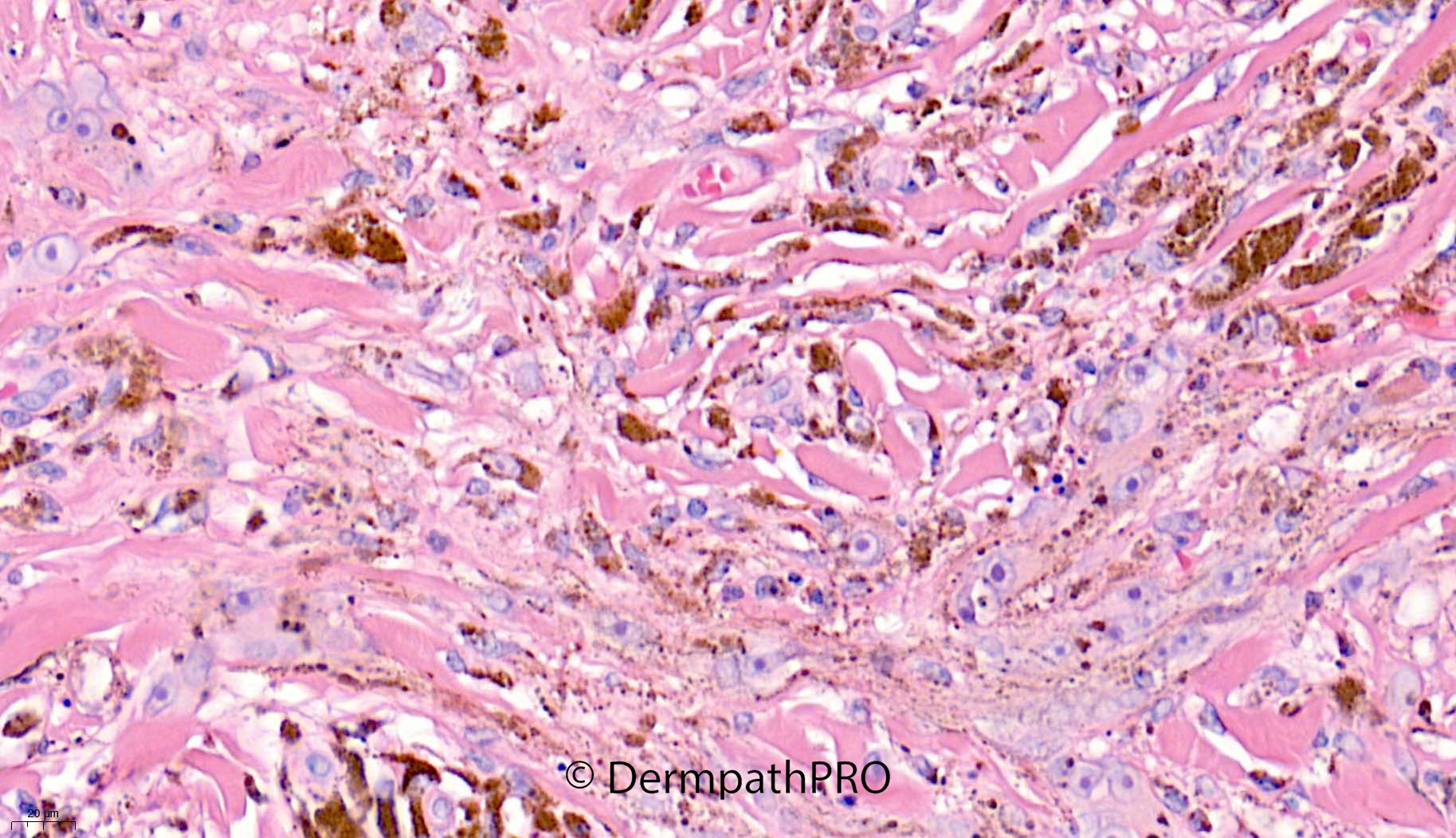
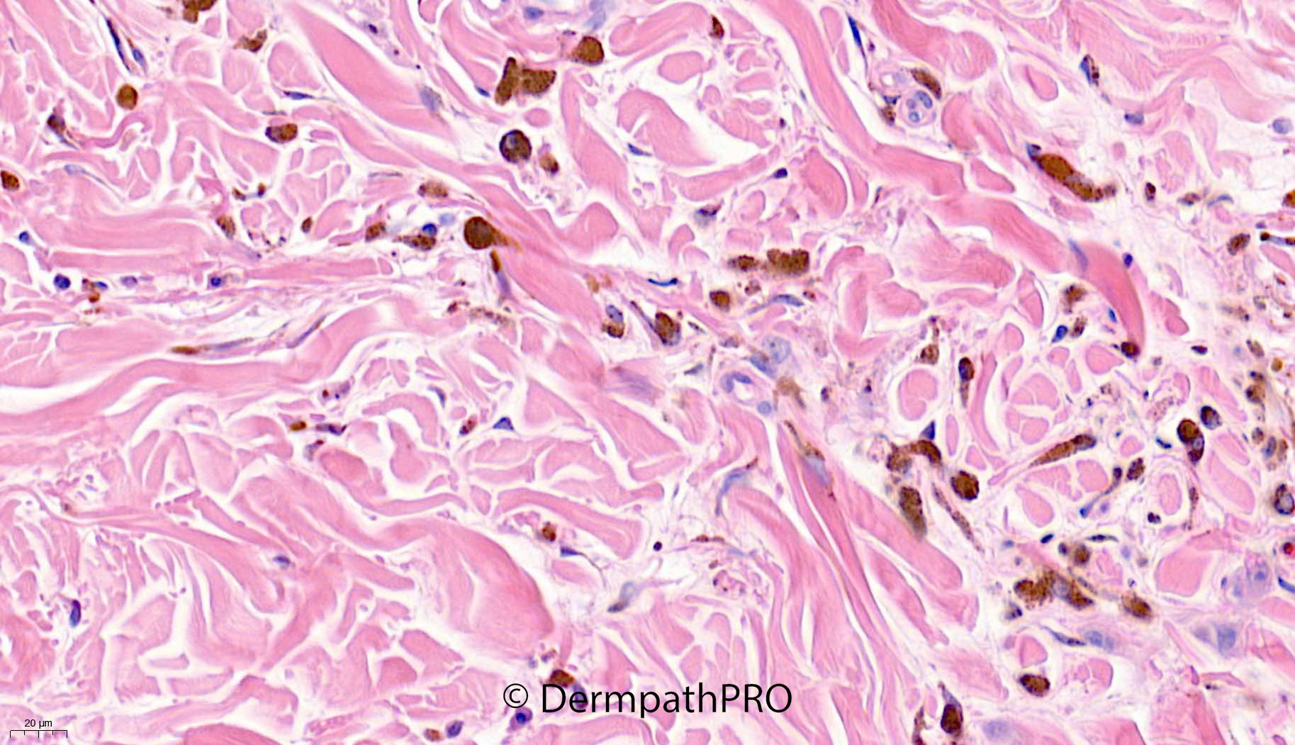
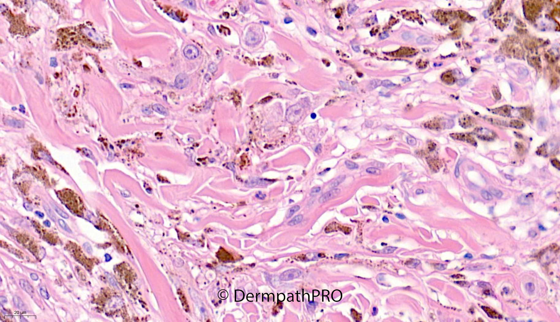
14-year-old girl, pigmented lesion right cheek


  Report Record

User Feedback


Epithelioid blue nevus VS PEM. Those large "RS-like" cells with prominent nucleoli are expected in PEM. PRKAR1A status for confirmation.

Share this comment


Link to comment
share_externally

Richard Logan

Posted

Cellular/epithelioid blue naevus.

Share this comment


Link to comment
share_externally

Meenakshi Batrani

Posted

On 03/03/2022 at 02:20, Shawn said:

Epithelioid blue nevus VS PEM. Those large "RS-like" cells with prominent nucleoli are expected in PEM. PRKAR1A status for confirmation.

Agree

Share this comment


Link to comment
share_externally

Favor PEM. Thought of DPN also, but, as pointed out by Shawn, RS-like cells are definitely in keeping with PEM. 

Share this comment


Link to comment
share_externally

Meenakshi Batrani

Posted

Challenging. The entire gamut of epithelioid blue nevus, PEM and BLITZ (Blue+Sptiz) or called as superficial variant of DPN by some, are overlapping. PRAKAR1 and Beta-Catenin will be helpful. 

Share this comment


Link to comment
share_externally

Saleem Taibjee

Posted

Yes, very well done. This is indeed pigmented epithelioid melanocytoma.

This case had already been signed out by another pathologist as blue naevus. I reviewed this case prior to MDT, when the small focus of epithelioid cells which show prominent nucleoi - 'fried eggs' - caught my attention, and which are highlighted above.

You can appreciate my excitement when I learned at the MDT that the patient has facial freckling and other pigmented lesions, and a history of angiomyxoma of the vulva previously, and the clinician was suspecting Carney complex. This information had not appeared on the original clinical request form. But, of course, on discovering this information, this puts this lesion more firmly into the PEM category. These are lesions which historically were called epithelioid blue naevus in the context of Carney, but this is no longer the appropriate term.

I show the Melan-A and S100 below. The Melan-A makes the fried egg cells really 'pop out'. The S100 also stains quite strongly. As you know this is indirect evidence that this is not a blue naevus (in which S100 is often diminished).

I have since heard that the patient now has genetically confirmed Carney complex.

I don't have access to IHC / tissue testing for PRKAR1A, and so have sent this case on to Arnaud de la Fouchardiere in Lyon; he has also confirmed loss of PRKAR1A on IHC.

BW
Saleem

58717_40.0xb Melan-A.jpg

58717_63.0x Melan-A.jpg

58717_40.0xb S100 labelled.jpg

Share this comment


Link to comment
share_externally

Great case, Saleem. And many learning points. Thanks.

Share this comment


Link to comment
share_externally


Join the conversation

You can post now and register later. If you have an account, sign in now to post with your account.

Guest
Add a comment...

×   Pasted as rich text.   Paste as plain text instead

  Only 75 emoji are allowed.

×   Your link has been automatically embedded.   Display as a link instead

×   Your previous content has been restored.   Clear editor

×   You cannot paste images directly. Upload or insert images from URL.

Loading...

×
×
  • Create New...