Jump to content
In this section we have spot diagnoses posted on a daily basis since June 2010, now over 4000! You can review the archived cases and read the suggested diagnoses by users and the final comment by the contributors.
Case are uploaded each week day by 10 am UK time with the correct diagnosis will generally be posted at 8 pm UK time. Why not view the most recent spot diagnosis and proffer a diagnosis?

Case Number : Case 3042 - 04 March 2022 Posted By: Dr. Richard Carr

Please read the clinical history and view the images by clicking on them before you proffer your diagnosis.
Submitted Date :
   (0 reviews)

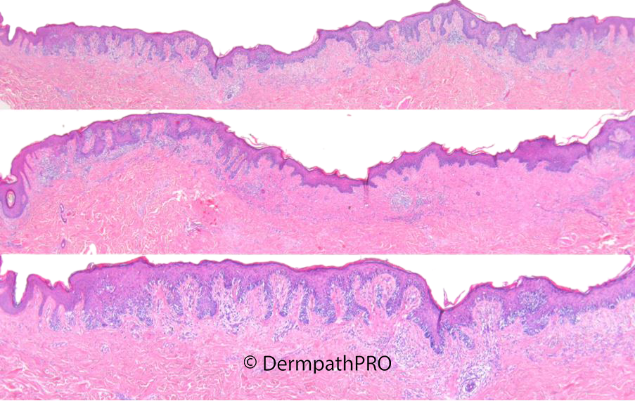
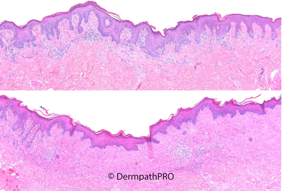
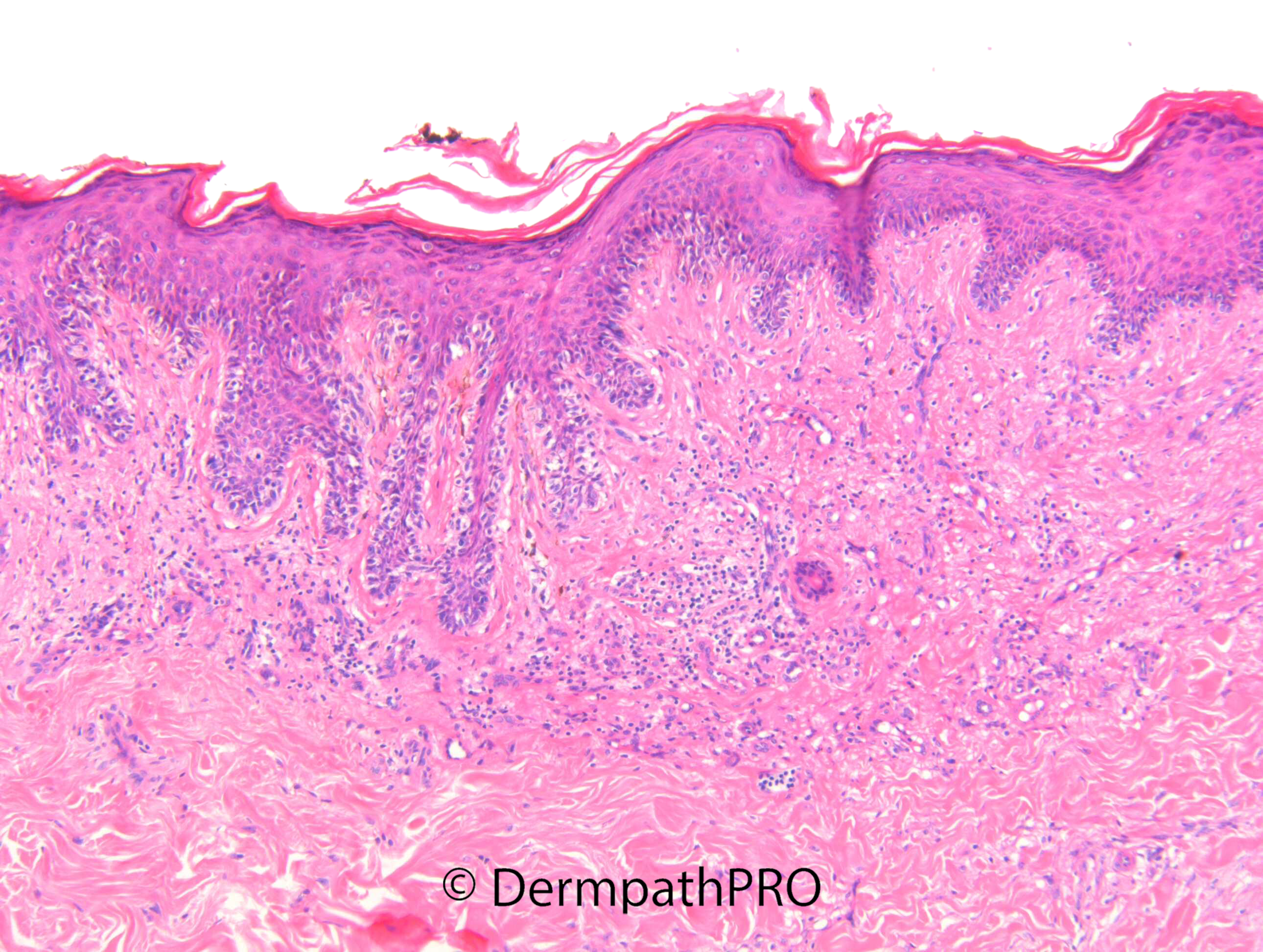
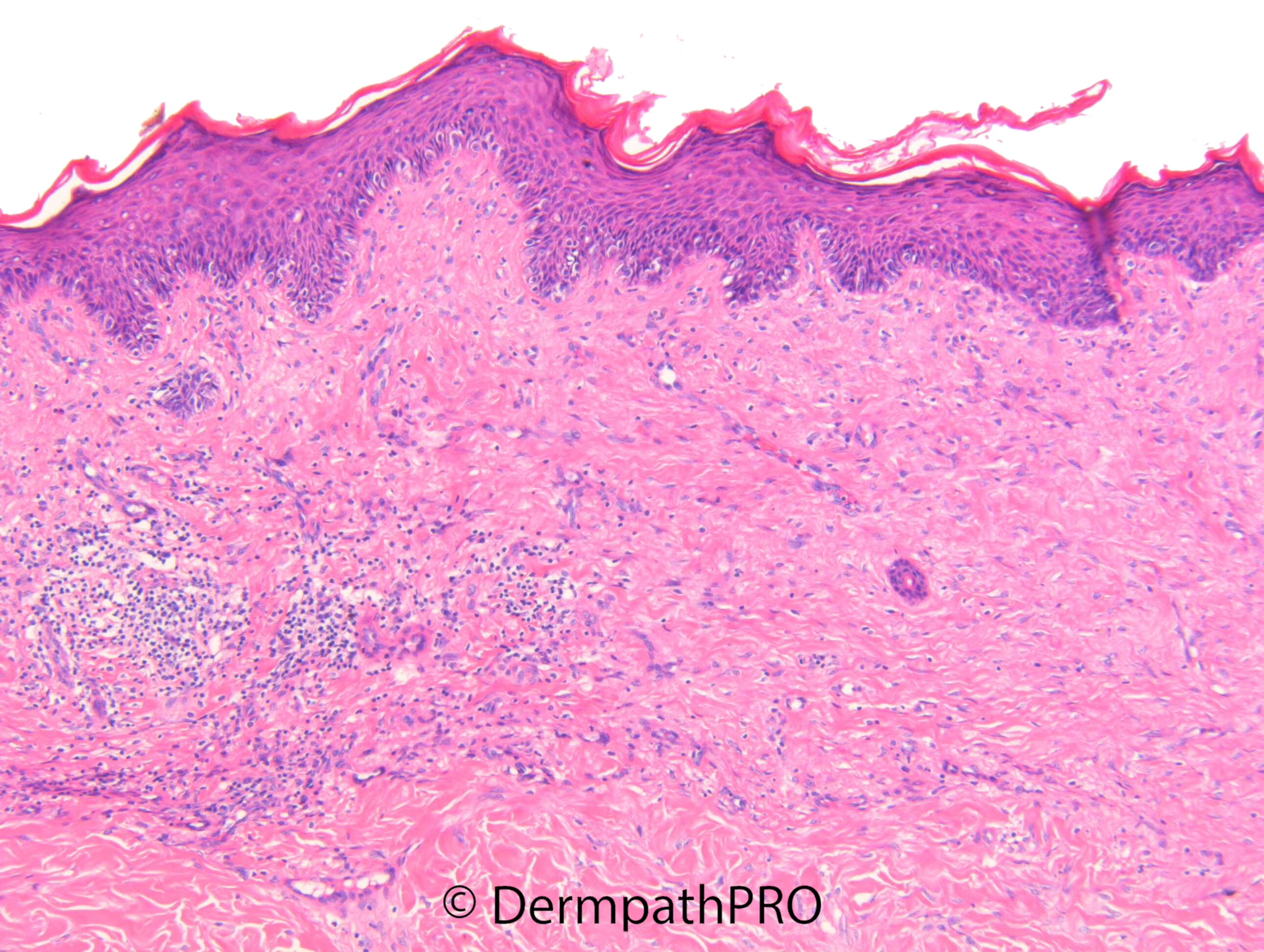
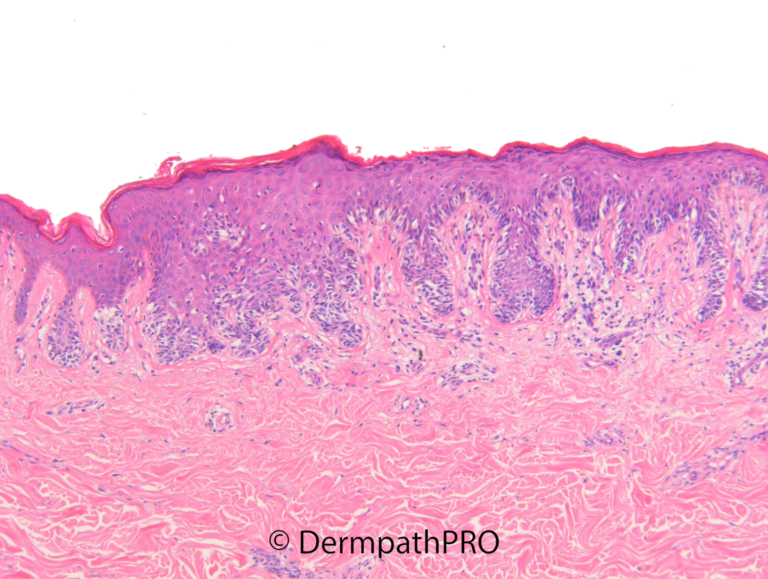
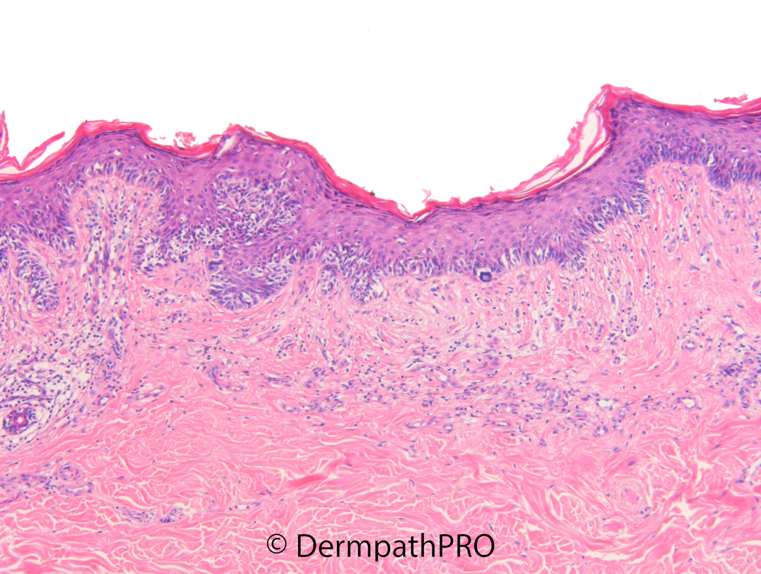
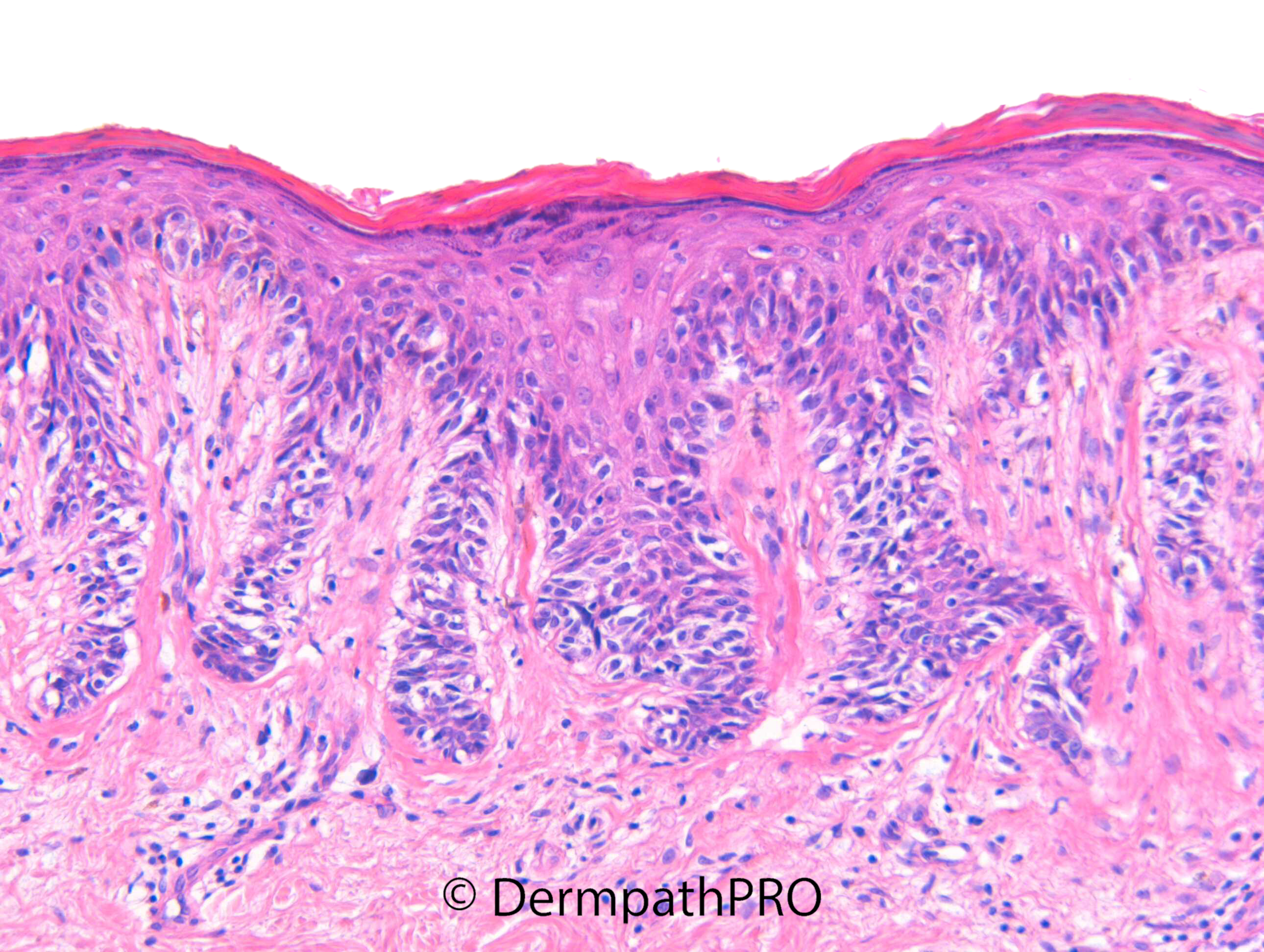
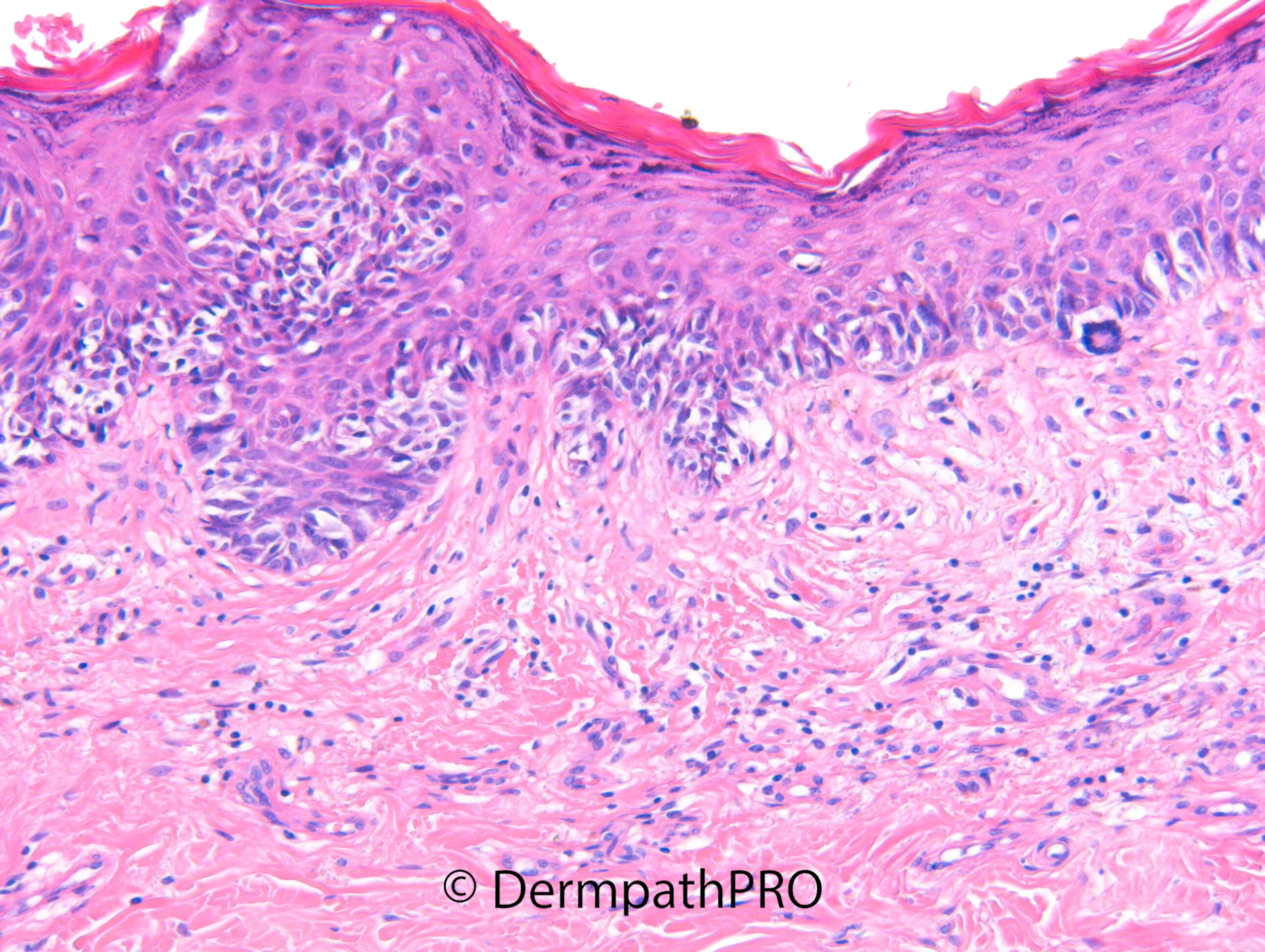
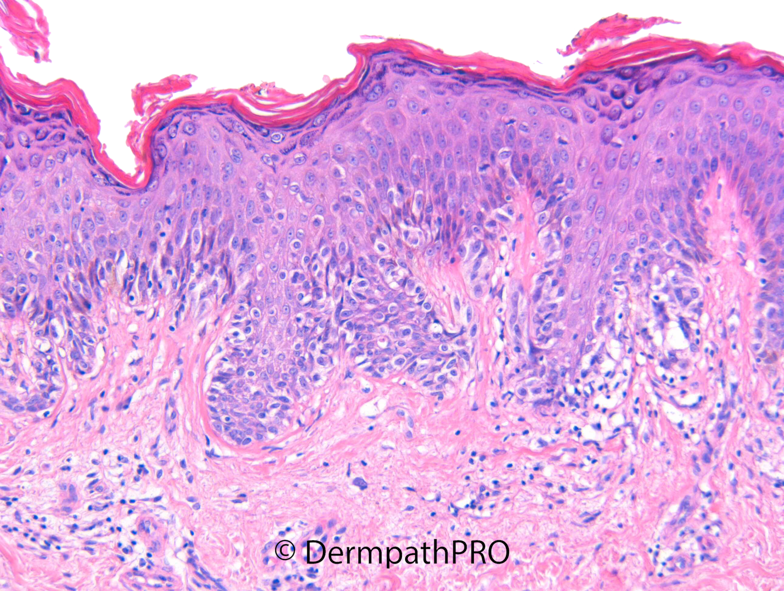
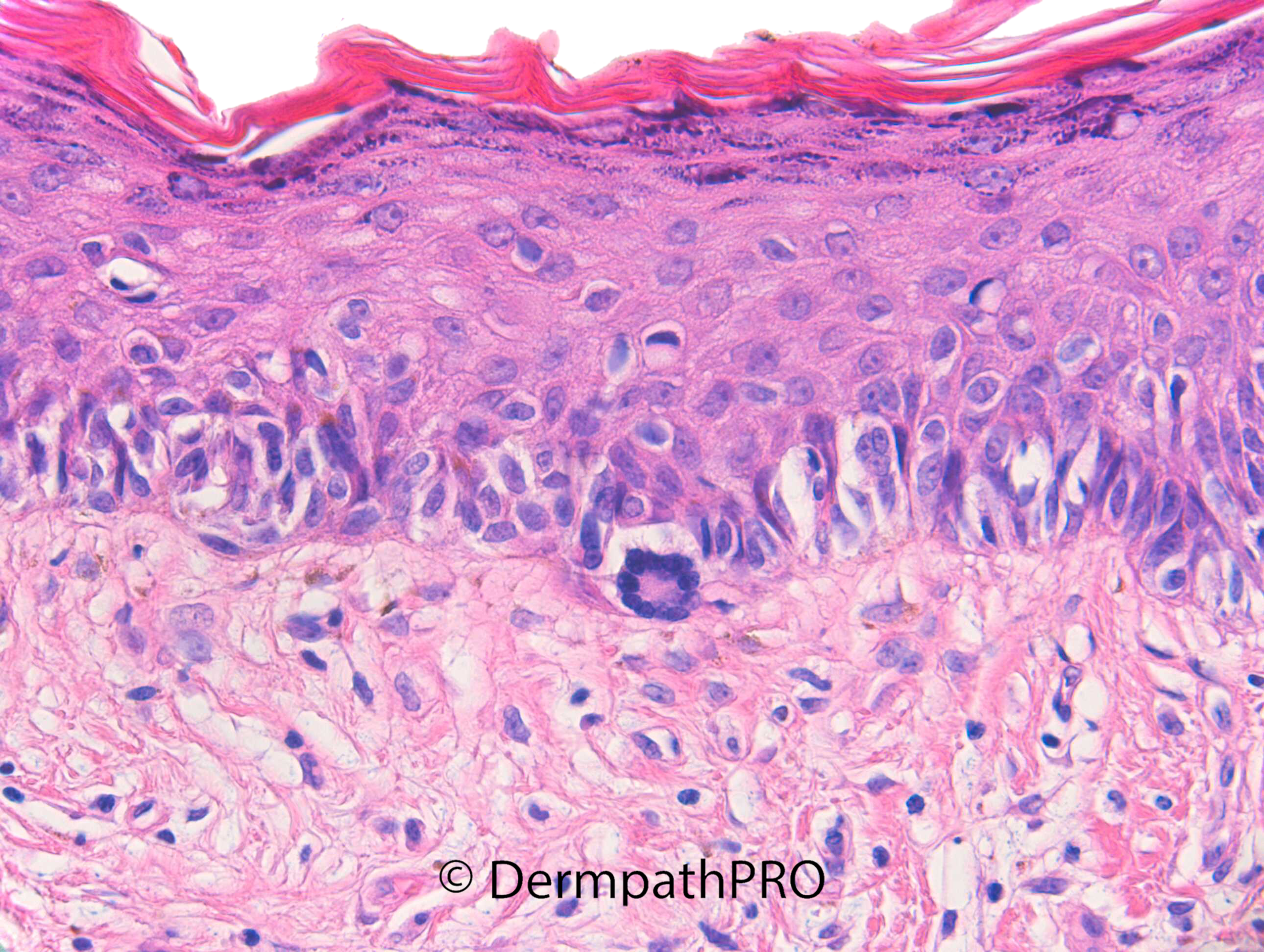
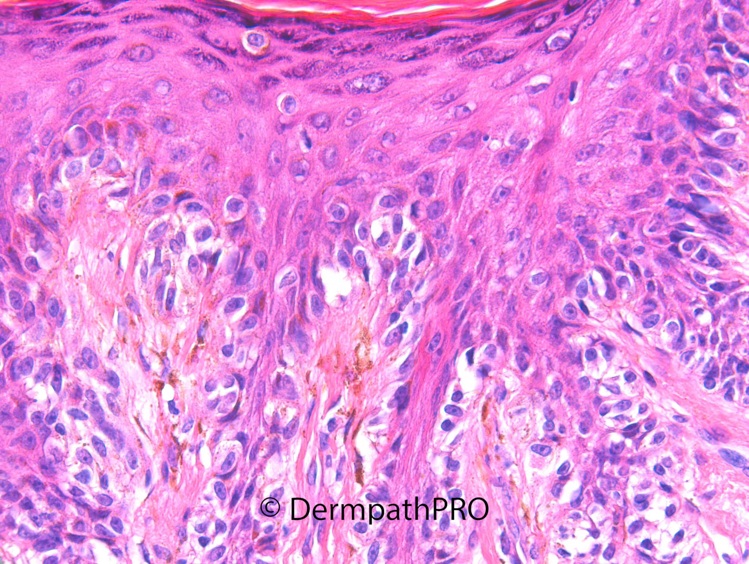
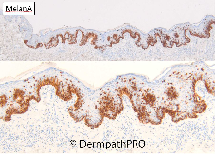
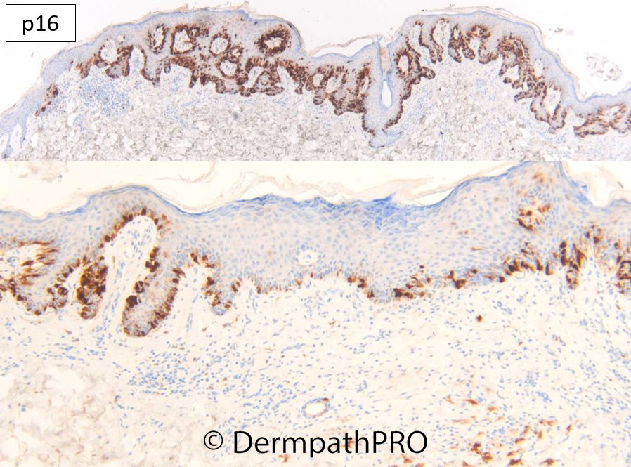
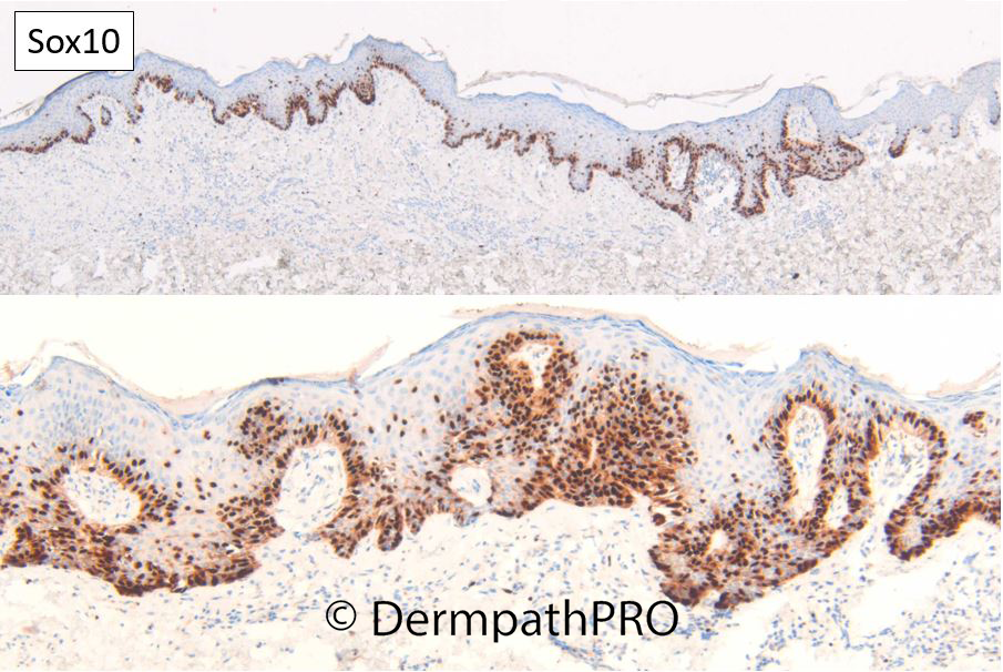
M45. Upper back. Erythematous lesion 15 x 10mm ?BCC


  Report Record

User Feedback


Atypical junctional proliferation over a dermal scar - It seems like the junctional proliferation extends past the edge of the scar, suggesting real MIS rather than just recurrence phenomenon.... Previous biopsy/trauma? 

Share this comment


Link to comment
share_externally

Meenakshi Batrani

Posted

Agree very broad and confluent proliferation suggestive of MMIS. 

Share this comment


Link to comment
share_externally

Meenakshi Batrani

Posted

No PRAME expression and p16 is retained, so may be a recurrent nevus simulating MMIS. Although, H&E was suspicious for MMIS, as the proliferation seem to go past the scar.

Share this comment


Link to comment
share_externally

Dr. Richard Carr

Posted

I favoured a mildly dysplastic naevus with rather confluent lentiginous pattern and pseudomelanomatous changes. I guess the important point is to ensure complete removal of this sort of challenging lesion which will garner a range of diagnostic opinions. The lesion lacks significant atypia and even the scatter of pagetoid cells are small with nuclei no larger than keratinocytes and with open chromatin. The multinucleate cell is rather typical of benign lesions. Pagetoid scatter seems to be very typical of melanoma but is clearly not entirely specific and not sensitive either. I'm still finding PRAME quite useful especially when it agrees with my favoured diagnosis!

Share this comment


Link to comment
share_externally


Join the conversation

You can post now and register later. If you have an account, sign in now to post with your account.

Guest
Add a comment...

×   Pasted as rich text.   Paste as plain text instead

  Only 75 emoji are allowed.

×   Your link has been automatically embedded.   Display as a link instead

×   Your previous content has been restored.   Clear editor

×   You cannot paste images directly. Upload or insert images from URL.

Loading...

×
×
  • Create New...