Jump to content
In this section we have spot diagnoses posted on a daily basis since June 2010, now over 4000! You can review the archived cases and read the suggested diagnoses by users and the final comment by the contributors.
Case are uploaded each week day by 10 am UK time with the correct diagnosis will generally be posted at 8 pm UK time. Why not view the most recent spot diagnosis and proffer a diagnosis?

Case Number : Case 3047 - 11 March 2022 Posted By: Dr. Richard Carr

Please read the clinical history and view the images by clicking on them before you proffer your diagnosis.
Submitted Date :
   (0 reviews)

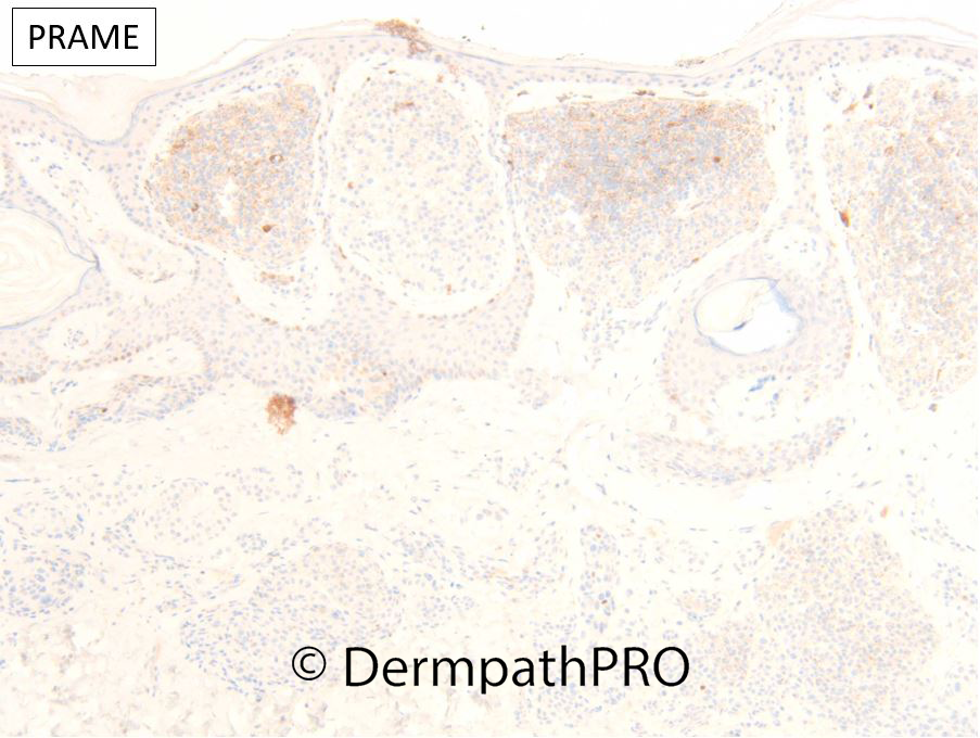
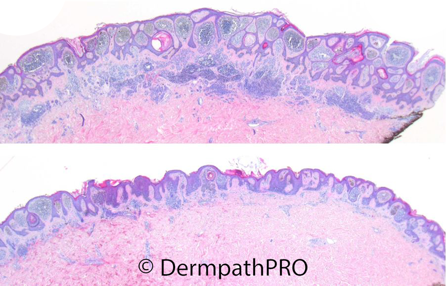
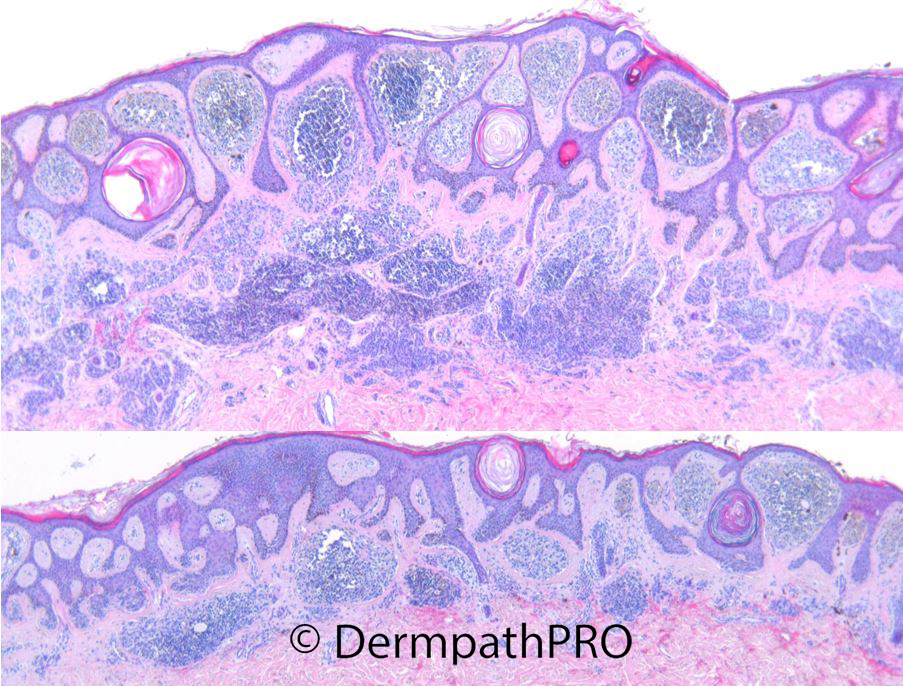
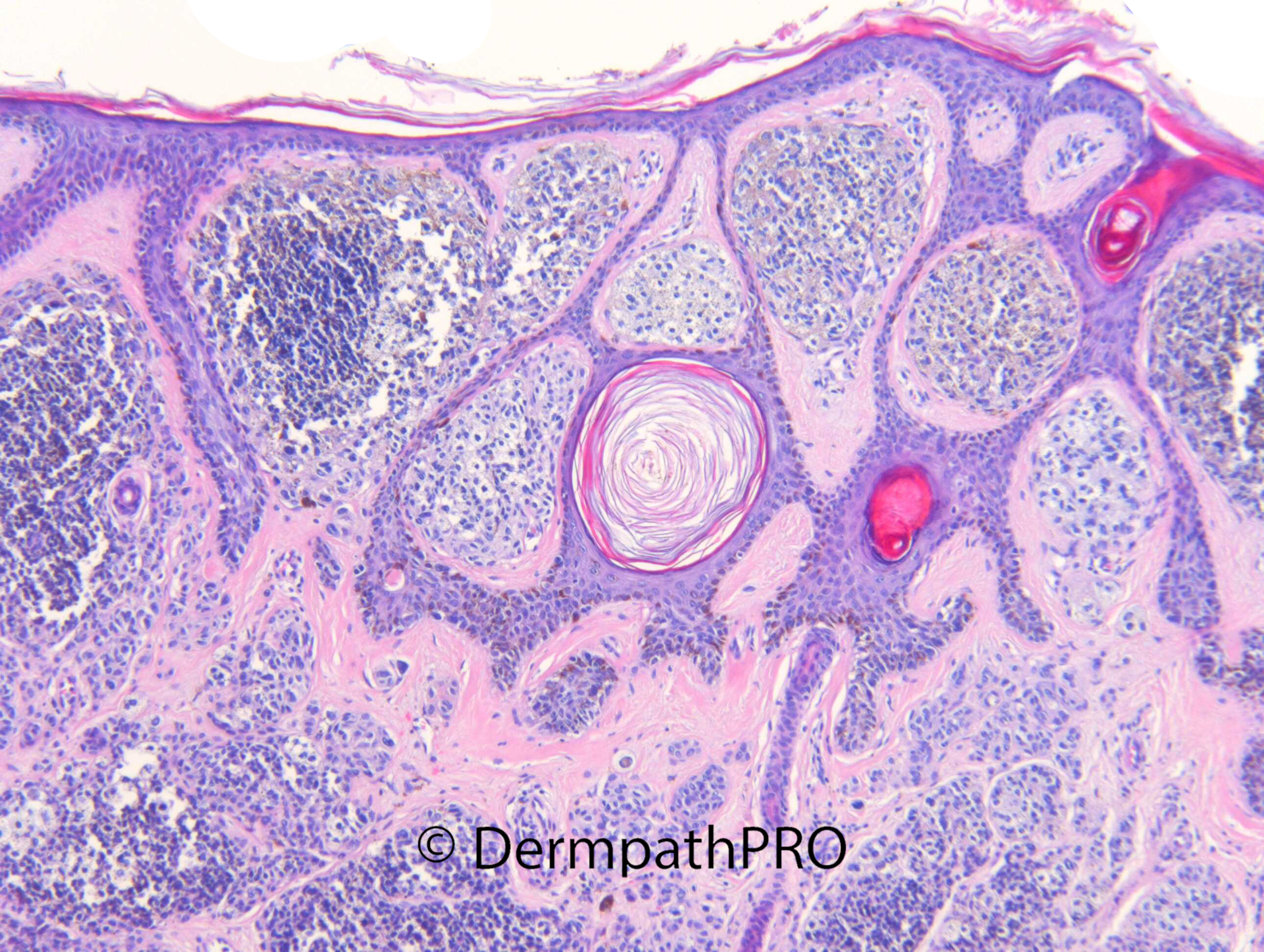
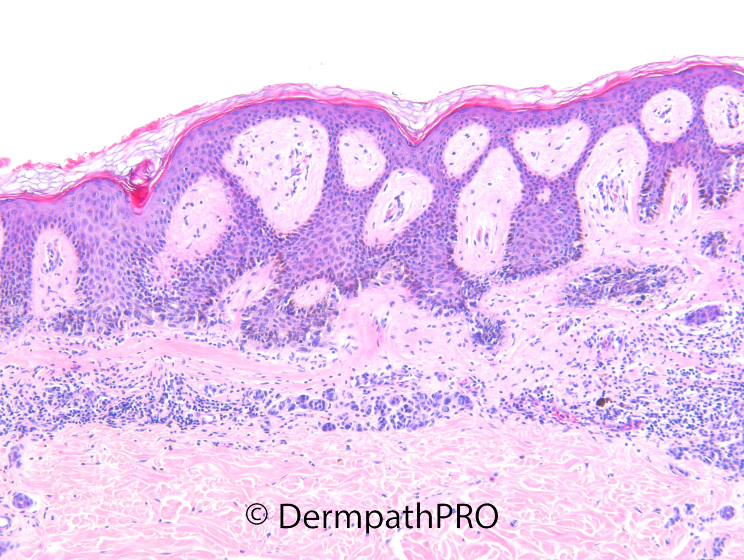
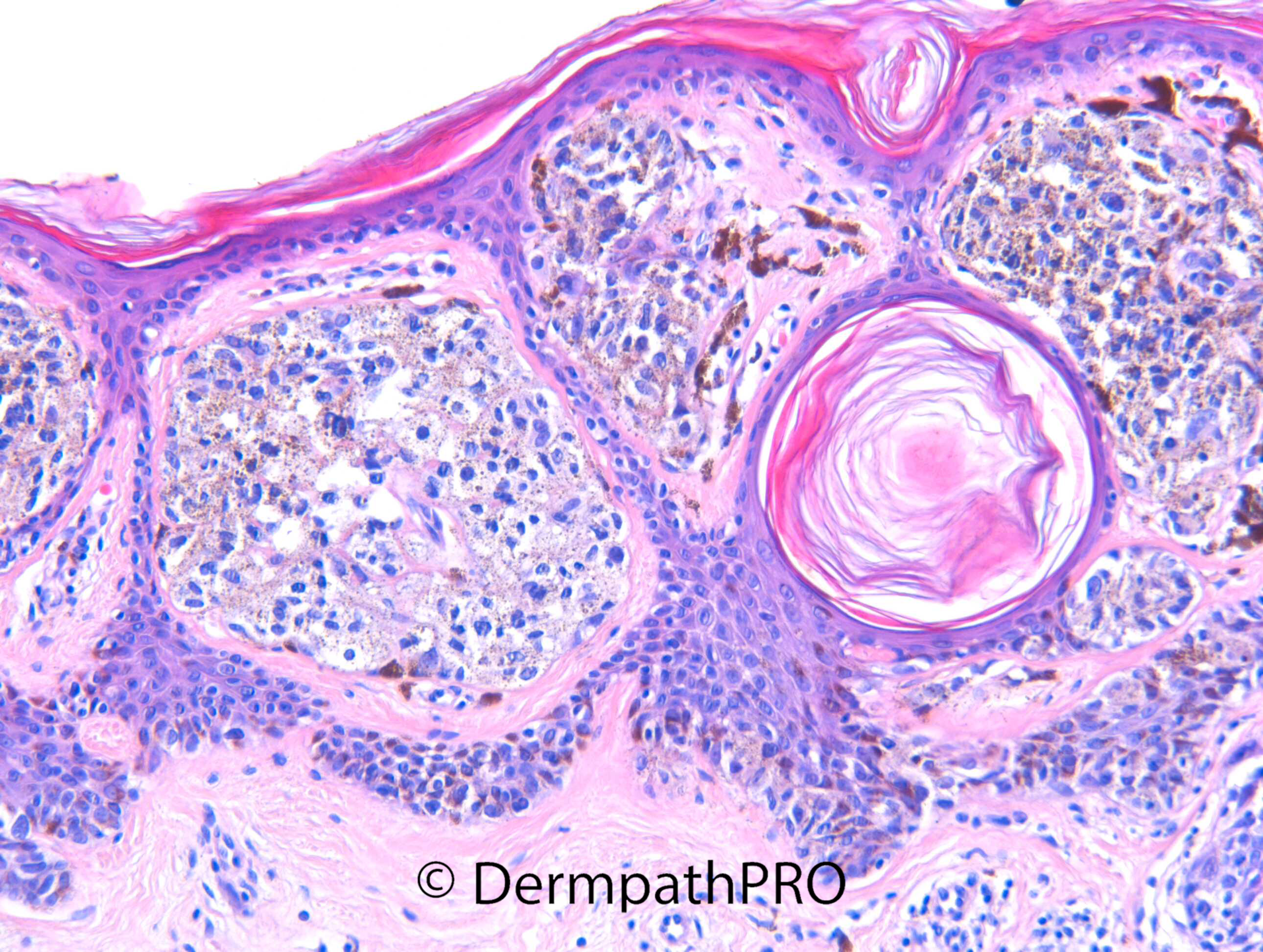
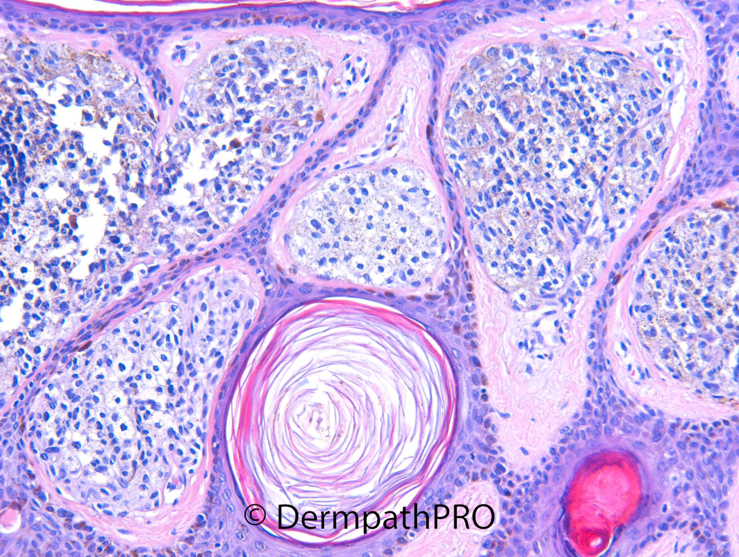
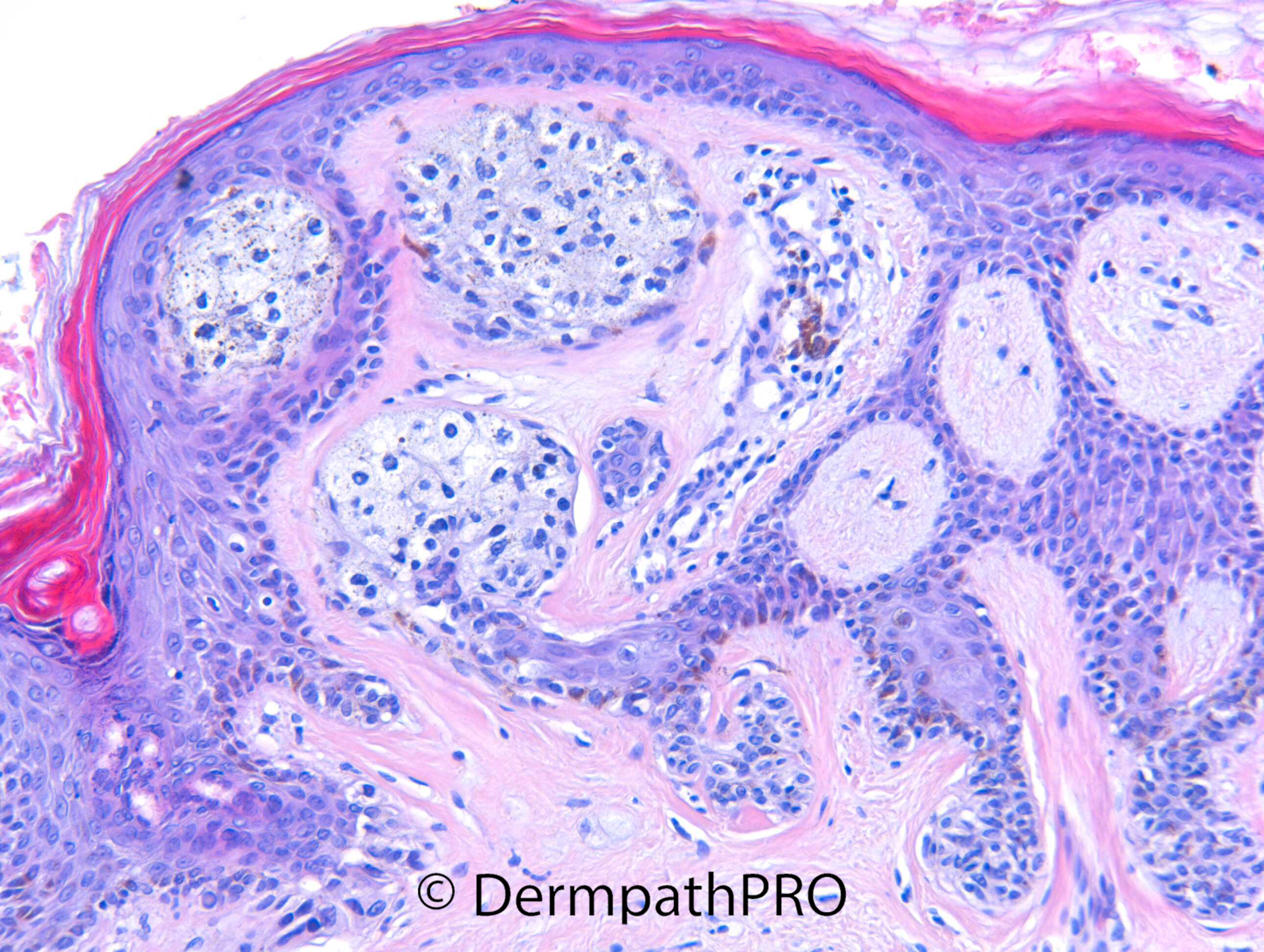
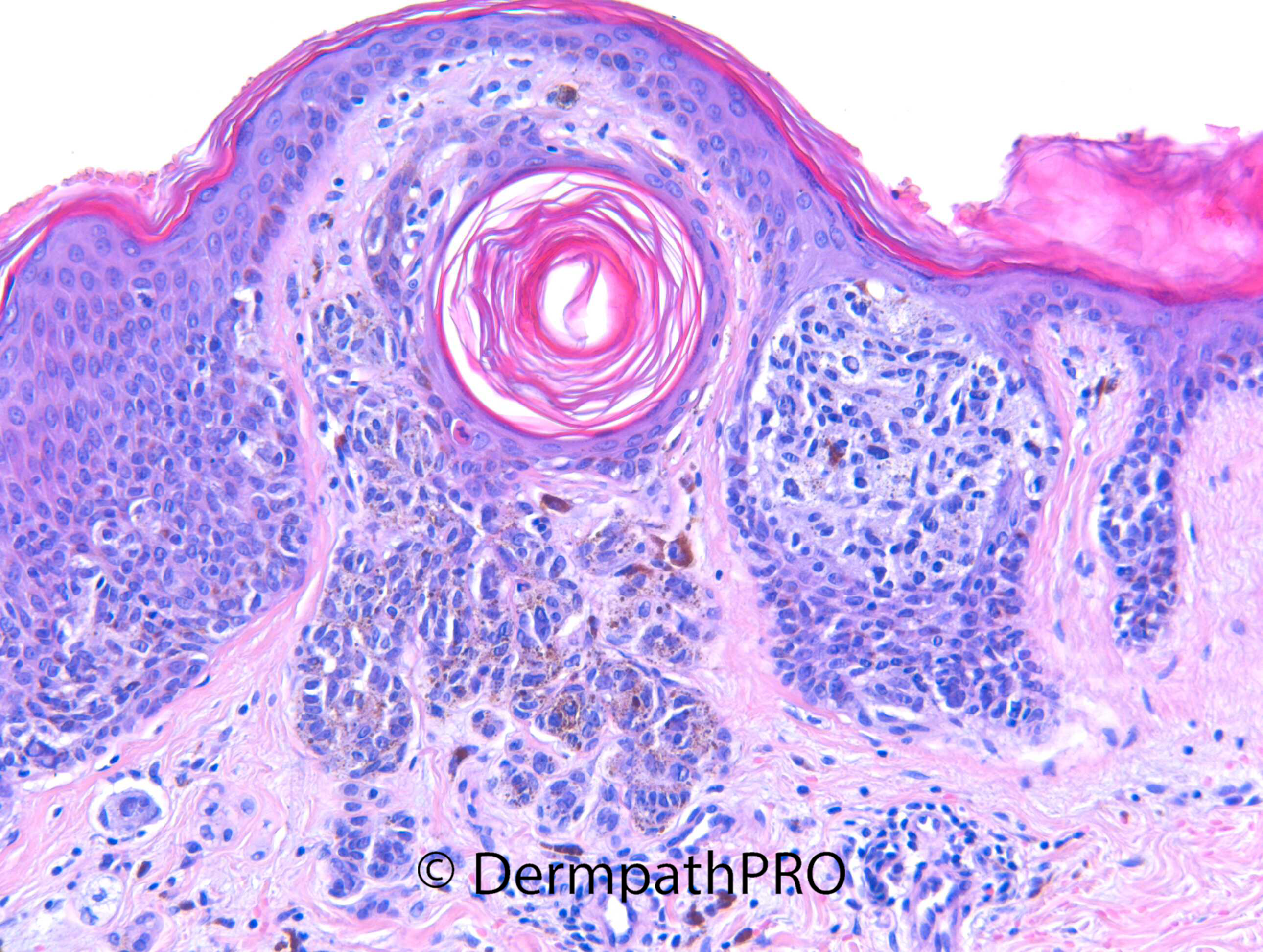
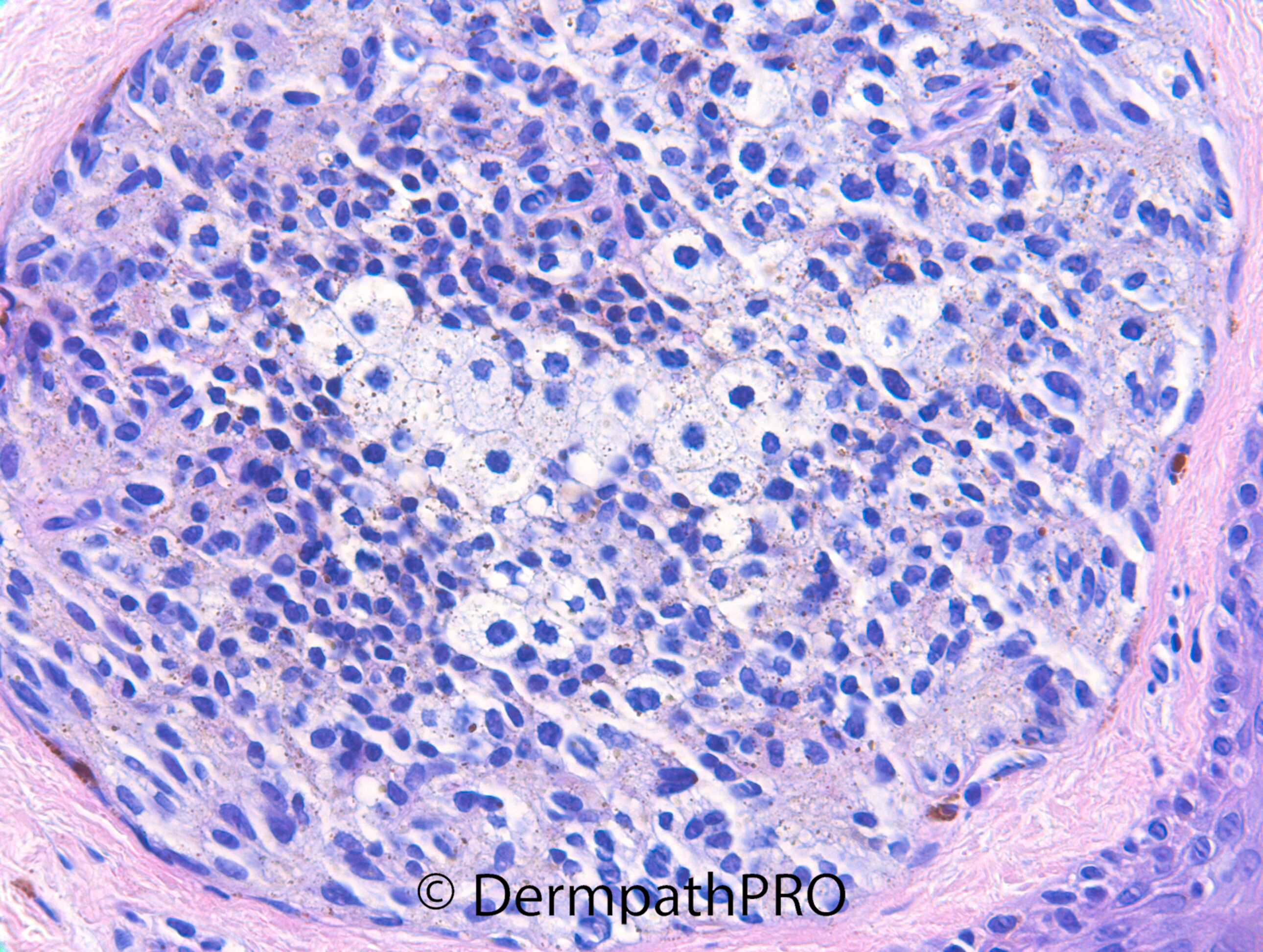
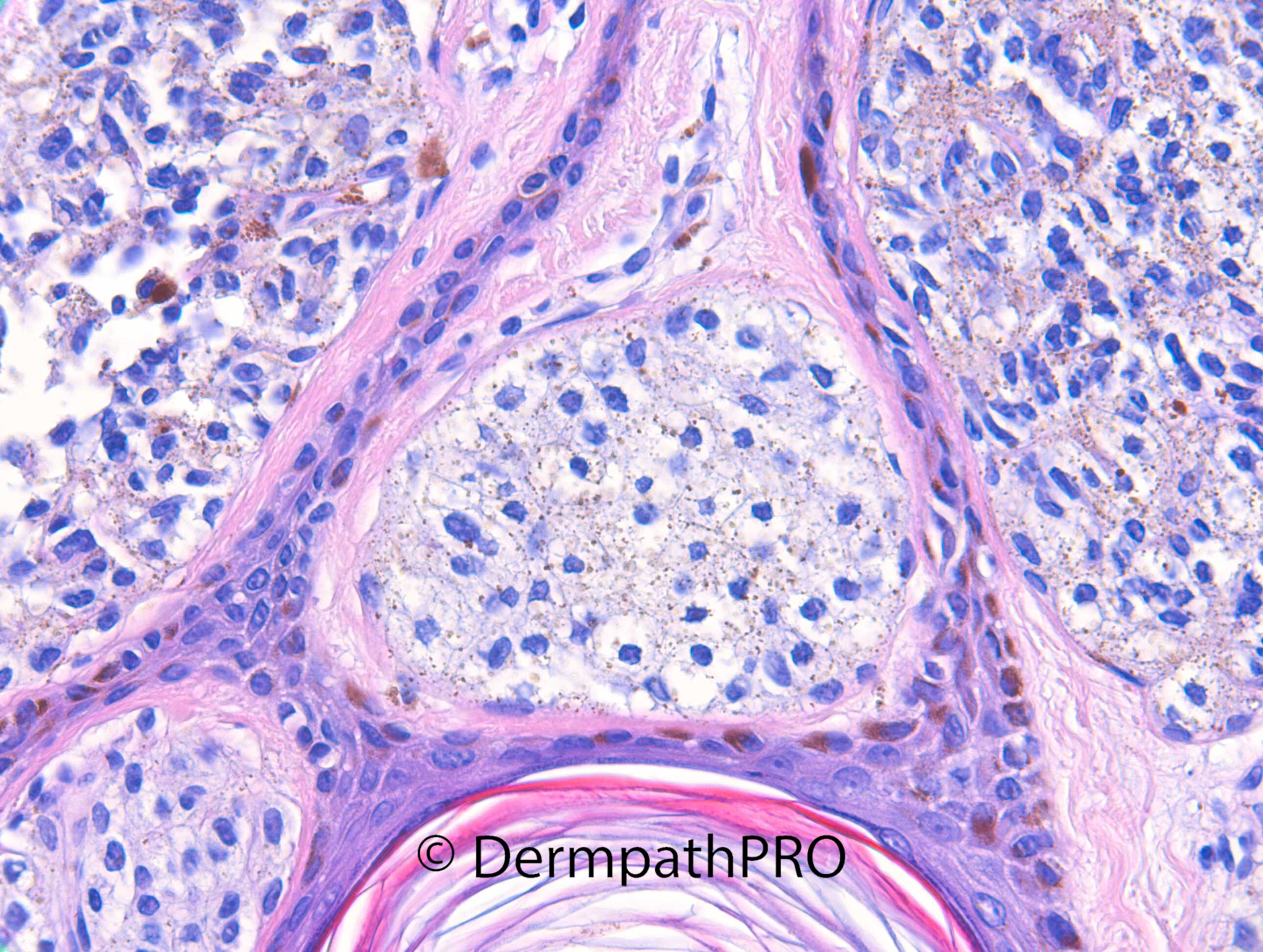
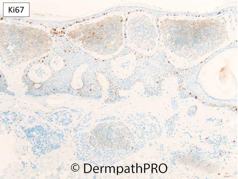
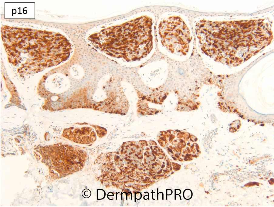
F40. Umbilicus. 1cm pigmented lesion, ?melanoma, intra-operative incision biopsy.


  Report Record

User Feedback


msofopoulos

Posted

Looks like compound nevus with site related atypia (special-site naevus of the umbilicus)

Share this comment


Link to comment
share_externally

20 hours ago, msofopoulos said:

Looks like compound nevus with site related atypia (special-site naevus of the umbilicus)

Agree.


Share this comment


Link to comment
share_externally

On 11/03/2022 at 11:41, msofopoulos said:

Looks like compound nevus with site related atypia (special-site naevus of the umbilicus)

Agree, IHC reassuring as well.

Share this comment


Link to comment
share_externally

Dr. Richard Carr

Posted

Yes special site (umbilicus) and balloon cell naevus. Ki67 low, p16 mosaic, PRAME negative.

Share this comment


Link to comment
share_externally


Join the conversation

You can post now and register later. If you have an account, sign in now to post with your account.

Guest
Add a comment...

×   Pasted as rich text.   Paste as plain text instead

  Only 75 emoji are allowed.

×   Your link has been automatically embedded.   Display as a link instead

×   Your previous content has been restored.   Clear editor

×   You cannot paste images directly. Upload or insert images from URL.

Loading...

×
×
  • Create New...