Jump to content
In this section we have spot diagnoses posted on a daily basis since June 2010, now over 4000! You can review the archived cases and read the suggested diagnoses by users and the final comment by the contributors.
Case are uploaded each week day by 10 am UK time with the correct diagnosis will generally be posted at 8 pm UK time. Why not view the most recent spot diagnosis and proffer a diagnosis?

Case Number : Case 3051 - 17 March 2022 Posted By: Saleem Taibjee

Please read the clinical history and view the images by clicking on them before you proffer your diagnosis.
Submitted Date :
   (0 reviews)

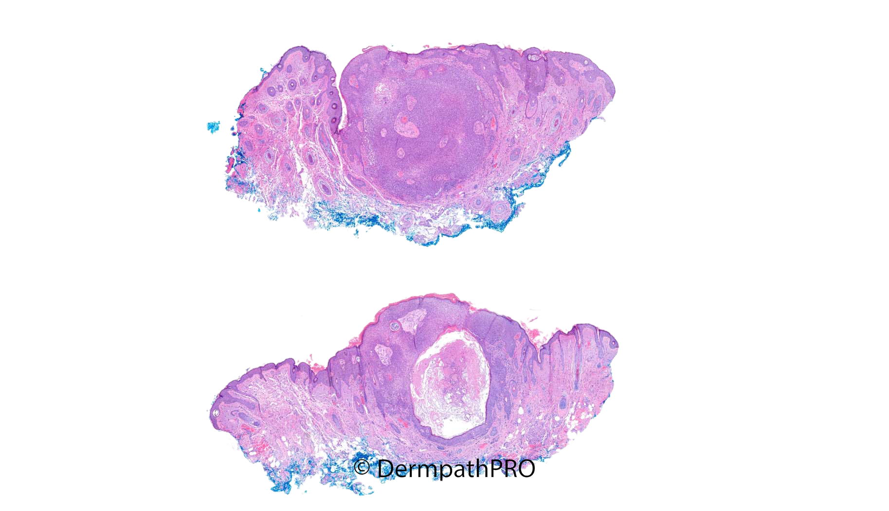
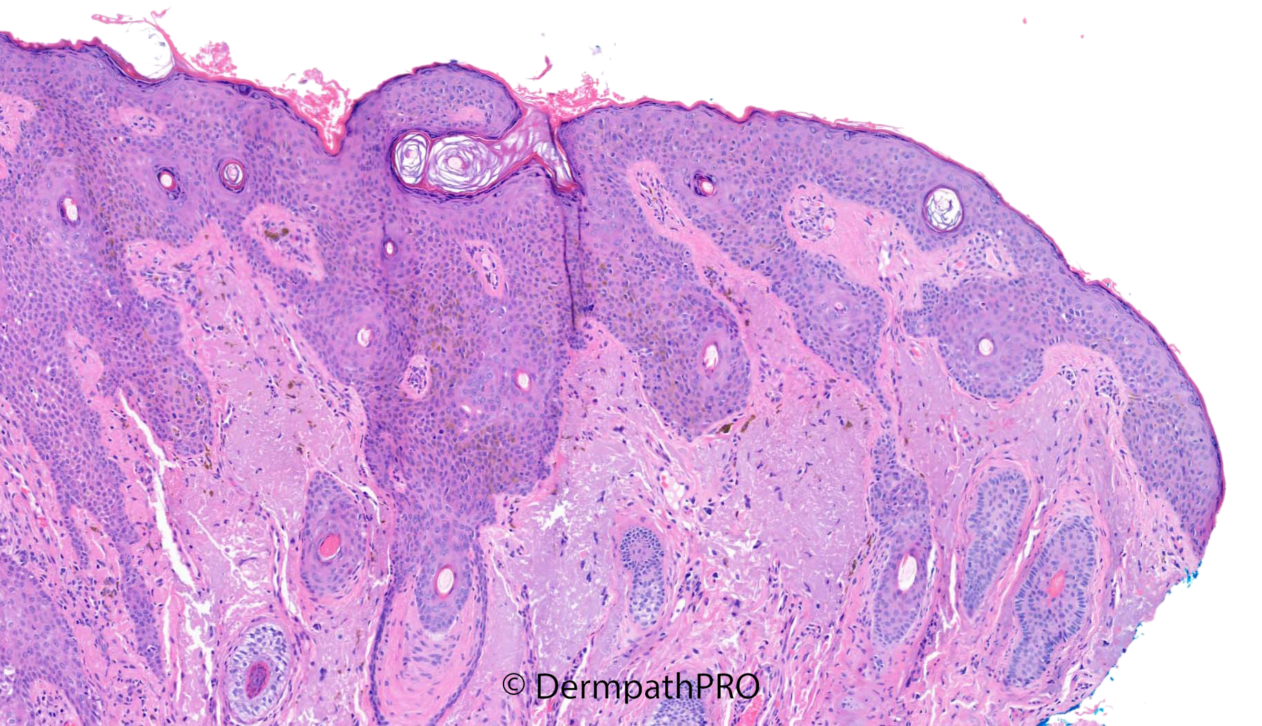
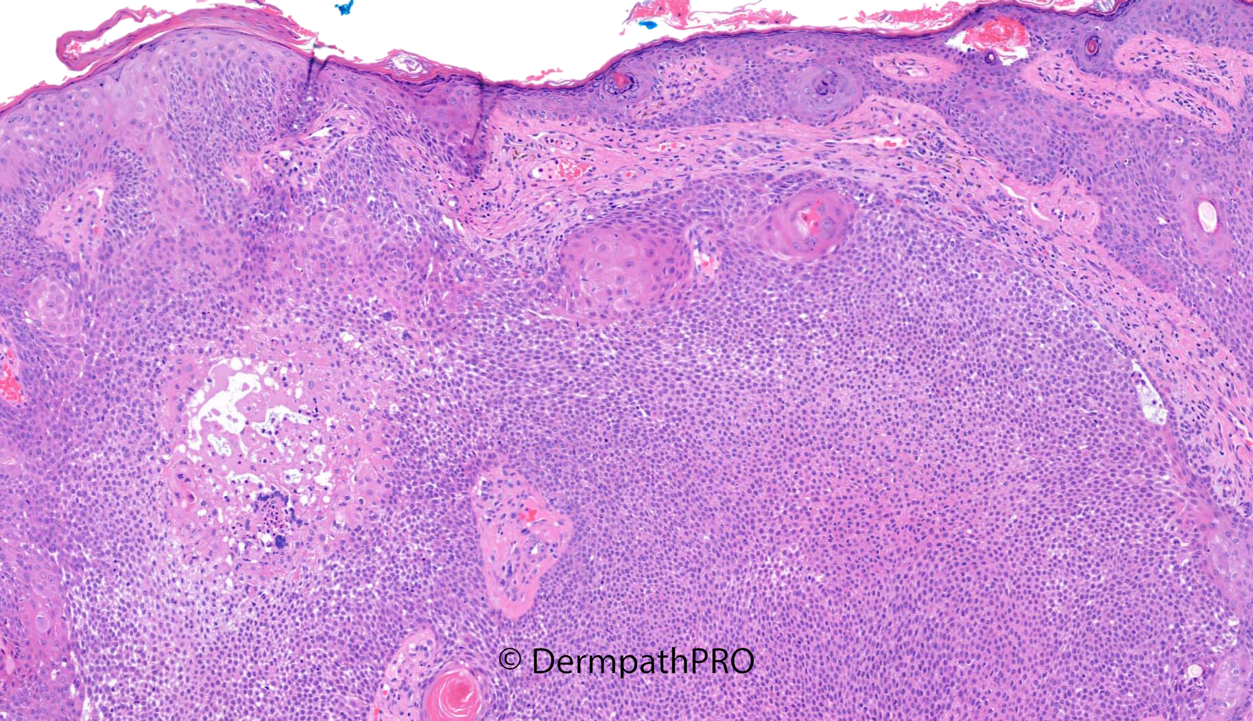
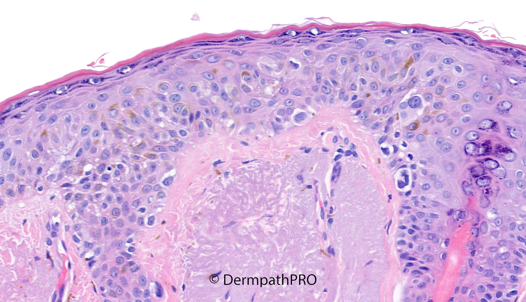
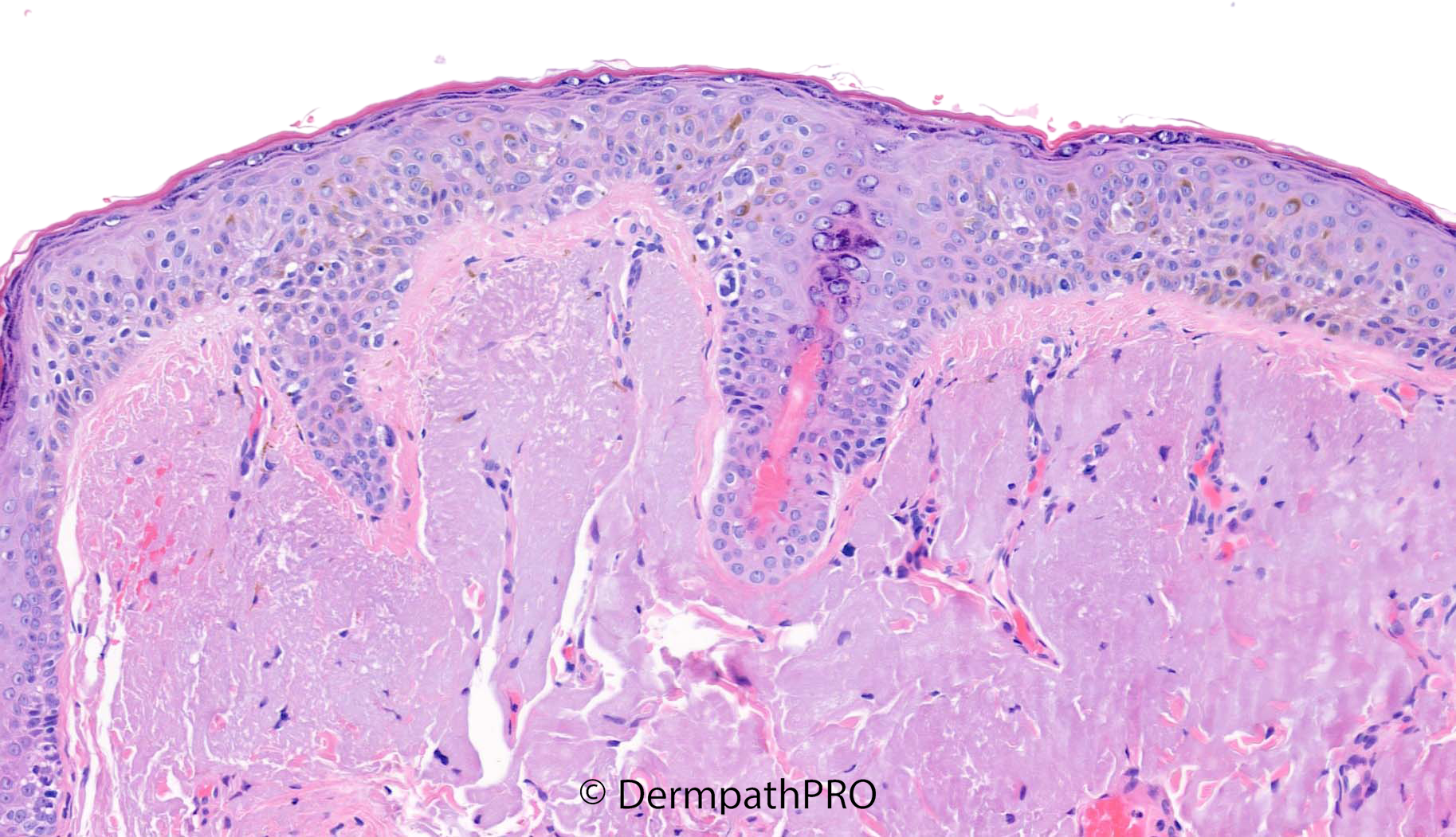
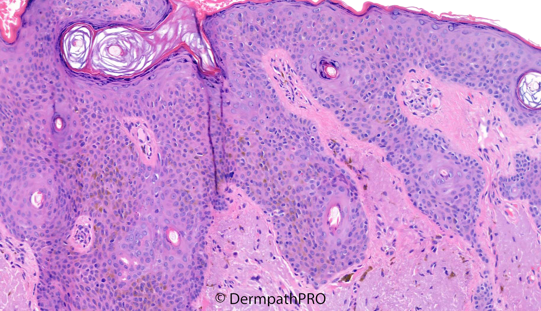
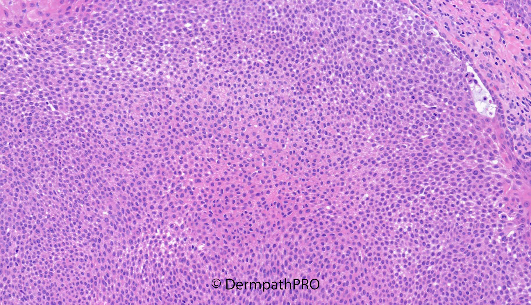
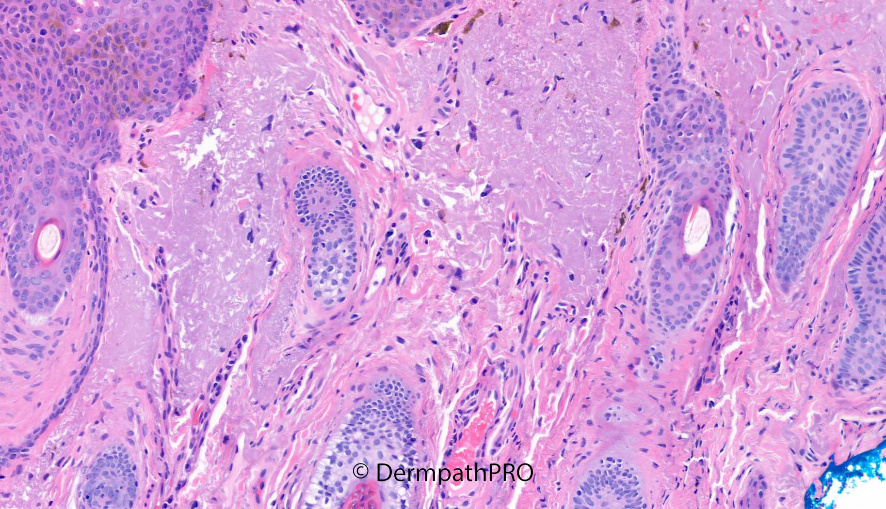
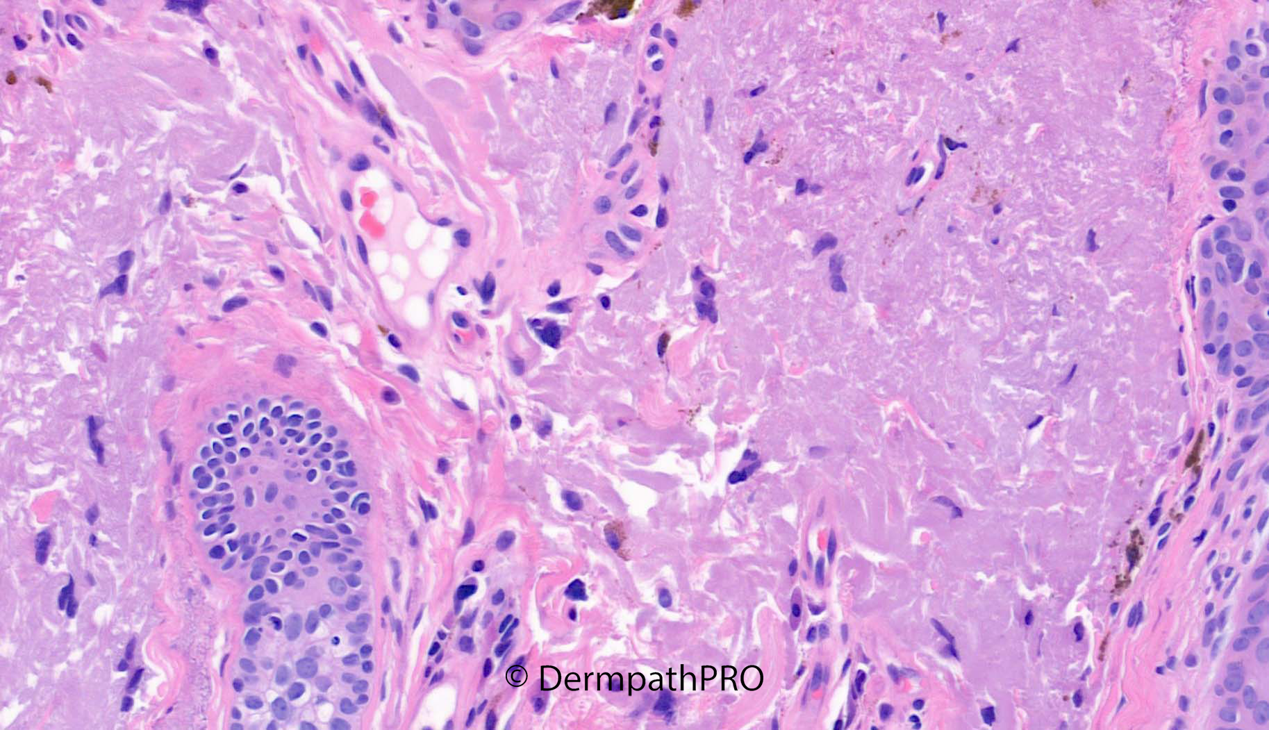
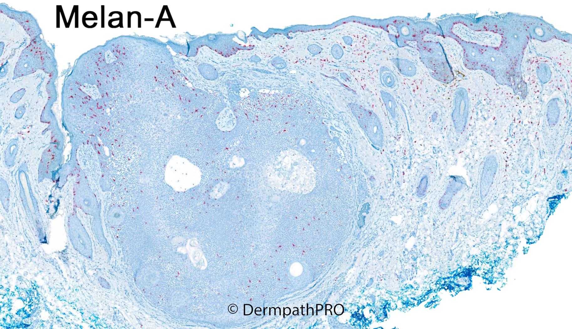
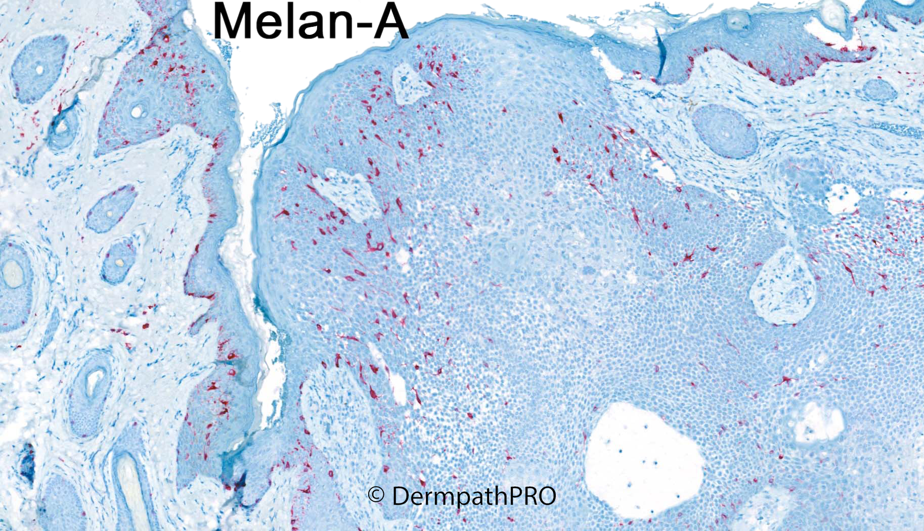
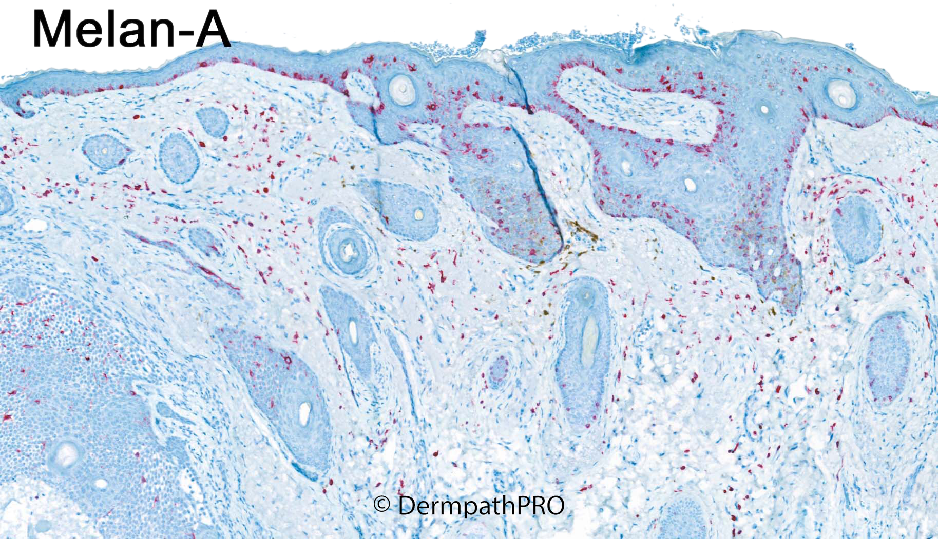
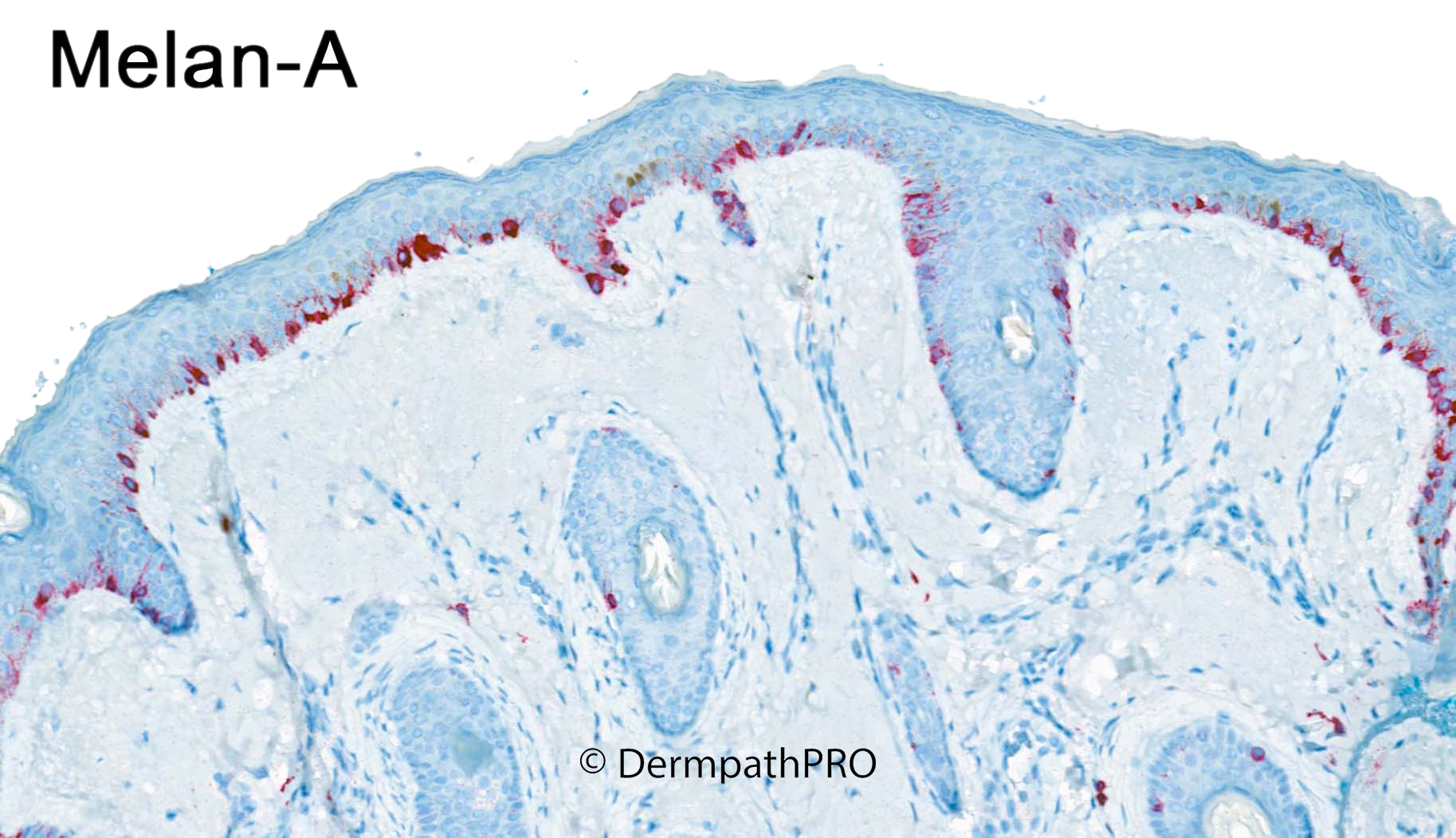
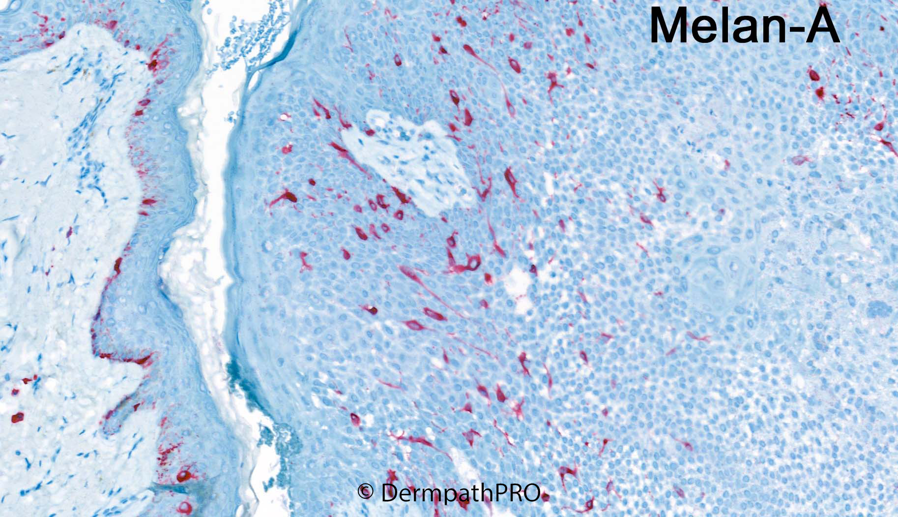
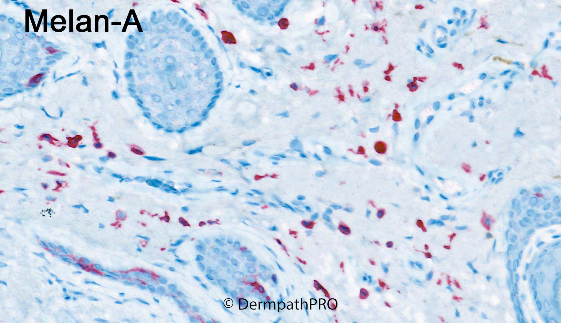
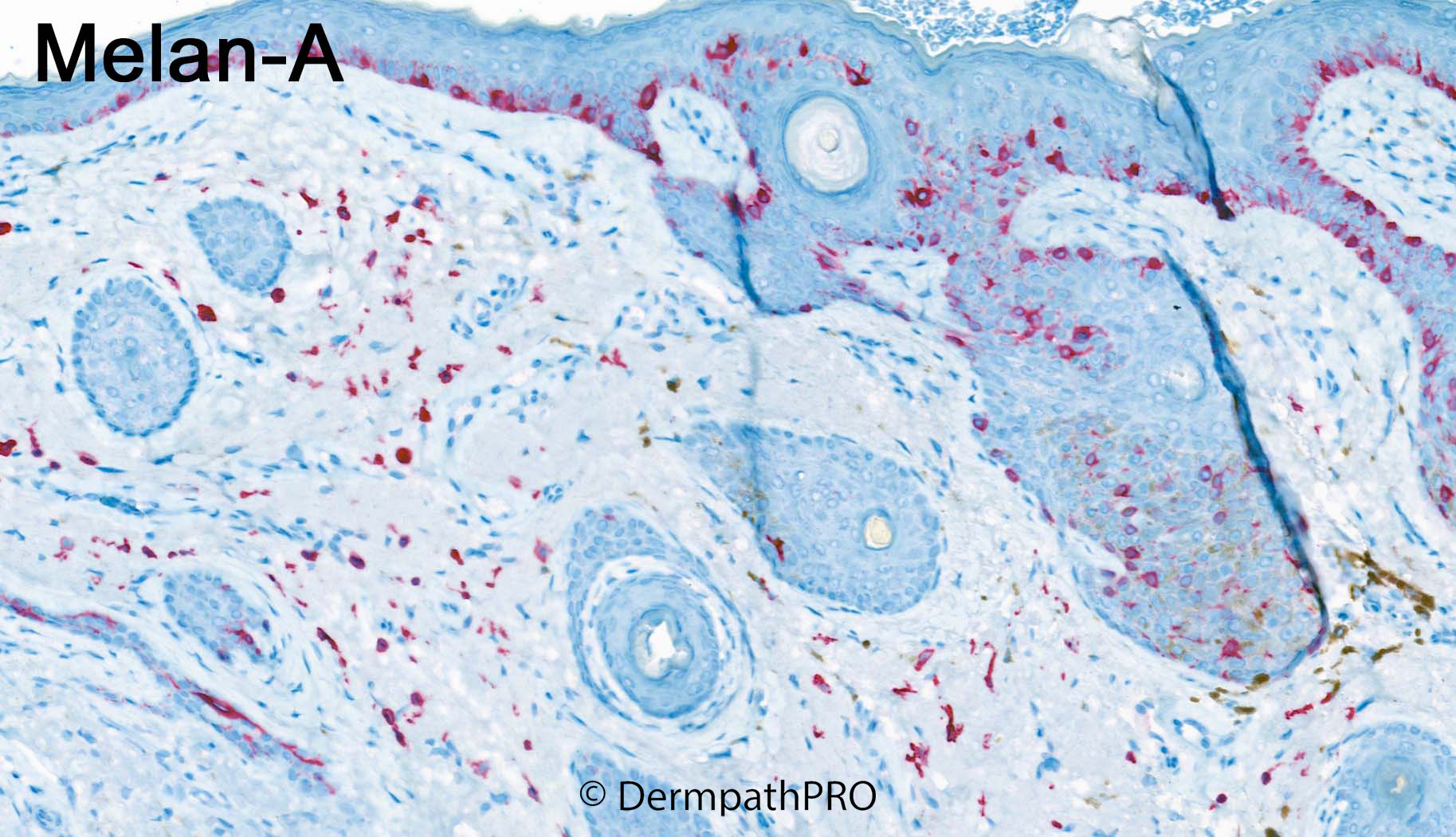
85F Excision left malar region ?lentigo maligna melanoma


  Report Record

User Feedback


Difficult... mostly not sure what to make of the dermal melan-A pos cells. Could represent dermal melanocytes that are just there singly for some reason or macrophages with melan-A reactivity. The rest looks like a pigmented poroma (+/- hidroacanthoma simplex) with melanocytic colonization. The junctional proliferation is bit atypical but at most, atypical melanocytic hyperplasia.


Share this comment


Link to comment
share_externally

Difficult for me too. ?A desmoplastic melanoma with associated  atypical intraepidermal lentiginous melanocytic hyperplasia colonizing an irritated seborrheic keratosis. PRAME?

Share this comment


Link to comment
share_externally

Meenakshi Batrani

Posted

Tough one. Looks like invasive lentigo maligna melanoma, and a nodule of poroma. Collision tumor or melanoma in situ colonizing poroma. 

Share this comment


Link to comment
share_externally

Saleem Taibjee

Posted

Yes, tricky case! I show this recent case above, but I also encountered another similar case last year, in which Melan-A showed rather unexpected dermal melanocytes (that was in a case of melanoma-in-situ).

I half-regretted requesting the Melan-A! There is an inverted follicular keratosis/irritated seb k (these can often look quite poroid), and the Melan-A picks out the reactive dendritic colonising melanocytes within this aspect i.e. 'melanoacanthoma'. Clearly this element accounts for the raised lesion which was noted clinically.

I had to look quite carefully on the H&E, and the area of junctional melanocytic hyperplasia in the adjacent epidermis was extremely focal, the images above depict the very worst area seen. Even in this area the epidermis is slightly acanthotic, and the melanocytes are scattered and not confluent. PRAME was negative. Hence I suspect a reactive solar melanocytic hyperplasia, and 'falling short of criteria for lentigo maligna' (the wording in my report).

The dermal melanocytes are very hard to see on the H&E, even when going back to the corresponding area picked out on the Melan-A (and also SOX-10). I cannot see any cytological atypia within the dermal cells. I really can't imagine that this is invasive melanoma, and the dermal melanocytes must surely be an incidental finding. 

However, there isn't very much in the literature about this sort of phenomenon. Nick Tiffin has brought to my attention this paper: Danga ME et al. Melan-A positive dermal cells in malignant melanoma in situ. J Cutan Pathol 2015;42:388-93, in which the authors described benign reactive dermal melanocytes as a not infrequent finding.

Of course, we can see genuine collision tumours, I have personally seen at least a couple of examples of lentigo maligna with BCC e.g. (one case seen with Richard Carr whilst in training) Taibjee SM et al. Lentigo maligna involving the tumour nests and stroma of a nodular basal cell carcinoma. Br J Dermatol 2007. However, I don't think the current case shown above is such an example.

BW, Saleem

Share this comment


Link to comment
share_externally


"I had to look quite carefully on the H&E, and the area of junctional melanocytic hyperplasia in the adjacent epidermis was extremely focal, the images above depict the very worst area seen. Even in this area the epidermis is slightly acanthotic, and the melanocytes are scattered and not confluent. PRAME was negative. Hence I suspect a reactive solar melanocytic hyperplasia, and 'falling short of criteria for lentigo maligna' (the wording in my report).

The dermal melanocytes are very hard to see on the H&E, even when going back to the corresponding area picked out on the Melan-A (and also SOX-10). I cannot see any cytological atypia within the dermal cells. I really can't imagine that this is invasive melanoma, and the dermal melanocytes must surely be an incidental finding."

Yep. So it’s convincing. Thanks, Saleem. 

Share this comment


Link to comment
share_externally


Join the conversation

You can post now and register later. If you have an account, sign in now to post with your account.

Guest
Add a comment...

×   Pasted as rich text.   Paste as plain text instead

  Only 75 emoji are allowed.

×   Your link has been automatically embedded.   Display as a link instead

×   Your previous content has been restored.   Clear editor

×   You cannot paste images directly. Upload or insert images from URL.

Loading...

×
×
  • Create New...