Jump to content
In this section we have spot diagnoses posted on a daily basis since June 2010, now over 4000! You can review the archived cases and read the suggested diagnoses by users and the final comment by the contributors.
Case are uploaded each week day by 10 am UK time with the correct diagnosis will generally be posted at 8 pm UK time. Why not view the most recent spot diagnosis and proffer a diagnosis?

Case Number : Case 3052 - 18 March 2022 Posted By: Dr. Richard Carr

Please read the clinical history and view the images by clicking on them before you proffer your diagnosis.
Submitted Date :
   (0 reviews)

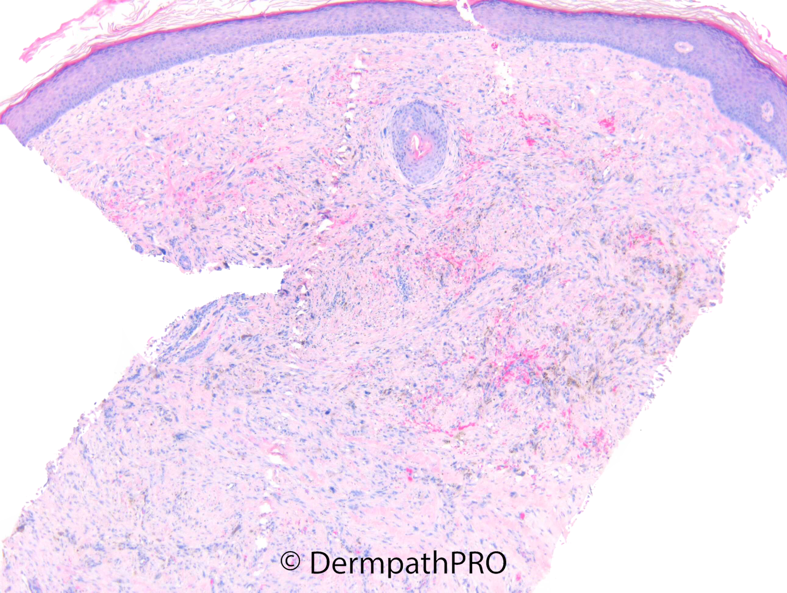
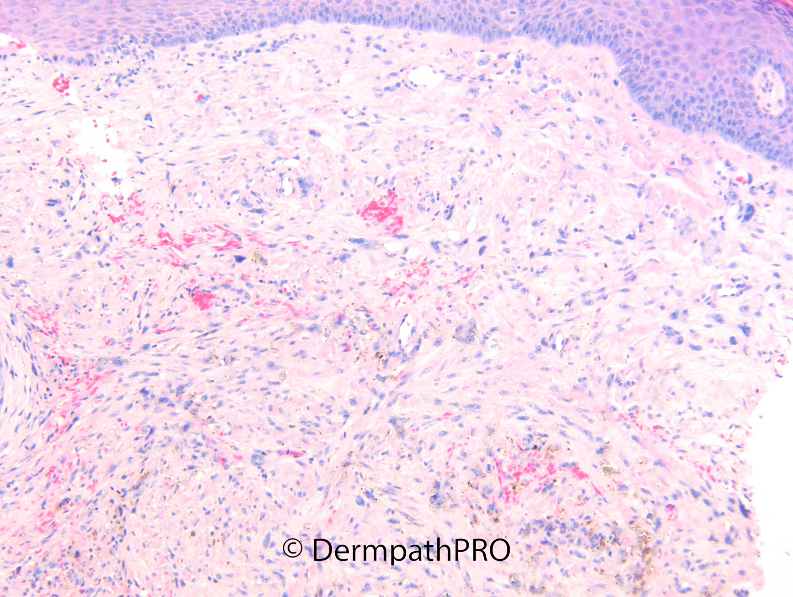
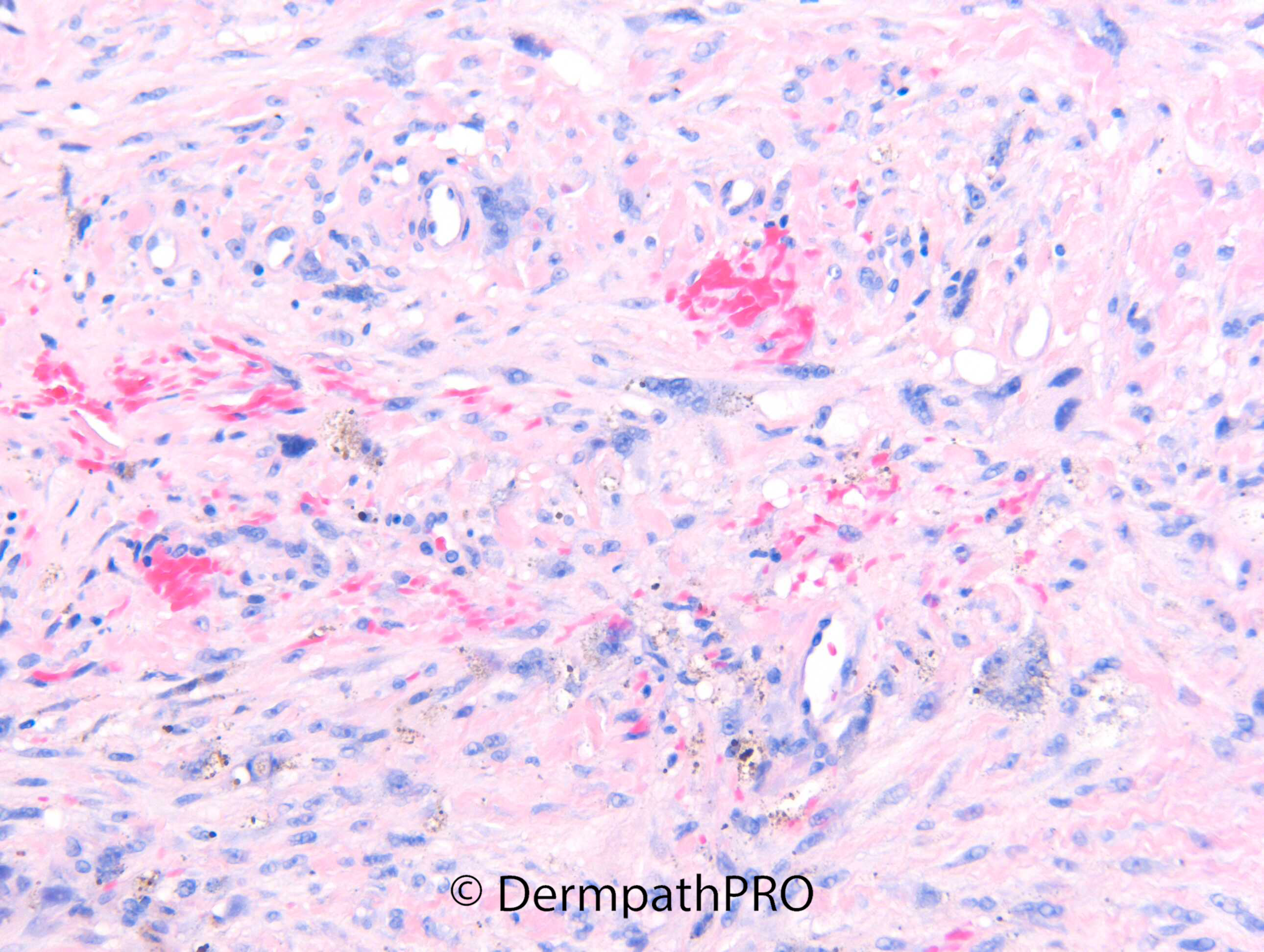
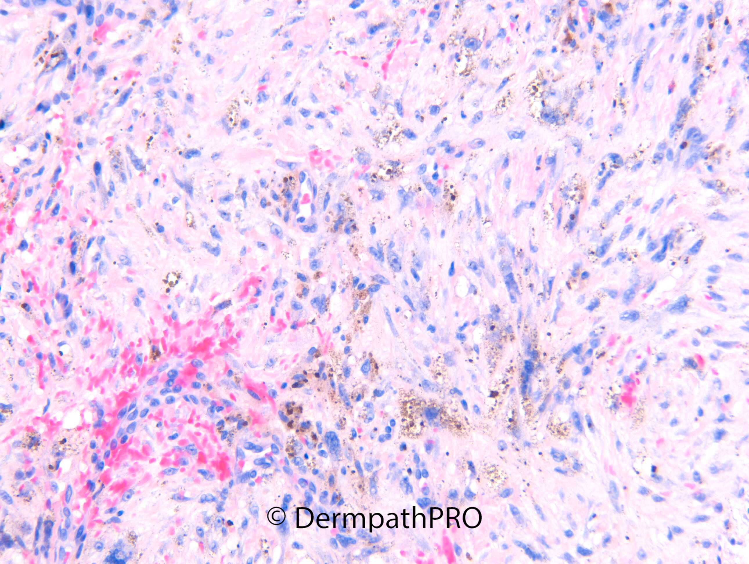
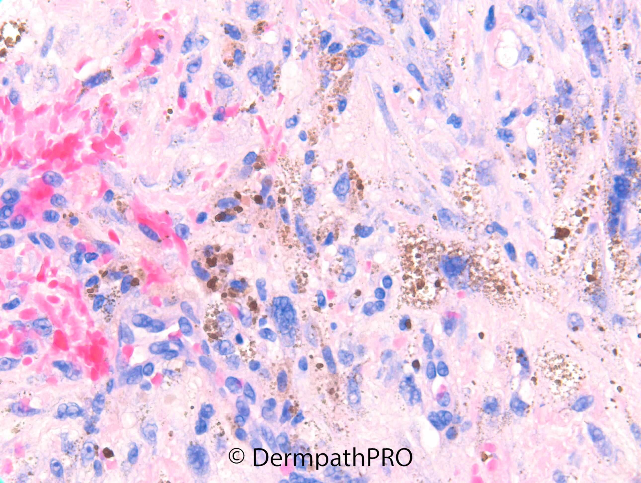
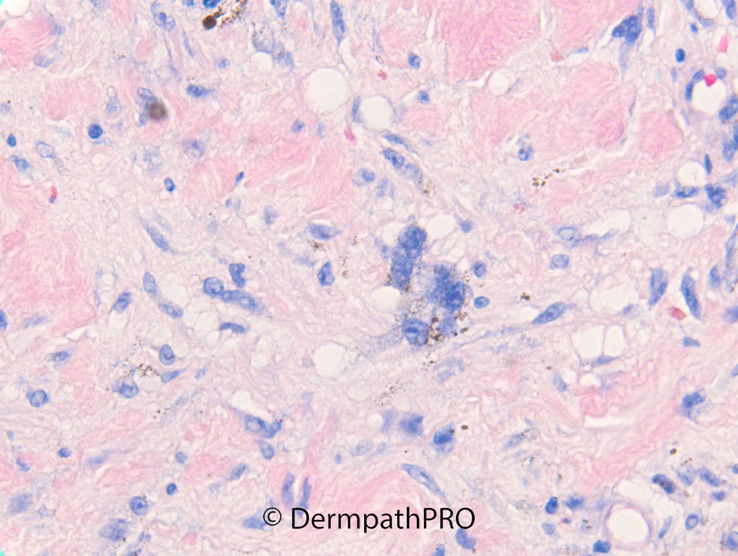
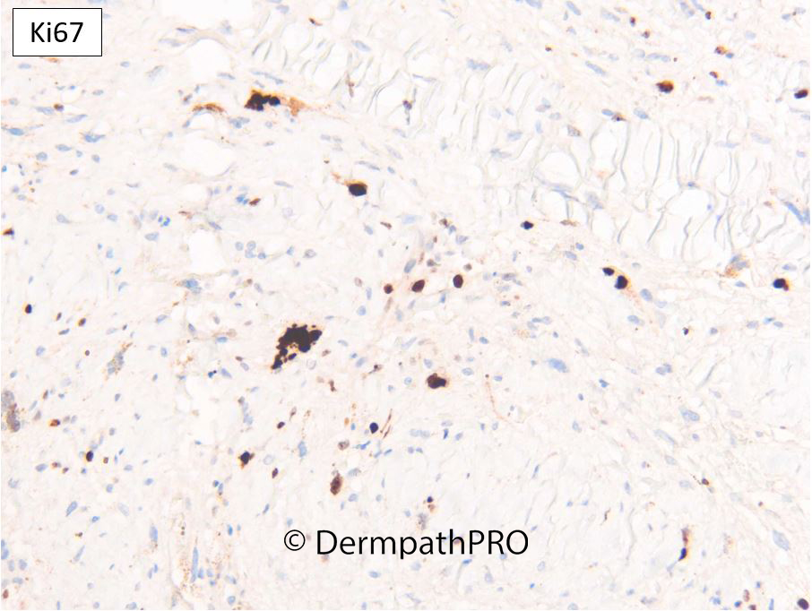
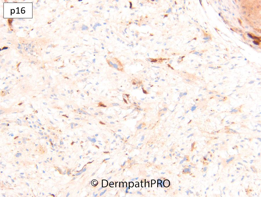
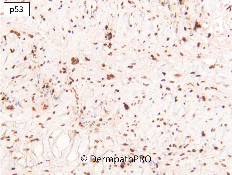
M75. Vertex scalp. 10mm purple lesion, incidental finding. ?BAK, ?angiosarcoma, ?AFX


  Report Record

User Feedback


Anil Patki

Posted

It's an infiltrative tumour of spindle cells with a hint of vasoformation and pigment which could be melanin or hemosiderin (taking into account the extravasated erythrocytes). Angiosarcoma and melanoma are the two top differentials. Immunos will be needed to arrive at the diagnosis.

Share this comment


Link to comment
share_externally

Favor AFX because of: 1-pigmented atypical multinucleated cells seems xantomatous/histiocytoid; 2-the epidermal basal focal atypical hyperplasia is keratocytic committed, not melanocytic, in keeping with AK, not with lentigo maligna. 

Share this comment


Link to comment
share_externally

Dr. Richard Carr

Posted

This is the xanthomatous variant of AFX with a lot of foam cells. Not that cellular or mitotic but p53 is stronly positive and highlights the lesion. p16 is null (>90% of cases I've tested are null). CD10 was diffusely positive. Other markers epithelial, melanocytic & vascular negative. I now do p53, p16 and Ki67 routinely for the lower cellularity and more mildly pleomorphic variants of AFX. p53 can be high or null. 

Share this comment


Link to comment
share_externally

msofopoulos

Posted

On 23/03/2022 at 14:58, Dr. Richard Carr said:

This is the xanthomatous variant of AFX with a lot of foam cells. Not that cellular or mitotic but p53 is stronly positive and highlights the lesion. p16 is null (>90% of cases I've tested are null). CD10 was diffusely positive. Other markers epithelial, melanocytic & vascular negative. I now do p53, p16 and Ki67 routinely for the lower cellularity and more mildly pleomorphic variants of AFX. p53 can be high or null. 

Have you ruled out melanoma with s100 or only by morphology? I think p53 can be pretty high on desmoplastic melanomas too. Great case with great photos!

Share this comment


Link to comment
share_externally

Dr. Richard Carr

Posted

Yes S100 negative. I wouldn't routinely add other melanocytic markers unless I was quite suspicious for melanoma as S100 is ~99% sensitive for melanoma.

My panel for a bog standard AFX is: CD10, S100, pan-keratin cocktails, p63. I've routinely been adding Ki67, p53 & p16 for the more indolent subtypes. I'll only add other differentiations specific markers e.g. vascular etc. if the case has some features suggestive. The treatment of all the differentials is excision with clear margins so in a resource poor setting arguable it's mainly academic as most lower grade, AFX-like spindle cell SCC and melanoma won't metastasise in any case.


Share this comment


Link to comment
share_externally


Join the conversation

You can post now and register later. If you have an account, sign in now to post with your account.

Guest
Add a comment...

×   Pasted as rich text.   Paste as plain text instead

  Only 75 emoji are allowed.

×   Your link has been automatically embedded.   Display as a link instead

×   Your previous content has been restored.   Clear editor

×   You cannot paste images directly. Upload or insert images from URL.

Loading...

×
×
  • Create New...