Jump to content
In this section we have spot diagnoses posted on a daily basis since June 2010, now over 4000! You can review the archived cases and read the suggested diagnoses by users and the final comment by the contributors.
Case are uploaded each week day by 10 am UK time with the correct diagnosis will generally be posted at 8 pm UK time. Why not view the most recent spot diagnosis and proffer a diagnosis?

Case Number : Case 3056 - 24 March 2022 Posted By: Saleem Taibjee

Please read the clinical history and view the images by clicking on them before you proffer your diagnosis.
Submitted Date :
   (0 reviews)

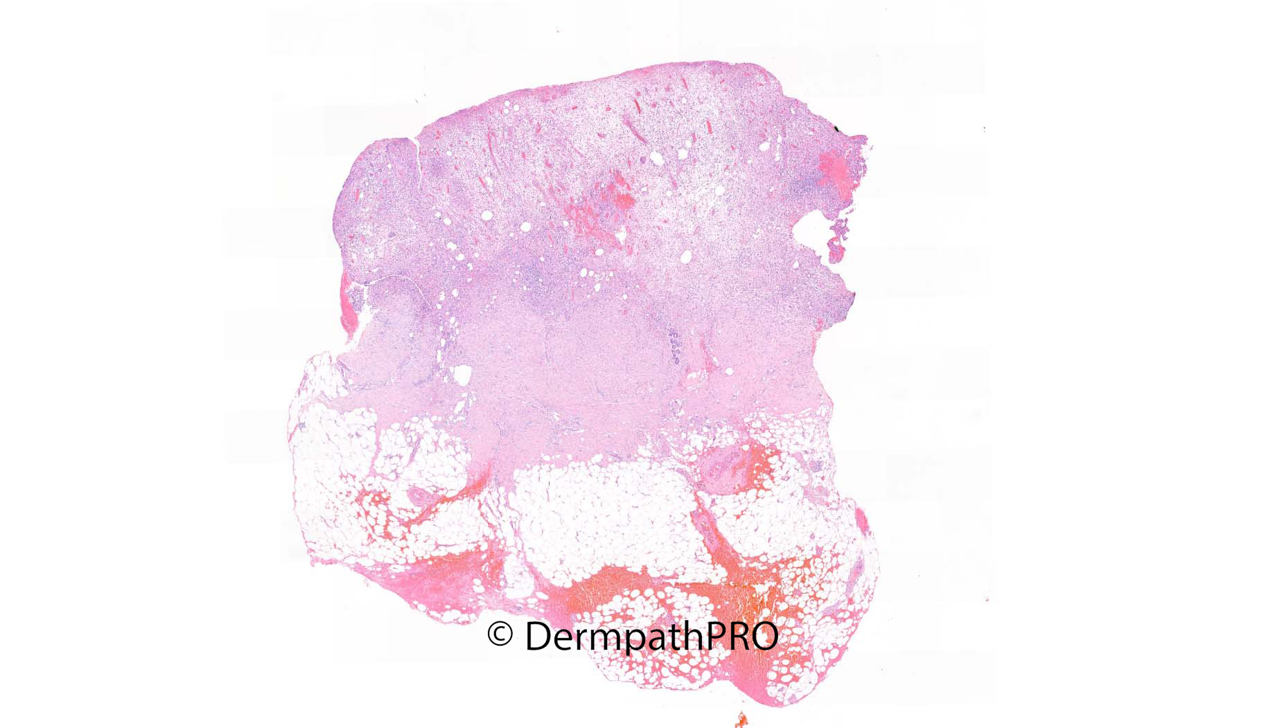
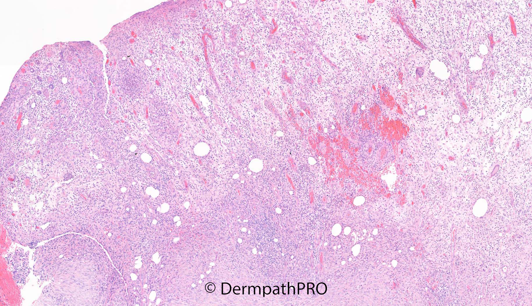
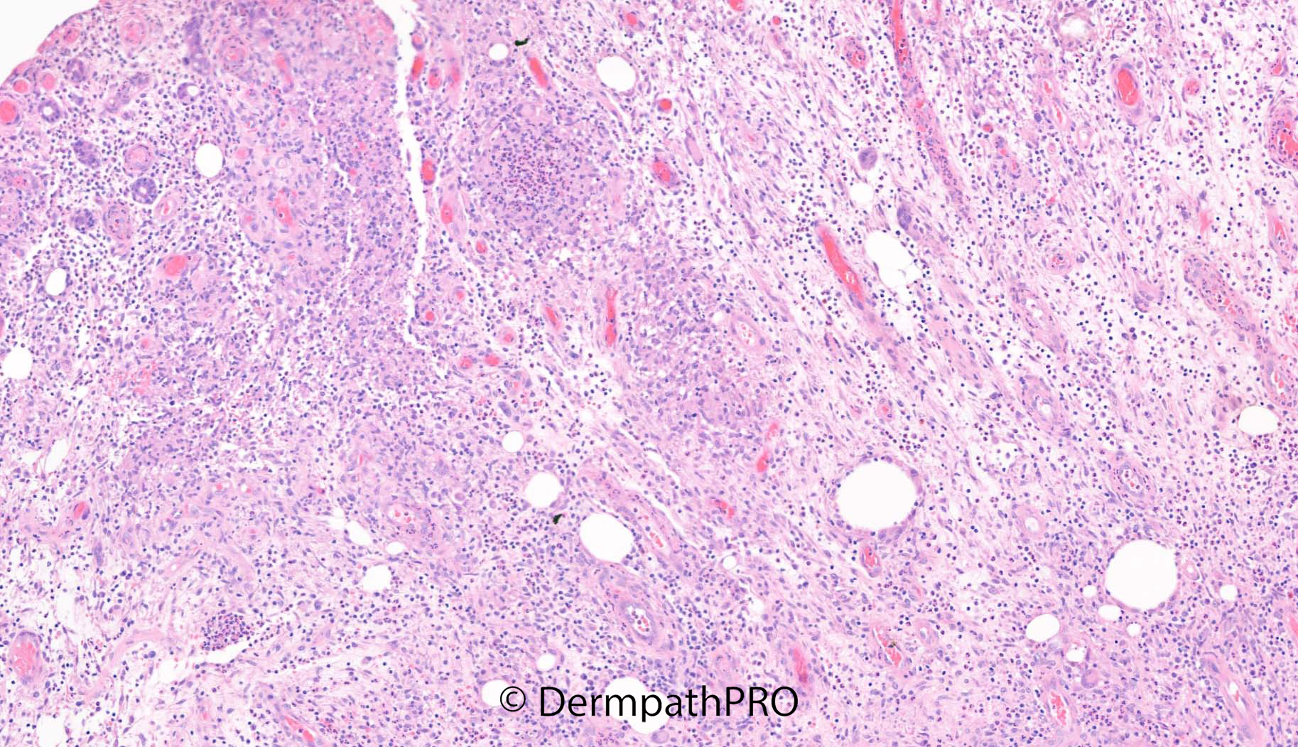
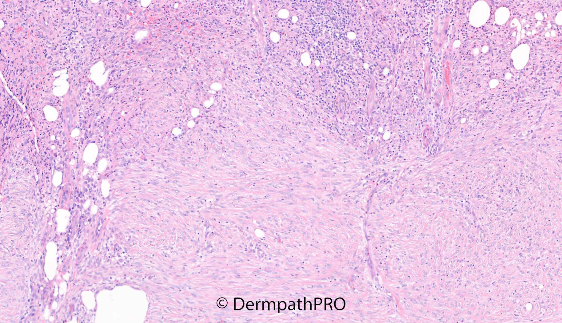
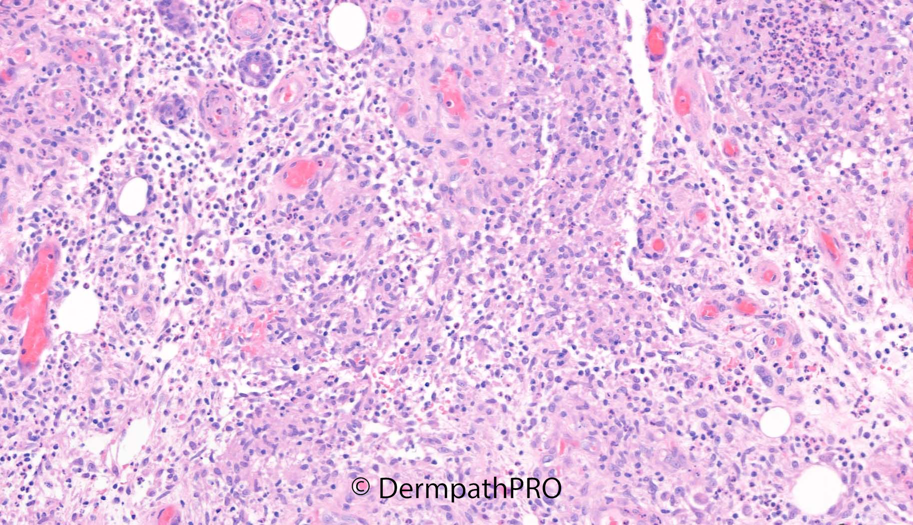
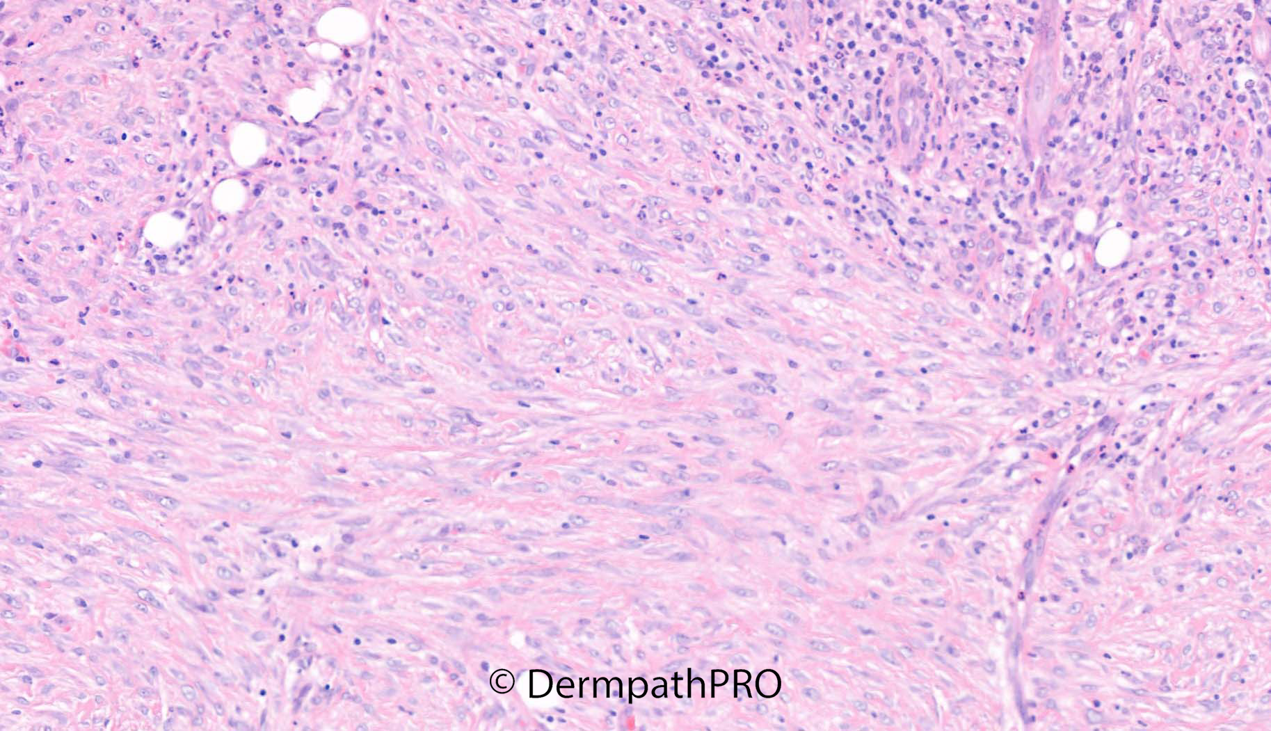
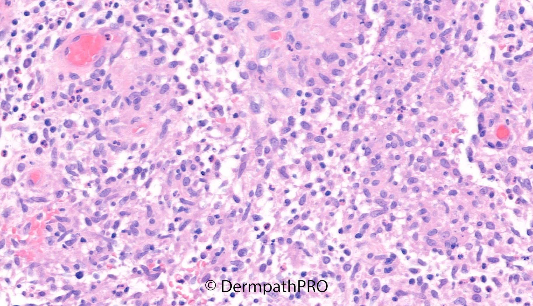
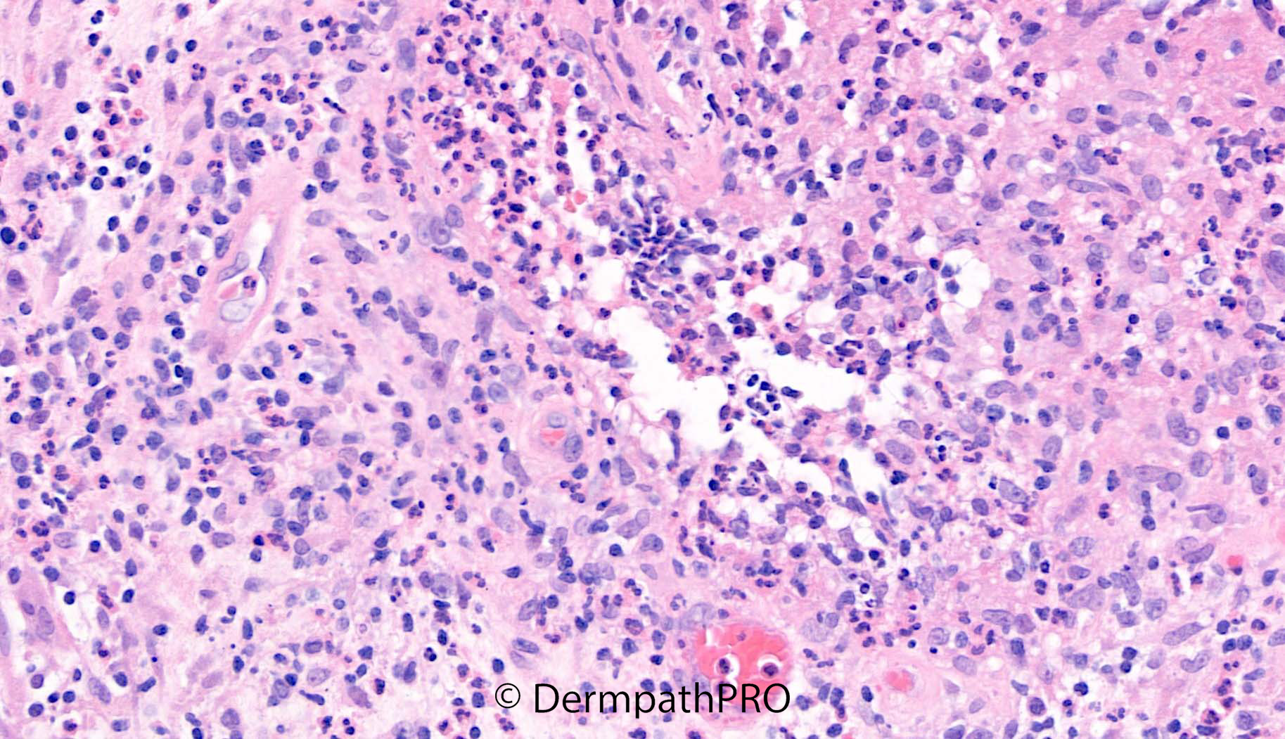
Punch biopsy left arm: 13F with ALL in remission. Erythema nodosum on shins. Now similar nodular lesions on chest and left arm ?erythema nodosum ?atypical infection ?leukaemia cutis


  Report Record

User Feedback


Looks like granulation tissue over a dermal scar with mixed inflammation - neutrophils + some mononuclear cells. Would determine the lineage of the mononuclear cells with CD3/20/34/68/MPO..correlate with prev ALL morphology

Share this comment


Link to comment
share_externally

Meenakshi Batrani

Posted

There is ulceration with scar and there appear to be suppurative granulomas. Would like to do special stains Fite and PAS to exclude mycobacterial or fungal infection. 

Share this comment


Link to comment
share_externally

Saman Fatah

Posted

Suppurative and granulomatous dermatitis with multiple vacuoles/circular spaces in the dermis. Infective aetiology requires exclusion first especially rapidly growing Non-Tuberculous Mycobacterial species including M. Chelonae. Some of the vacuoles is occasionally heaped with organism but ultimately tissue culture is  required for confirmation. 
Was the patient on oral prednisolone or or immunosuppressive medication?

If triple tissue cultures is sterile then neutrophilic diseases is probably in next line of DDx if clinical picture fits.

Share this comment


Link to comment
share_externally

Saleem Taibjee

Posted

Great responses as usual! Saman, you've even correctly identified the organism. This was disseminated Mycobacterium chelonae infection, arising at the site of a central line insertion.

It was a good personal learning case for me.

I initially signed out the case as features of granulation tissue. When we reviewed the case at MDT shortly afterwards, including the clinical features in which the suspicion of one of the senior dermatologists for atypical infection became apparent , I requested special stains. The vast majority of the histology is granulation tissue, but I have picked out the very focal suppurative granulomatous elements on retrospective review of the histology and shown these in the above last 2 images.

Even on the ZN, acid-fast bacilli were only apparent in a small detached fragment in the biopsy after careful scrutiny (see below).

The message is a low threshold for special stains for organisms in any immunocompromised patient, and a careful look at the ZN (all parts of the section) on high power.

BW, Saleem

09959_2.0x ZN.jpg09959_63.0x ZNb.jpg

09959_63.0x ZNc.jpg


09959_63.0x ZN.jpg

Share this comment


Link to comment
share_externally

Saman Fatah

Posted

Thanks Saleem for the update. The histology of these NTM infections can be very variable and subtle compared to classic dermpath/dermatology teaching and textbook description. As you rightly mentioned one has to be always vigilant about infective causes in immunocompromised hosts. M. Chelonae is ubiquitous in the environment and relatively common isolate in the UK compared to other species (it was just an educated guess based on most likely scenario). Dr Carr has a special phrase that he has used for a similar situation but unfortunately I can not recall it well at present.

Share this comment


Link to comment
share_externally


Join the conversation

You can post now and register later. If you have an account, sign in now to post with your account.

Guest
Add a comment...

×   Pasted as rich text.   Paste as plain text instead

  Only 75 emoji are allowed.

×   Your link has been automatically embedded.   Display as a link instead

×   Your previous content has been restored.   Clear editor

×   You cannot paste images directly. Upload or insert images from URL.

Loading...

×
×
  • Create New...