Jump to content
In this section we have spot diagnoses posted on a daily basis since June 2010, now over 4000! You can review the archived cases and read the suggested diagnoses by users and the final comment by the contributors.
Case are uploaded each week day by 10 am UK time with the correct diagnosis will generally be posted at 8 pm UK time. Why not view the most recent spot diagnosis and proffer a diagnosis?

Case Number : Case 3061 - 31 March 2022 Posted By: Saleem Taibjee

Please read the clinical history and view the images by clicking on them before you proffer your diagnosis.
Submitted Date :
   (0 reviews)

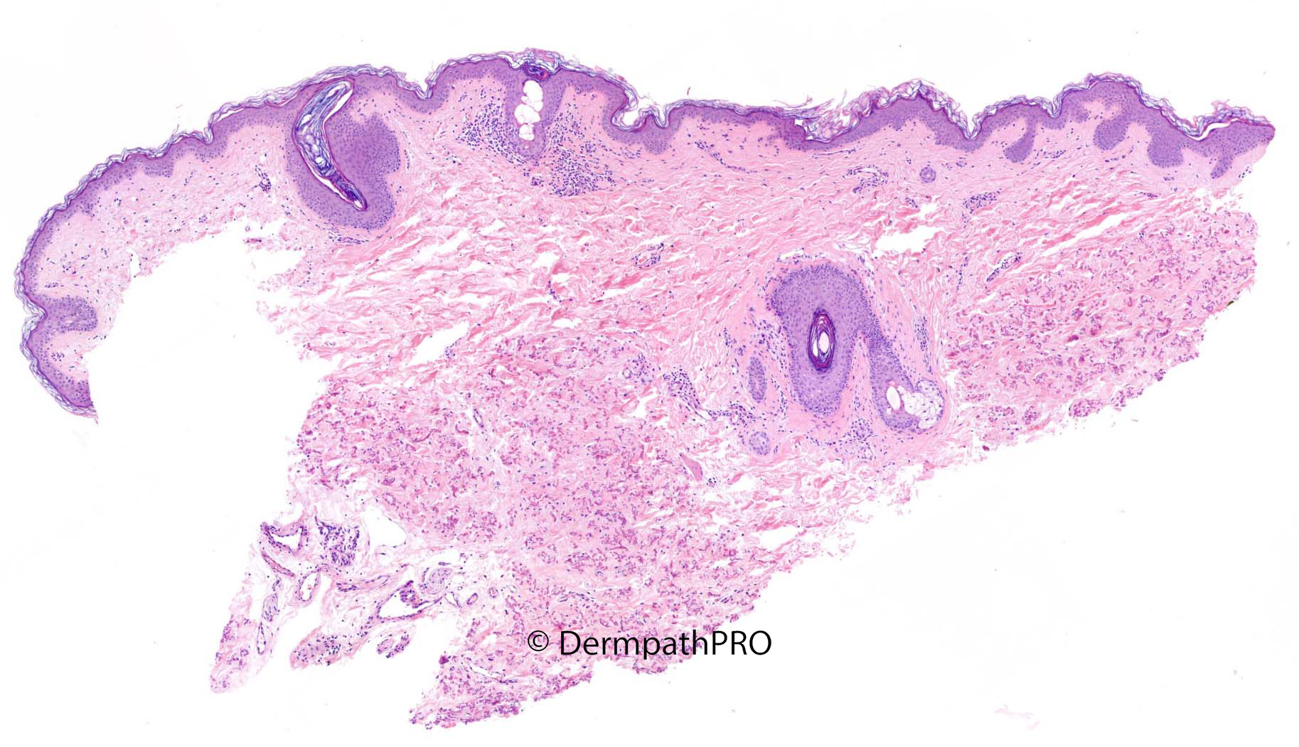
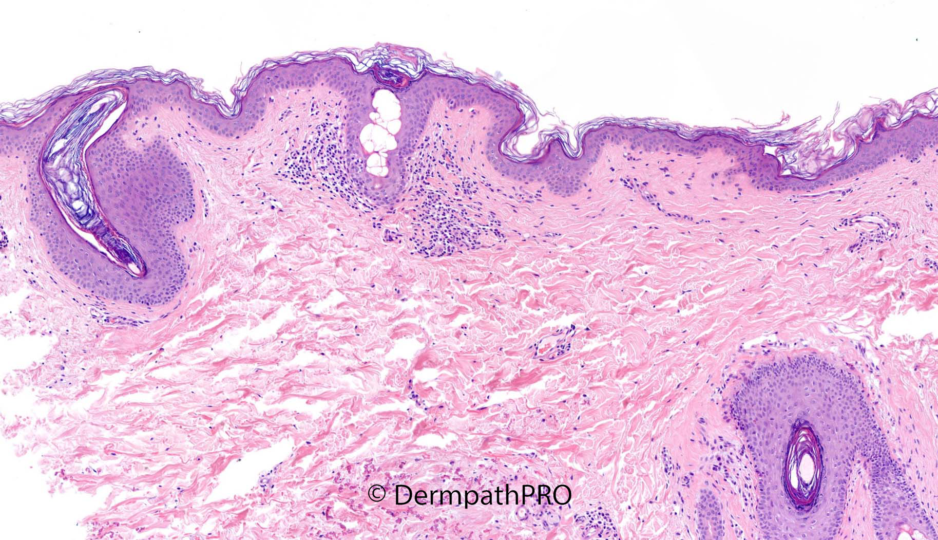
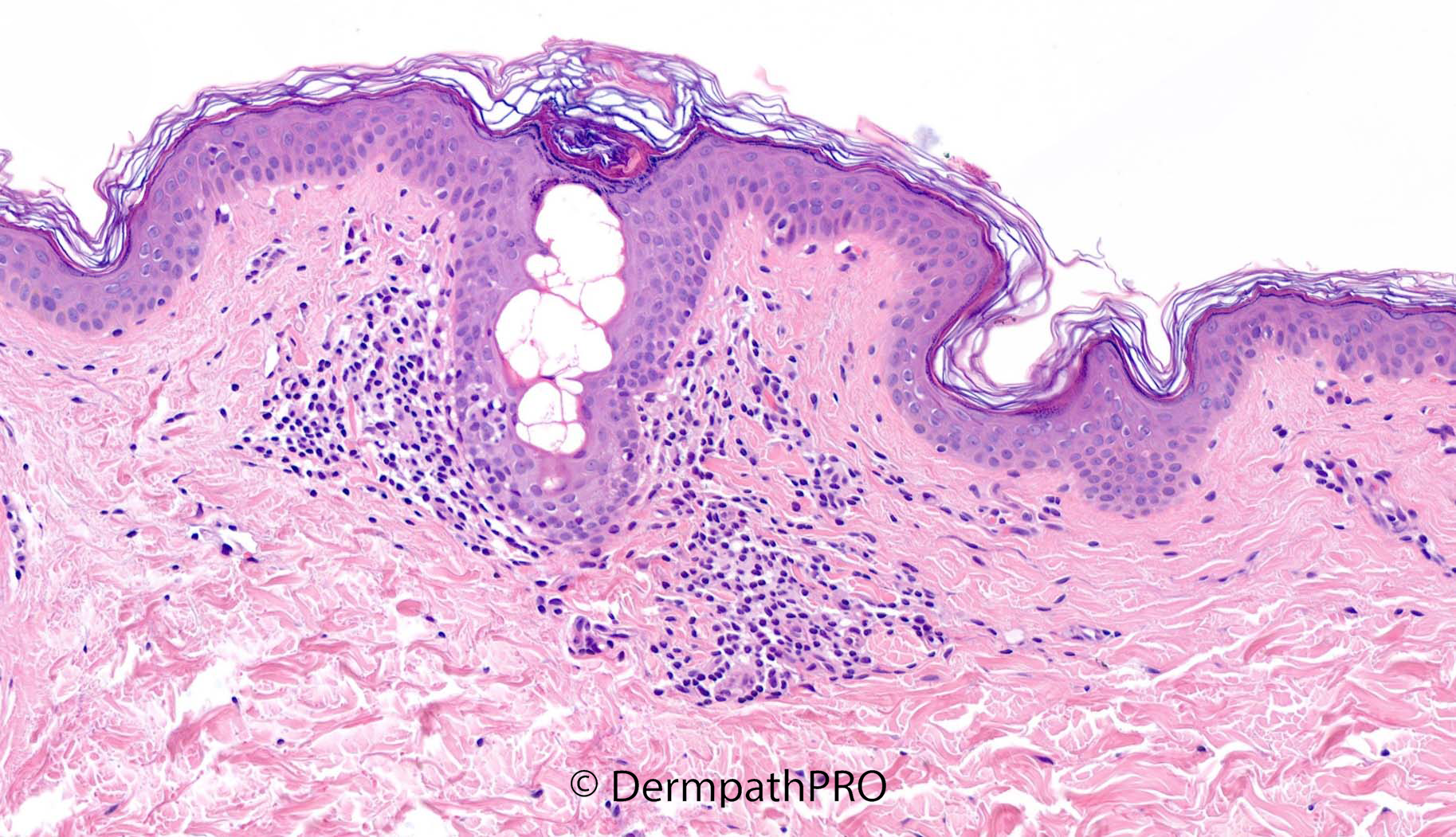
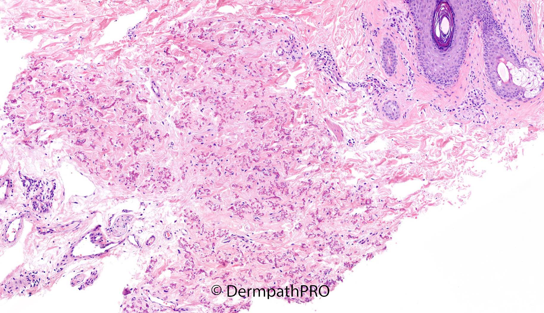
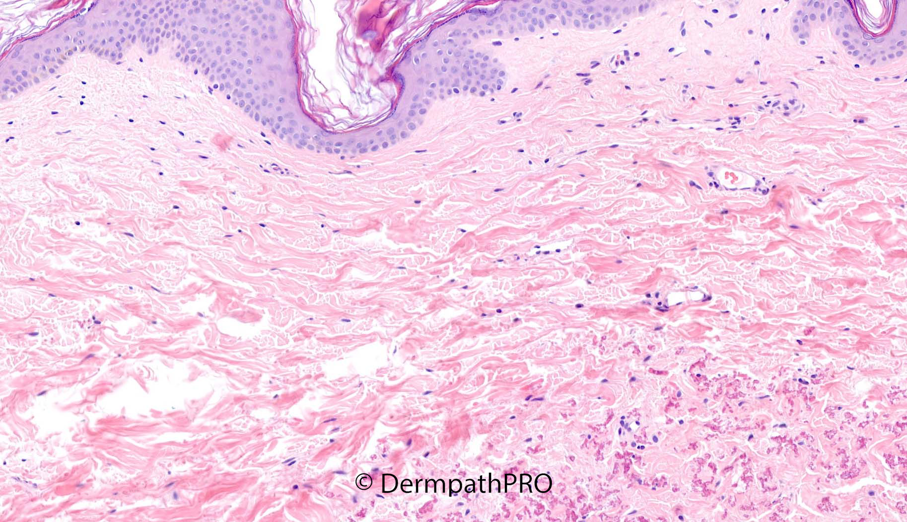
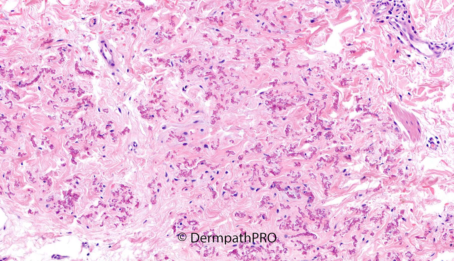
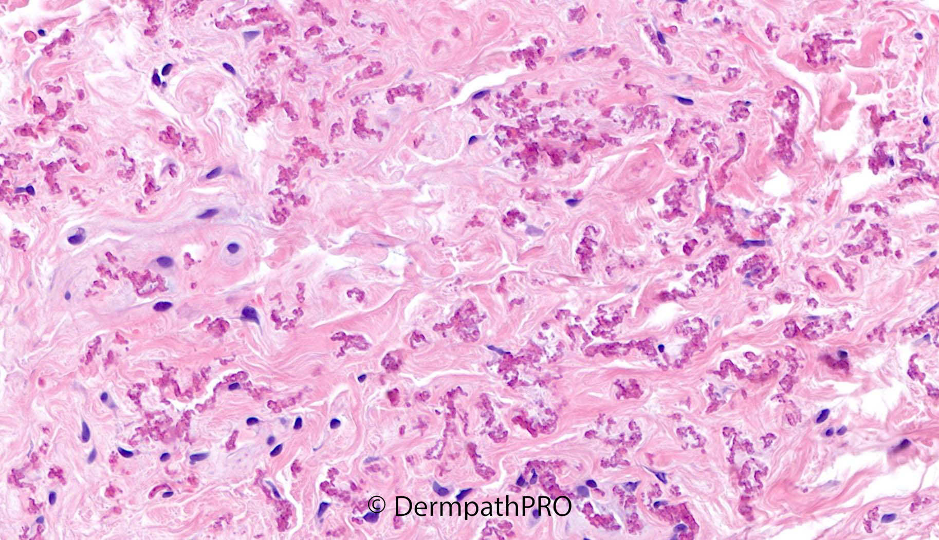
36F punch biopsy left axilla, 3-4 years duration ?morphoea ?Lichen sclerosus


  Report Record

User Feedback


daniellindsay

Posted (edited)

PXE

Also ?mast cells in superficial dermis, would do mast cell markers and correlate clinically

Edited by daniellindsay

Share this comment


Link to comment
share_externally

Anil Patki

Posted

Two interesting features are seen in this case. The intraepidermal portion of a sweat duct is blocked by a keratotic plug. The dilated intraepidermal sweat duct is surrounded by lymphocytic infiltrate resulting is some spongiosis. The gland does not seem to be associated with a follicle and maybe it's an apoeccrine gland in the axilla.

The second feature is the presence of crumpled elastic fibres in the lower dermis. Fox Fordyce disease and pseudoxanthoma elasticum are the two conditions that come to my mind.

Share this comment


Link to comment
share_externally

I don’t see the a perifollicular foam cell infiltrate, that is a distinct histopathologic feature of FFD. But agree with Anil. 

Share this comment


Link to comment
share_externally

Krishnakumar subramanian

Posted

fox Fordyce disease with PXE  changes in the dermis

Share this comment


Link to comment
share_externally

Saleem Taibjee

Posted

Yes, well done, you all spotted the PXE-like features. This was not suggested clinically but is striking histologically. There is clumping and fragmentation of elastic fibres within the mid-to-deep reticular dermis. This is further highlighted by the EVG stain, with calcification of the fibres confirmed on von Kossa stain, shown below.

I hadn't considered Fox-Fordyce disease, but it is an excellent thought, even if it does not correlate so well with the clinical suggestion. Although there was no active lichenoid inflammation, in areas I did wonder if the upper dermis is rather featureless with possible subtle hyalinisation. The EVG also suggested that elastic fibres may be diminished (but not completely absent) in the papillary dermis. Hence, I did wonder if there might also be subtle quiescent lichen sclerosus.
Interestingly, McKee's Pathology of the Skin indicates that PXE-like changes have been described in inflammatory dermatoses including both lichen sclerosus and morphoea, and some of these patients may be heterozygous for mutations in ABCC6 (e.g. Bowen AR et al. J Cutan Pathol 2007;34(10):777-81). There is a noteworthy case of a patient with confirmed PXE showing clinical features of morphoea (Duschner N et al. Am J Dermatopathol 2020:42:e57-58). This may be relevant to the current case, in which perhaps there is subtle lichen sclerosus.

63180_5.0x EVG labelled.jpg

63180_20.0x EVG  labelled.jpg

63180_20.0x Von Kossa  labelled.jpg

Share this comment


Link to comment
share_externally


Join the conversation

You can post now and register later. If you have an account, sign in now to post with your account.

Guest
Add a comment...

×   Pasted as rich text.   Paste as plain text instead

  Only 75 emoji are allowed.

×   Your link has been automatically embedded.   Display as a link instead

×   Your previous content has been restored.   Clear editor

×   You cannot paste images directly. Upload or insert images from URL.

Loading...

×
×
  • Create New...