Jump to content
In this section we have spot diagnoses posted on a daily basis since June 2010, now over 4000! You can review the archived cases and read the suggested diagnoses by users and the final comment by the contributors.
Case are uploaded each week day by 10 am UK time with the correct diagnosis will generally be posted at 8 pm UK time. Why not view the most recent spot diagnosis and proffer a diagnosis?

Case Number : Case 3090 - 06 May 2022 Posted By: Dr. Richard Carr

Please read the clinical history and view the images by clicking on them before you proffer your diagnosis.
Submitted Date :
   (0 reviews)

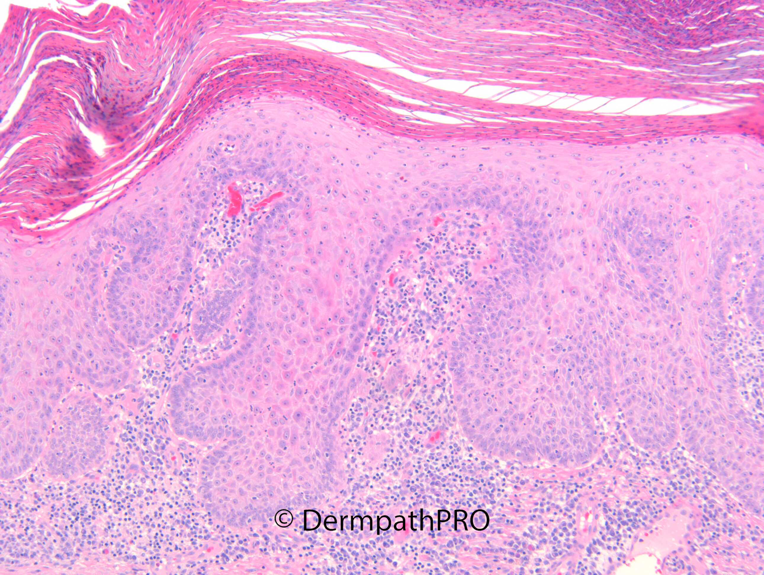
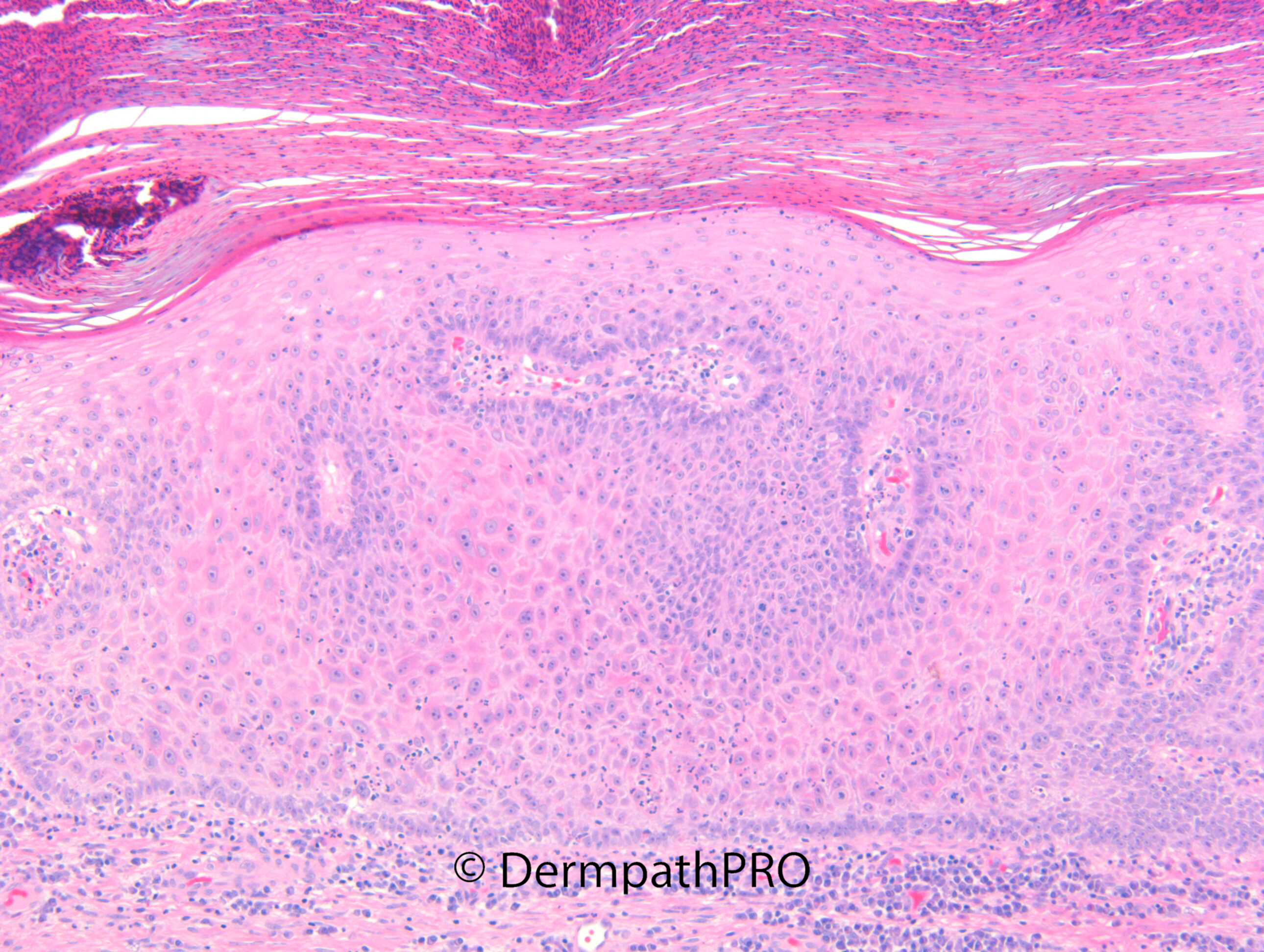
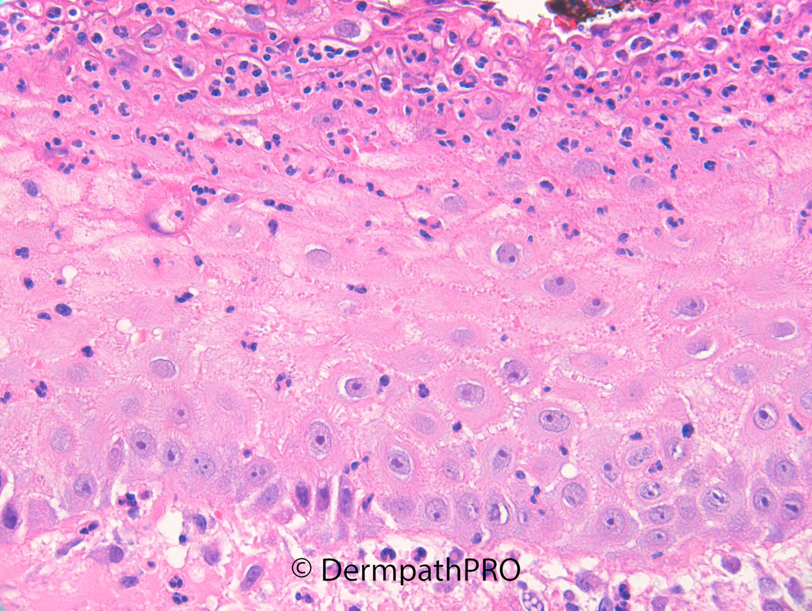
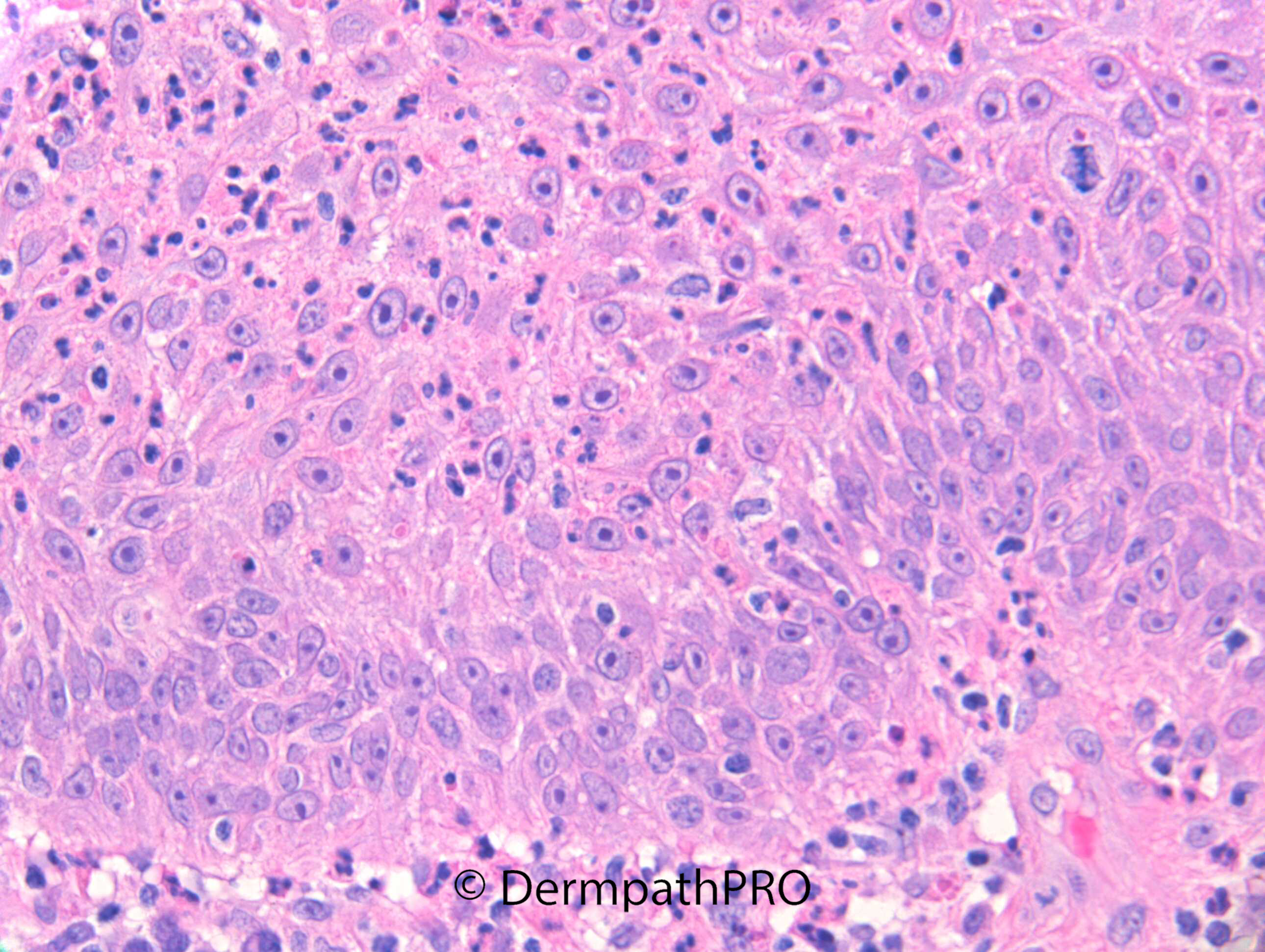
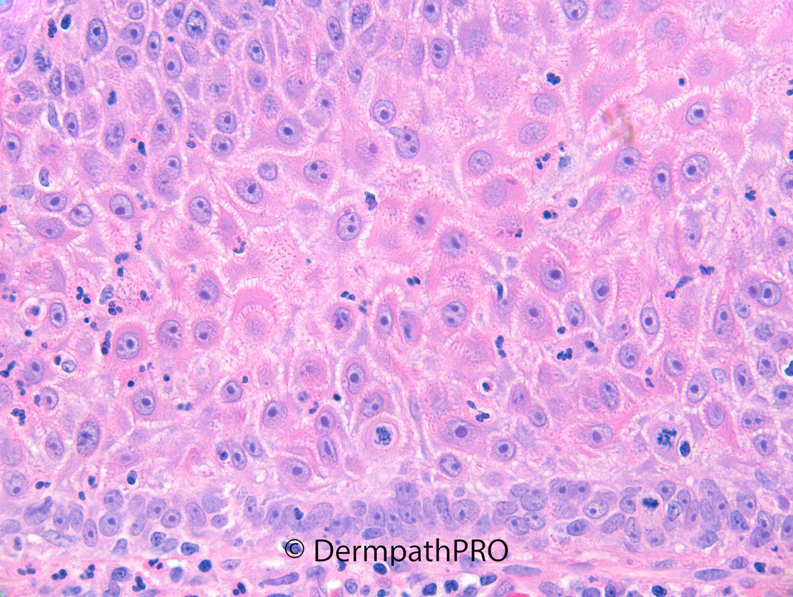
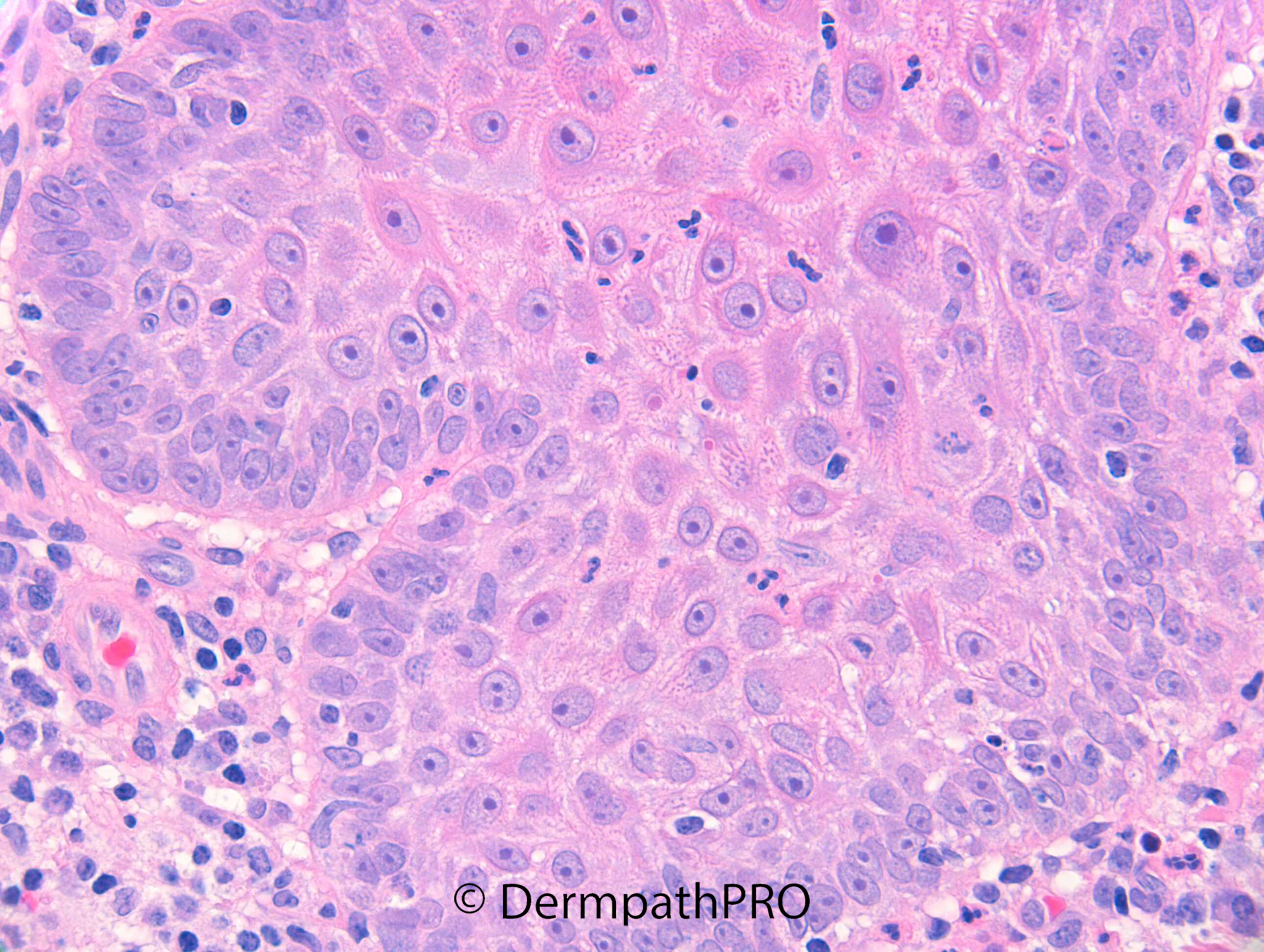
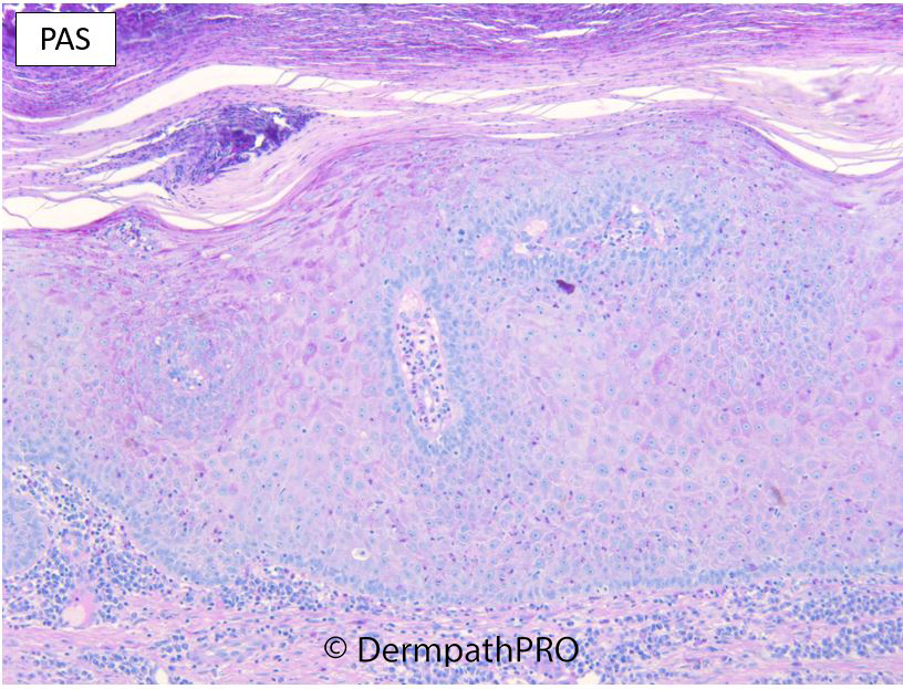
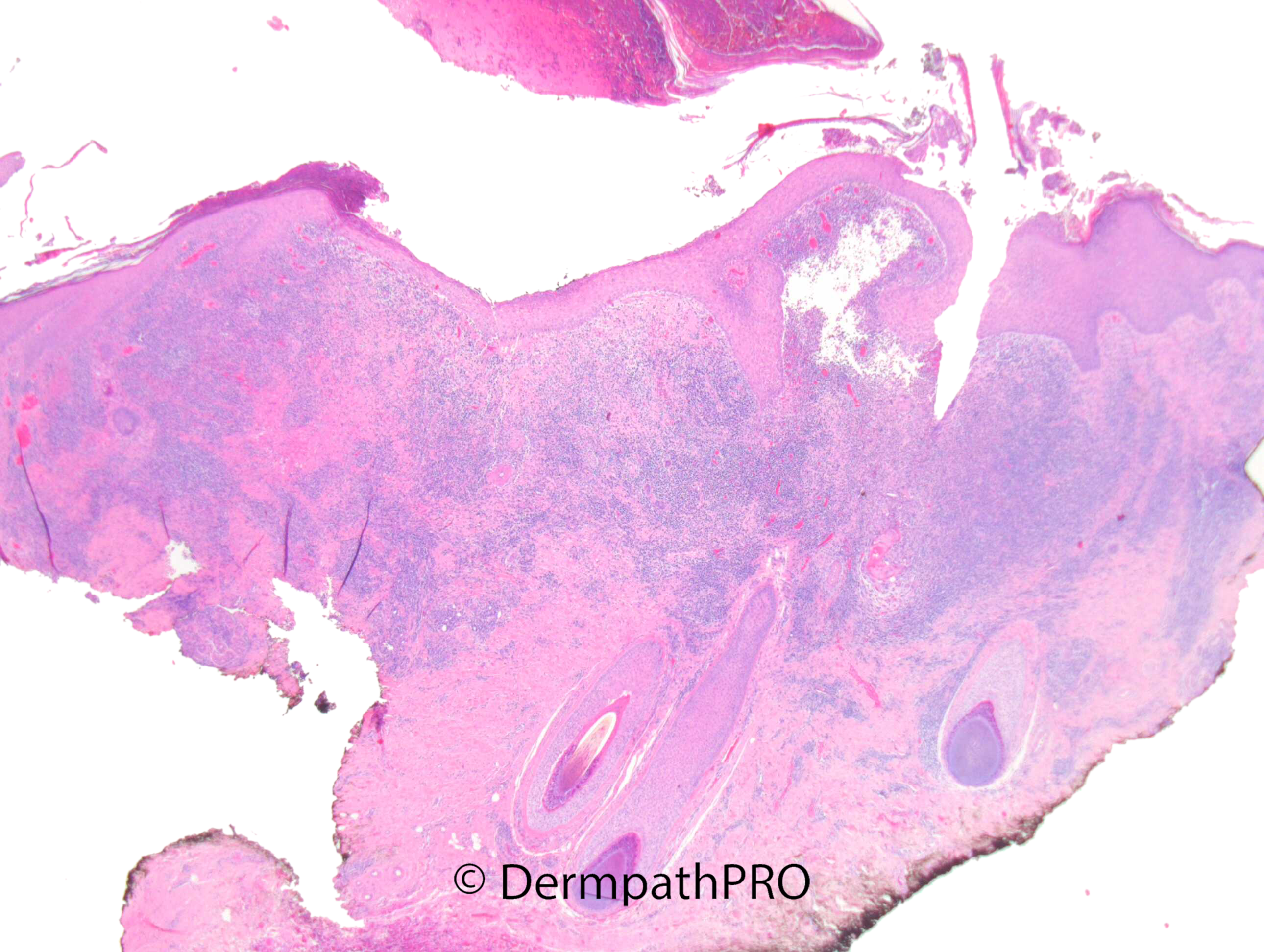
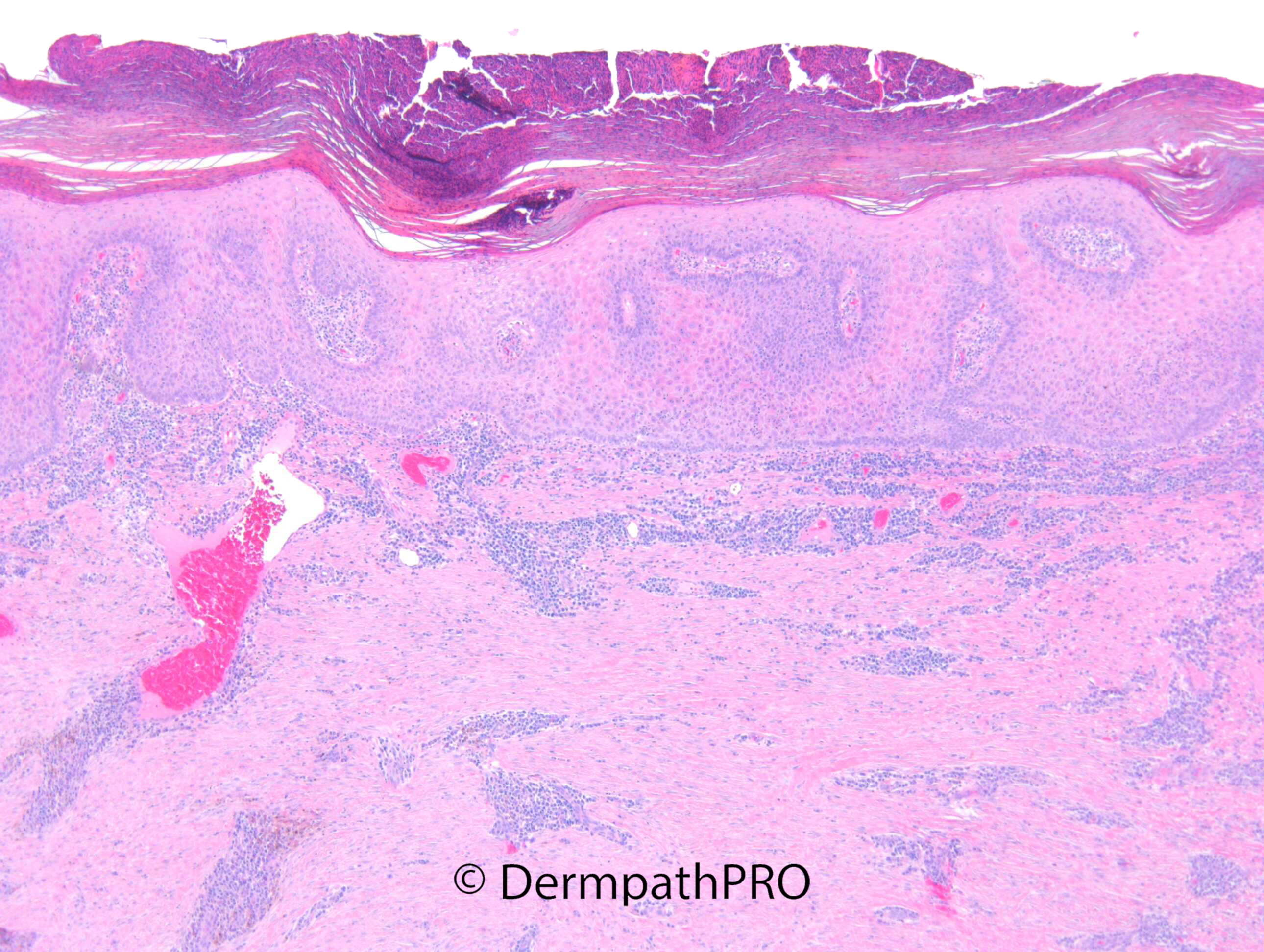
M62. Past h/o NMSCs treated in NHS in London. >12/12 h/o lesion right cheek, increasing in size, Previous biopsy 10 months ago showed inflammation only. Lesion now 2cm indurated crusted nodule ?SCC ?BCC ?inflamed squamoproliferative lesion.

  Report Record

User Feedback


Meenakshi Batrani

Posted

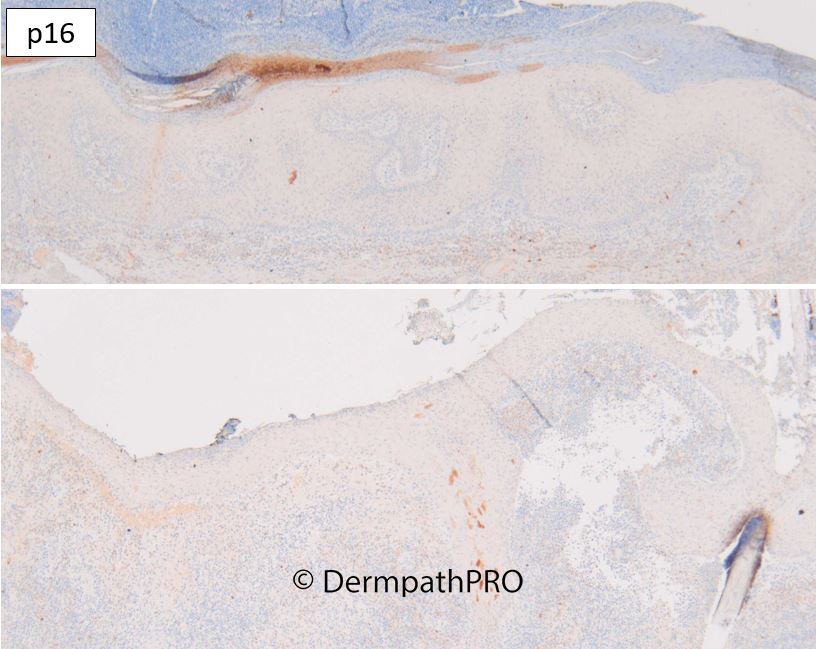
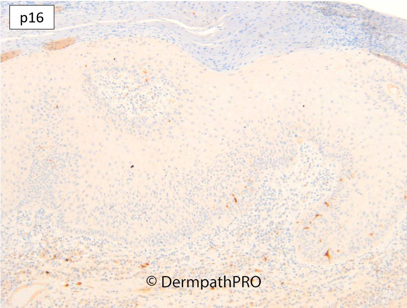
Histology is good for clear cell acanthoma. Don't know what to make of loss of p16.

Share this comment


Link to comment
share_externally

Richard Logan

Posted

Whilst some of the changes in the epidermis are reminiscent of those seen in clear cell acanthoma, there are a number of other features that put me off that diagnosis.  In the views shown, we do not see any evidence of a sharp edge to the lesion.  The degree of inflammation underneath and also the thickness of the inflammatory scab are much greater than might be expected.  Also I have never heard of a clear cell acanthoma on the face, especially one which is 2cm in diameter.

Having given all these reasons why I don't think it is a clear cell acanthoma, I can't give a definite alternative diagnosis.  I would want to exclude an infective cause, especially with all those neutrophils around.  The possibility of halogenoderma also crossed my mind.  There are some mitotic figures in the basal layer, but I don't think this is a malignant lesion, just a very active, inflammatory process.

Share this comment


Link to comment
share_externally

Agree with Richard, dermis looks scarred, consistent with hx of prior Mohs. Favor a reactive epidermal proliferation over site of scar.

Share this comment


Link to comment
share_externally

Dr. Richard Carr

Posted

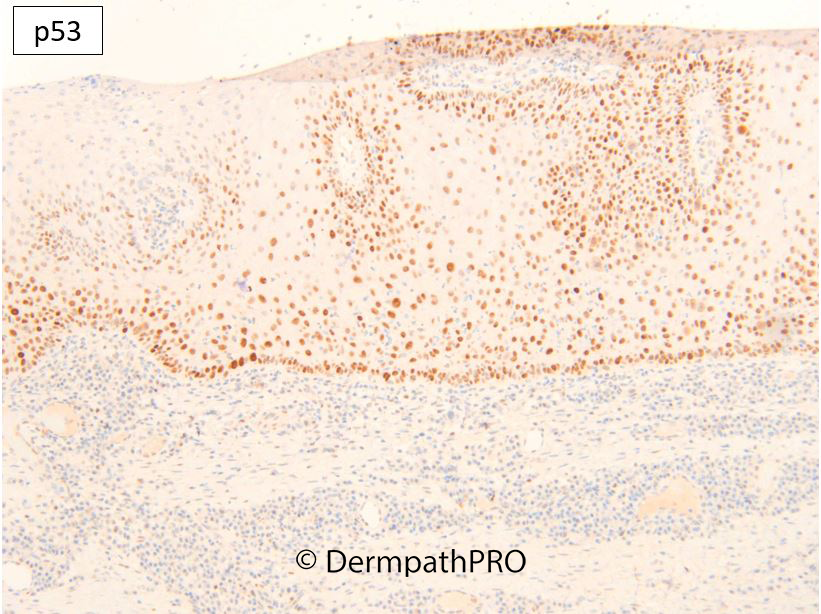
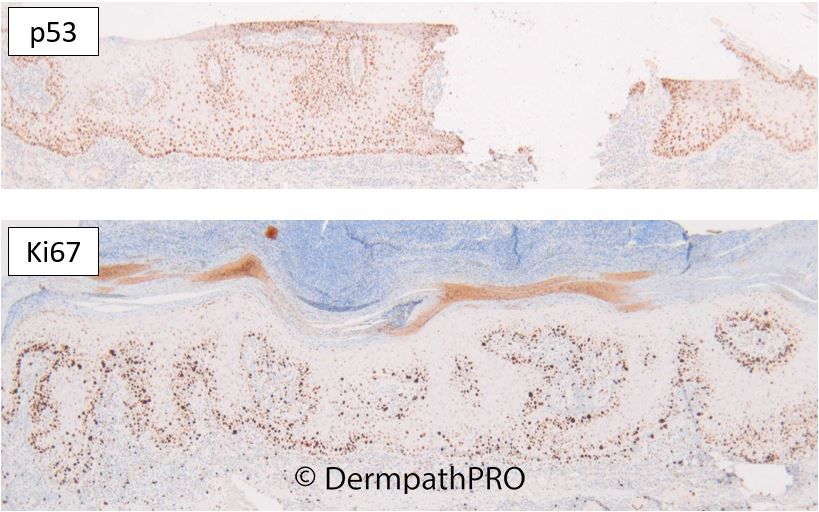
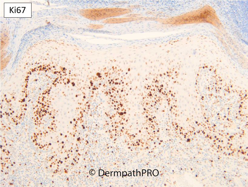
I believe this fits best with so-called "psoriasiform keratosis" and just like epidermolytic hyperkeratosis, focal acantholytic dyskeratosis, porokeratosis probably represents a predominently maturational defect but it is highly interesting the lesion is null for p16 something we've found almost specific for malignancy although I'm guessing there may be lesions equivalent to atypical Spitz tumours that might have a p16 null pattern but not be frankly malignant (e.g. the mitotic sebaecoma-like tumour I showed recently). We've discussed that there are many concepts to be determined. Certainly you will find a nice discussion of psoriasiform keratosis and the DDx with clear cell acanthoma in the references below. Richard has actually summarised these above! I suggested they manage this lesion as actinic keratosis. I'm assuming the scarring was due to the previous biopsy. There was also a folliculitis (image 1) which I presume is secondary to the "keratosis". I think psoriasiform keratosis, like so-called lichenoid keratosis, may prove to be a mixed bag of lesions but it's a nice label to put on a lesion like this and it appears it has not been reported since 2010 so I'd guess it's under-recognised although it's taken me a few years to find one myself!

Walsh SN, Hurt MA, Santa Cruz DJ. Psoriasiform keratosis. Am J Dermatopathol. 2007 Apr;29(2):137-40. doi: 10.1097/01.dad.0000246177.63145.b3. PMID: 17414434.

Mutasim DF. Psoriasiform keratosis: a lesion mimicking psoriasis. Am J Dermatopathol. 2007 Oct;29(5):482-4. doi: 10.1097/DAD.0b013e3181468e4e. PMID: 17890921.

Sezer E, Böer A, Falk T. Identification of human papilloma virus type 6 in psoriasiform keratosis. Am J Dermatopathol. 2010 Jul;32(5):492-4. doi: 10.1097/DAD.0b013e3181c62245. PMID: 20442638.


Share this comment


Link to comment
share_externally

Meenakshi Batrani

Posted

21 hours ago, Dr. Richard Carr said:

I believe this fits best with so-called "psoriasiform keratosis" and just like epidermolytic hyperkeratosis, focal acantholytic dyskeratosis, porokeratosis probably represents a predominently maturational defect but it is highly interesting the lesion is null for p16 something we've found almost specific for malignancy although I'm guessing there may be lesions equivalent to atypical Spitz tumours that might have a p16 null pattern but not be frankly malignant (e.g. the mitotic sebaecoma-like tumour I showed recently). We've discussed that there are many concepts to be determined. Certainly you will find a nice discussion of psoriasiform keratosis and the DDx with clear cell acanthoma in the references below. Richard has actually summarised these above! I suggested they manage this lesion as actinic keratosis. I'm assuming the scarring was due to the previous biopsy. There was also a folliculitis (image 1) which I presume is secondary to the "keratosis". I think psoriasiform keratosis, like so-called lichenoid keratosis, may prove to be a mixed bag of lesions but it's a nice label to put on a lesion like this and it appears it has not been reported since 2010 so I'd guess it's under-recognised although it's taken me a few years to find one myself!

Walsh SN, Hurt MA, Santa Cruz DJ. Psoriasiform keratosis. Am J Dermatopathol. 2007 Apr;29(2):137-40. doi: 10.1097/01.dad.0000246177.63145.b3. PMID: 17414434.

Mutasim DF. Psoriasiform keratosis: a lesion mimicking psoriasis. Am J Dermatopathol. 2007 Oct;29(5):482-4. doi: 10.1097/DAD.0b013e3181468e4e. PMID: 17890921.

Sezer E, Böer A, Falk T. Identification of human papilloma virus type 6 in psoriasiform keratosis. Am J Dermatopathol. 2010 Jul;32(5):492-4. doi: 10.1097/DAD.0b013e3181c62245. PMID: 20442638.


Agree goes more with psoriasiform keratosis than CCA. I had somewhat similar case, and had referred to the same three articles. In my case I favoured CCA, but agree based on distinguishing features provided in these articles, this case fits more with PK. Because of histological overlap Sezer E, Böer A had speculated that PK may be a variant or an early stage of CCA. Moreover there is some debate regarding clear cell acanthoma vs clear cell acanthosis as well. Interestingly, authors the below article  performed p16 stain and found the staining to be erratic, although no further details are provided.  

Zedek DC, Langel DJ, White WL. Clear-cell acanthoma versus acanthosis: a psoriasiform reaction pattern lacking tricholemmal differentiation. Am J Dermatopathol. 2007 Aug;29(4):378-84. 10.1097/DAD.0b013e31806f46f2.

Share this comment


Link to comment
share_externally


Join the conversation

You can post now and register later. If you have an account, sign in now to post with your account.

Guest
Add a comment...

×   Pasted as rich text.   Paste as plain text instead

  Only 75 emoji are allowed.

×   Your link has been automatically embedded.   Display as a link instead

×   Your previous content has been restored.   Clear editor

×   You cannot paste images directly. Upload or insert images from URL.

Loading...

×
×
  • Create New...