Jump to content
In this section we have spot diagnoses posted on a daily basis since June 2010, now over 4000! You can review the archived cases and read the suggested diagnoses by users and the final comment by the contributors.
Case are uploaded each week day by 10 am UK time with the correct diagnosis will generally be posted at 8 pm UK time. Why not view the most recent spot diagnosis and proffer a diagnosis?

Case Number : Case 3094 - 12 May 2022 Posted By: Saleem Taibjee

Please read the clinical history and view the images by clicking on them before you proffer your diagnosis.
Submitted Date :
   (0 reviews)

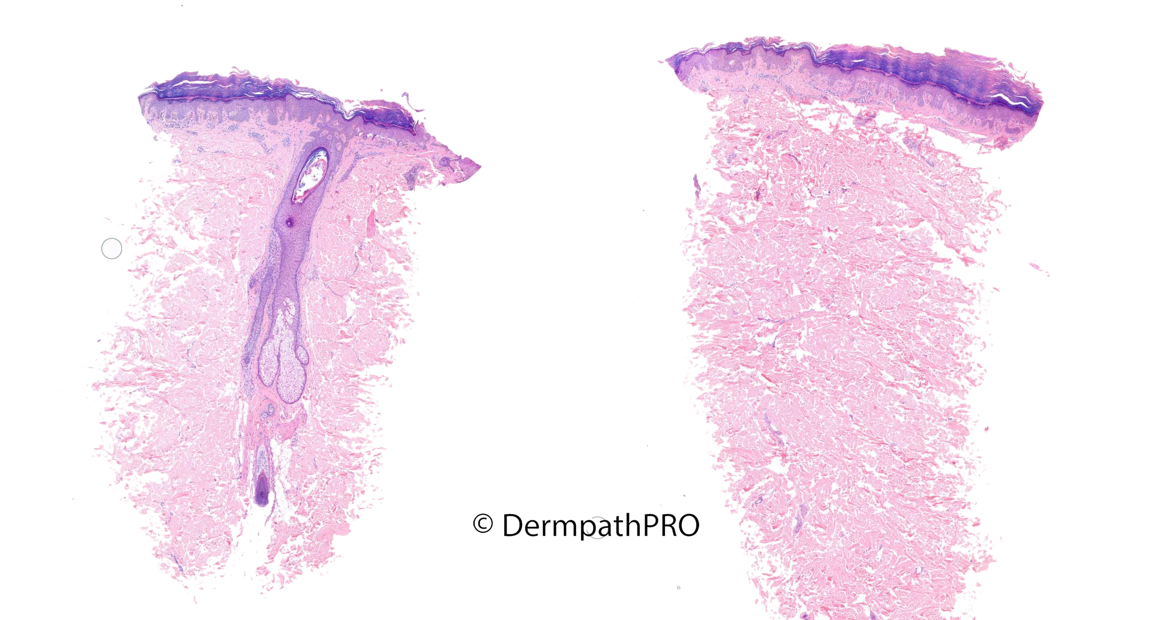
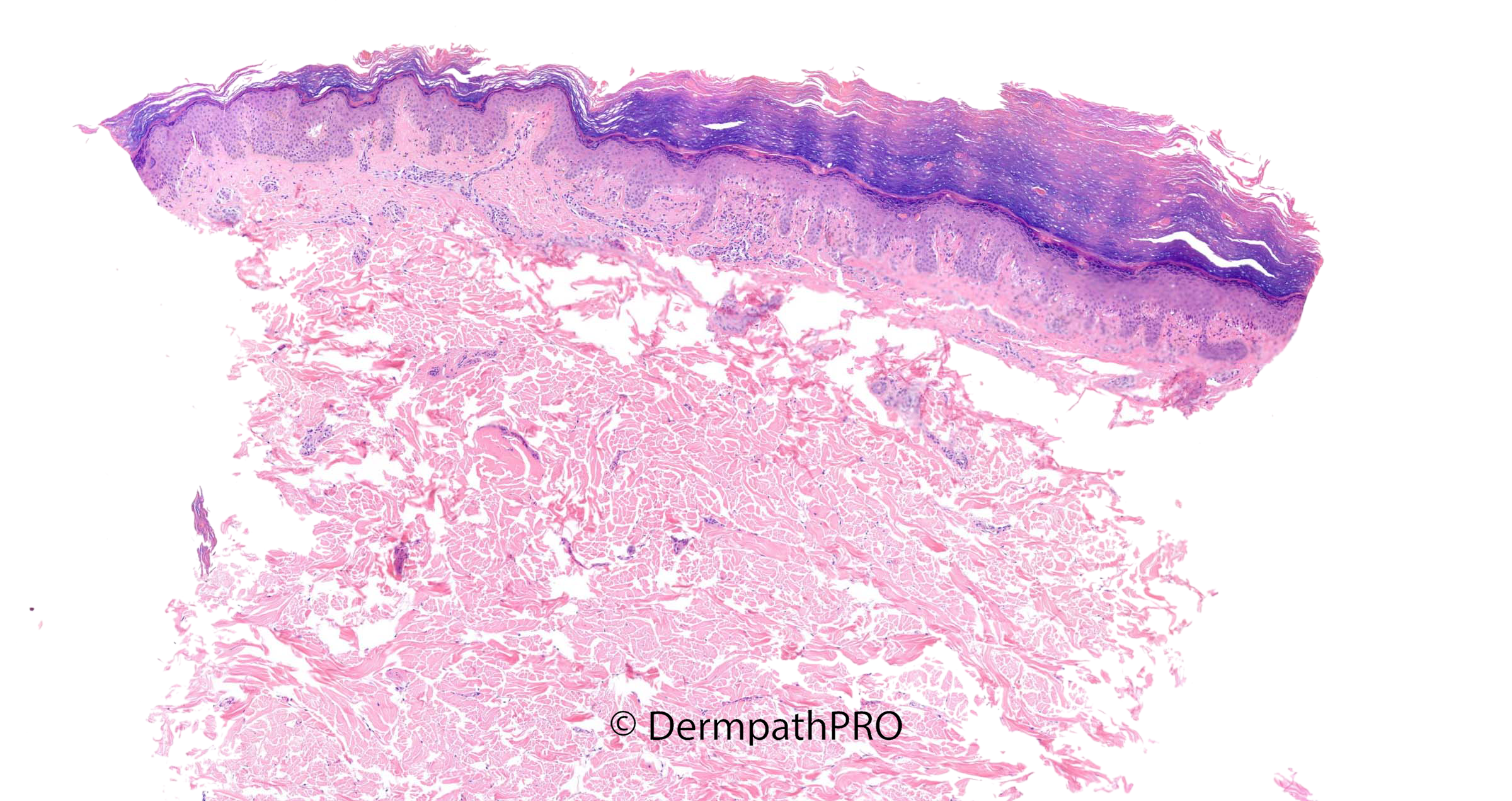
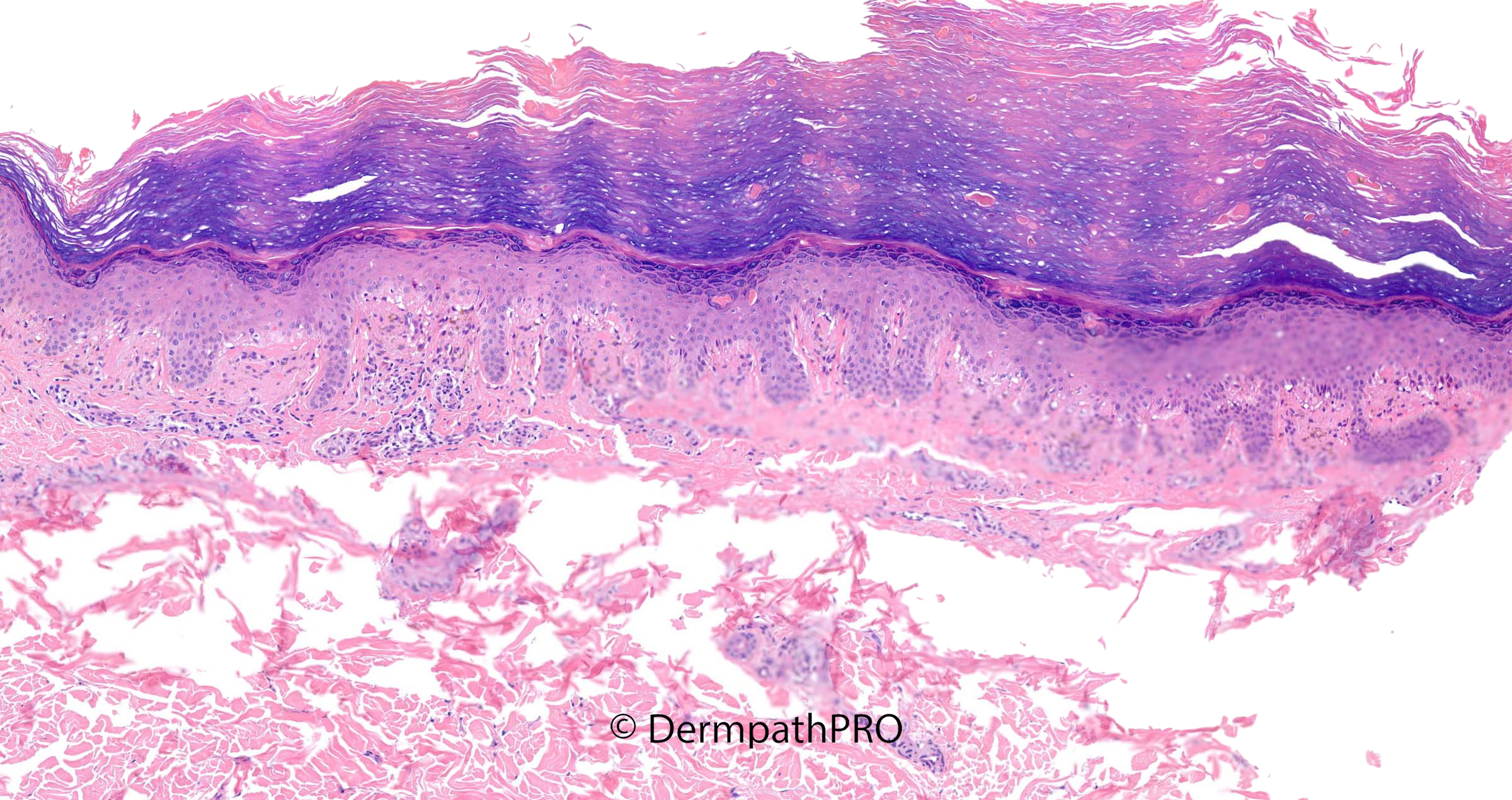
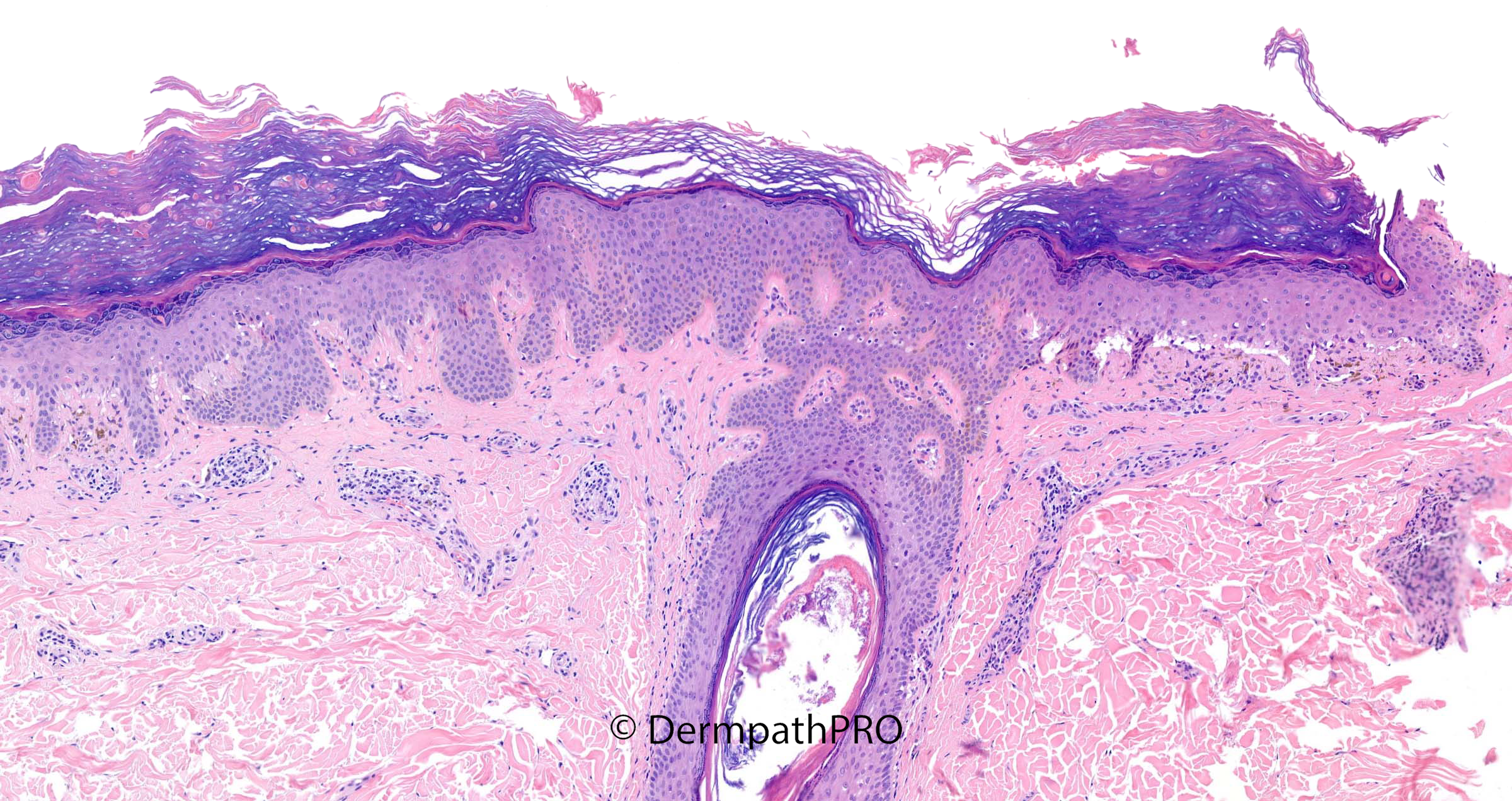
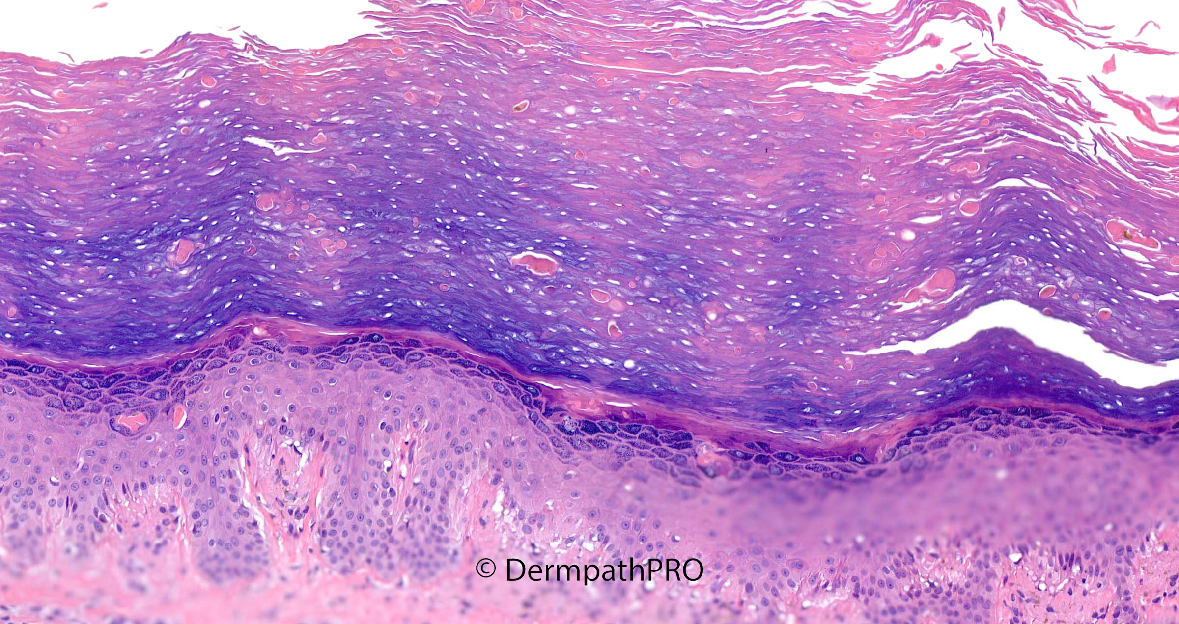
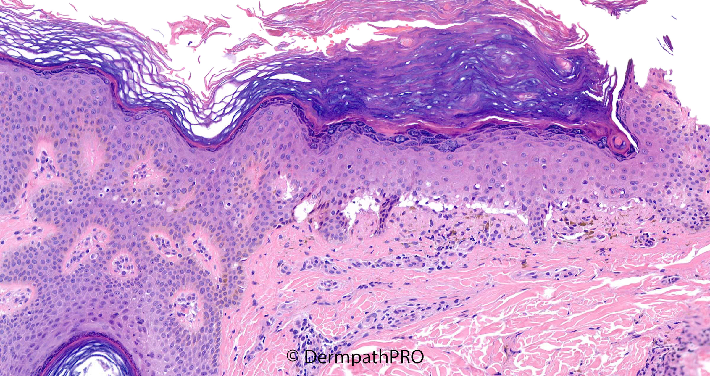
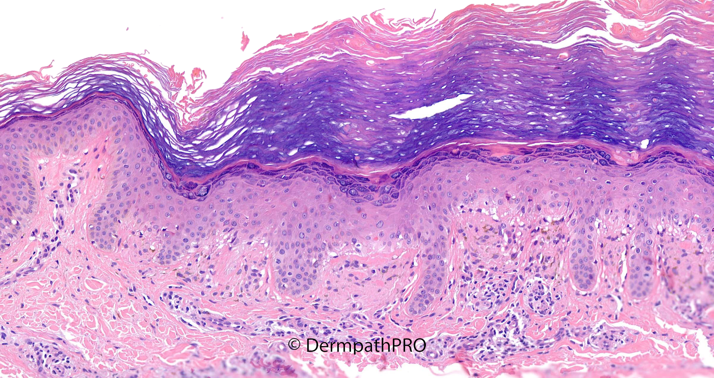
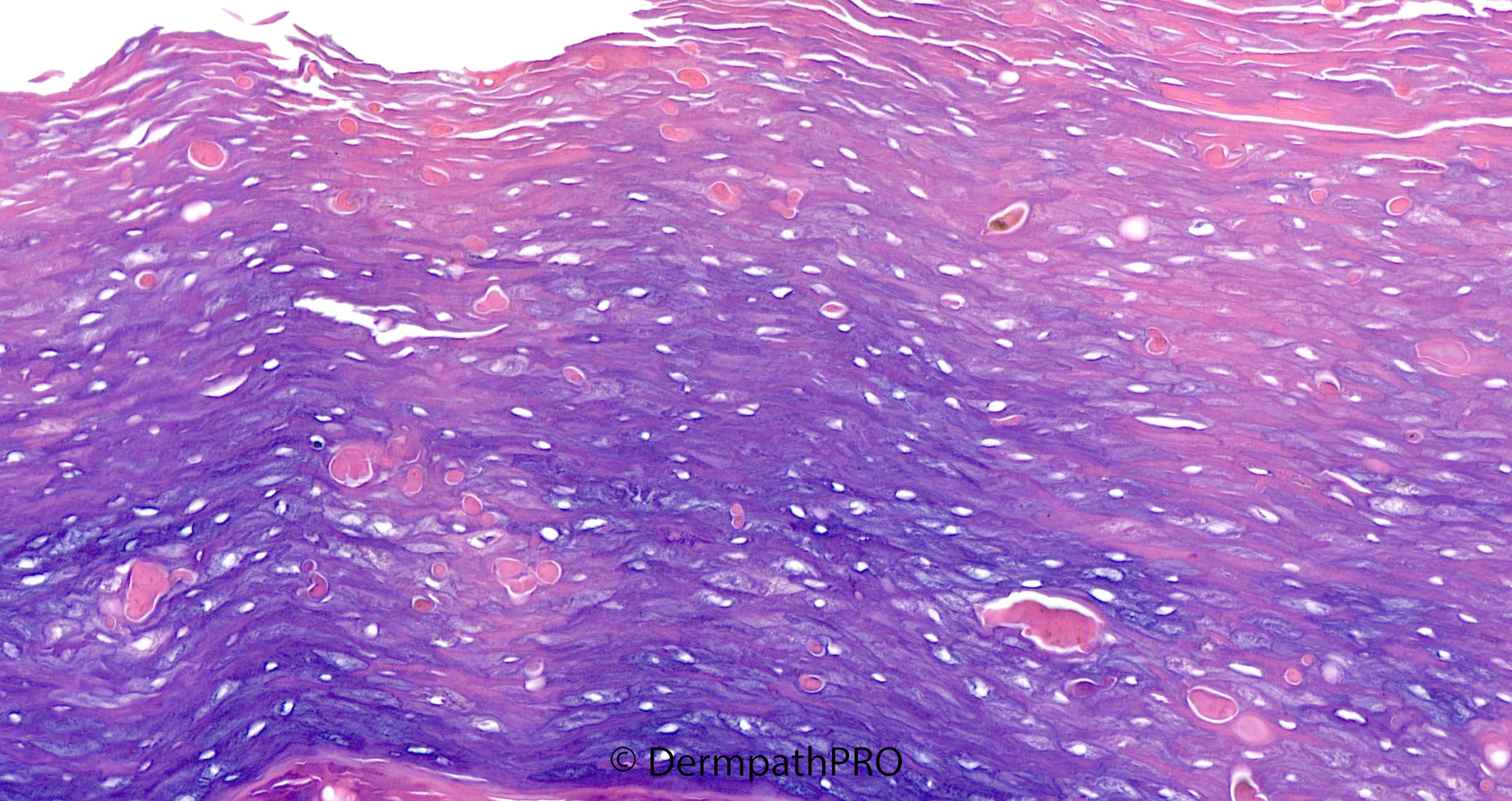
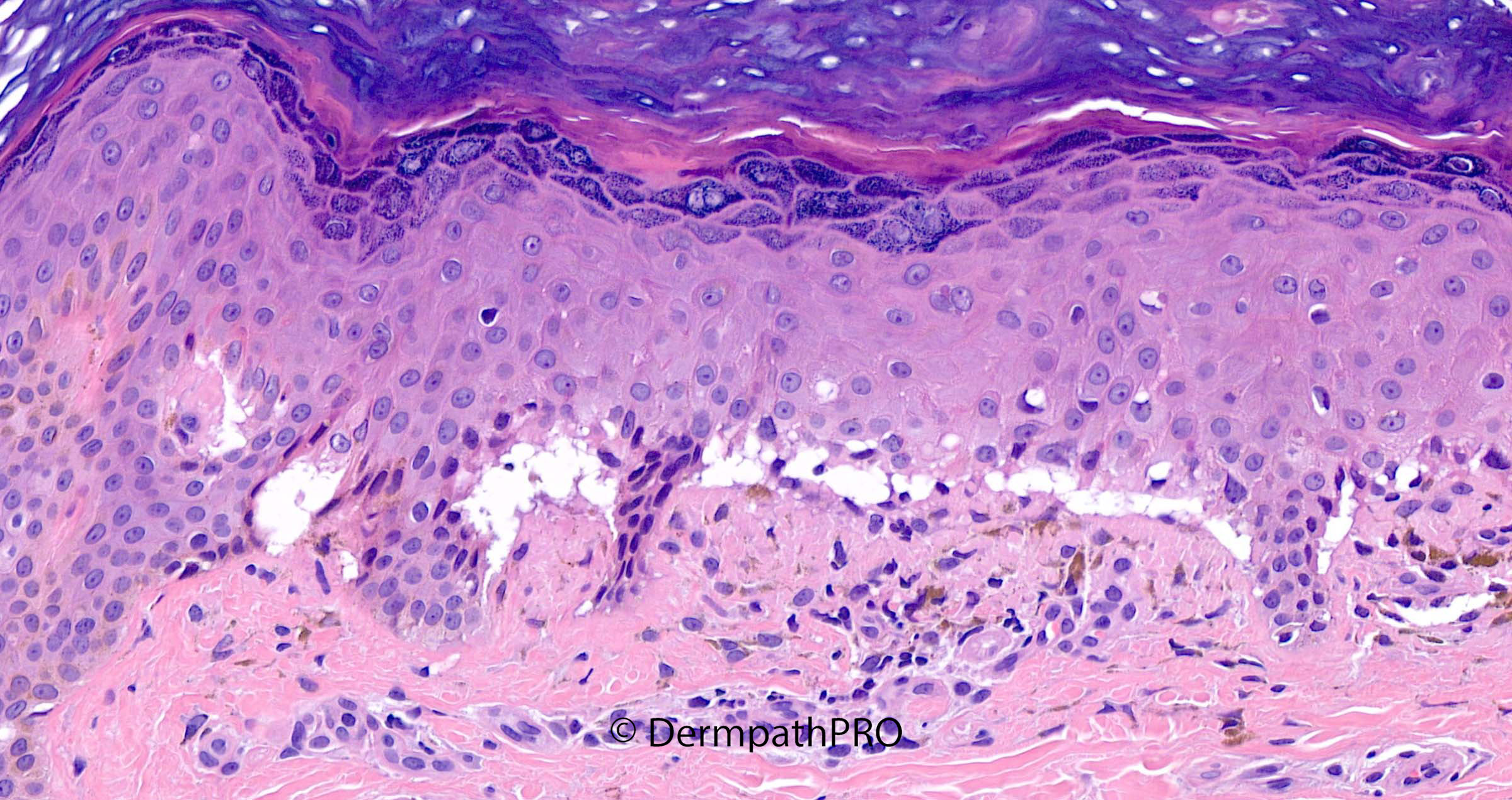
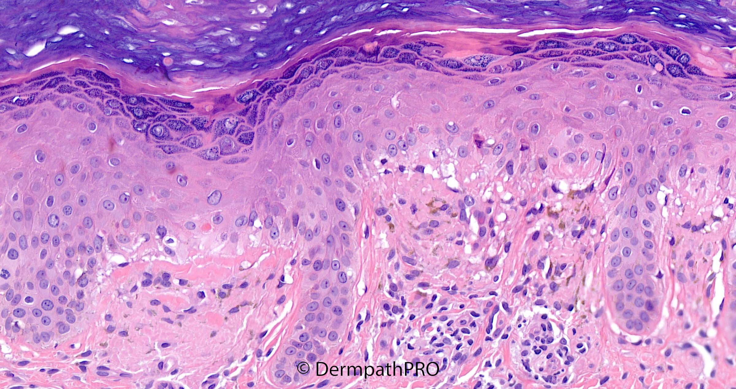
25M Punch biopsy upper back. 2-year history. Widespread prominent hair follicles on arms and thighs


  Report Record

User Feedback


Anil Patki

Posted

Lichen amyloidosus or primary cutaneous amyloidosis with ? transepidermal elimination of amyloid.

Share this comment


Link to comment
share_externally

Agree with cutaneous amyloidosis, throw in a CK5/6?

Share this comment


Link to comment
share_externally

Eman El-Nabarawy

Posted

High up dyskeratotic keratinocytes in the epidermis and stratum corneum r a clue to Adult onset Still's disease, persistent lesions, with reported cases of follicular accentuation. Still's disease shows focal interface tissue reaction but here the interface reaction is so severe for Still's disease with ?fibrin deposition under the epidermis! Could this be overlap of AOSD with SLE, which is also reported?

Share this comment


Link to comment
share_externally

Richard Logan

Posted

These histological changes are really striking.  If I may make a clinic-pathological comment, the “prominence” of the follicles must be because the interfollicular epithelium is so grossly hyper-keratotic that the uninvolved follicles are visually accentuated.  There is certainly a vacuolar interface reaction going on with transepidermal elimination of amorphous pink globules, which must be amyloid.  I don’t recall seeing such an appearance before, even in this compendium of exotica!

Share this comment


Link to comment
share_externally

vincenzo

Posted

3 hours ago, Eman El-Nabarawy said:

High up dyskeratotic keratinocytes in the epidermis and stratum corneum r a clue to Adult onset Still's disease, persistent lesions, with reported cases of follicular accentuation. Still's disease shows focal interface tissue reaction but here the interface reaction is so severe for Still's disease with ?fibrin deposition under the epidermis! Could this be overlap of AOSD with SLE, which is also reported?

The transepidermal globular bodies look like dyskeratotic cells...so Still disease could be a good spot. 

Share this comment


Link to comment
share_externally

Saleem Taibjee

Posted

Very interesting comments.I believe that this a striking example of lichen (cutaneous) amyloidosis, and indeed that was the clinician's suggested diagnosis.

But I too had not previously come across an example with such a degree of transepidermal elimination which was one reason for sharing this case with you.

Another interesting aspect which is also alluded to in the discussion is the suggestion of interface reaction with basal vacuolar degeneration and subepidermal clefting. In fact, I have seen this before in lichen amyloid, to the point where is mimics a genuine interface inflammatory process such as lichen planus or lupus. But having now seen this on a few occasions I have concluded that it can be part of the constellation of histological features in lichen amyloidosis, and presumably the mechanical rubbing and trauma induces this appearance.

Lastly I wanted to mention my experience with the keratin stains in cutaneous amyloid. It seems quite variable which keratin stain is the best in staining the amyloid (as you all know, Congo Red is rather unreliable in this setting). I show the staining pattern below for a variety of keratin stains in this case. 34BE12 was clearly the superior stain in this case. CK5/6 wasn't that great. So a message is to consider requesting more than one keratin stain when assessing for cutaneous amyloid.

BW, Saleem

image.jpeg

image.jpeg

image.jpeg

image.jpeg

image.jpeg


image.jpeg

Share this comment


Link to comment
share_externally


Join the conversation

You can post now and register later. If you have an account, sign in now to post with your account.

Guest
Add a comment...

×   Pasted as rich text.   Paste as plain text instead

  Only 75 emoji are allowed.

×   Your link has been automatically embedded.   Display as a link instead

×   Your previous content has been restored.   Clear editor

×   You cannot paste images directly. Upload or insert images from URL.

Loading...

×
×
  • Create New...