Jump to content
In this section we have spot diagnoses posted on a daily basis since June 2010, now over 4000! You can review the archived cases and read the suggested diagnoses by users and the final comment by the contributors.
Case are uploaded each week day by 10 am UK time with the correct diagnosis will generally be posted at 8 pm UK time. Why not view the most recent spot diagnosis and proffer a diagnosis?

Case Number : Case 4005 - 27 May 2022 Posted By: Dr. Richard Carr

Please read the clinical history and view the images by clicking on them before you proffer your diagnosis.
Submitted Date :
   (0 reviews)

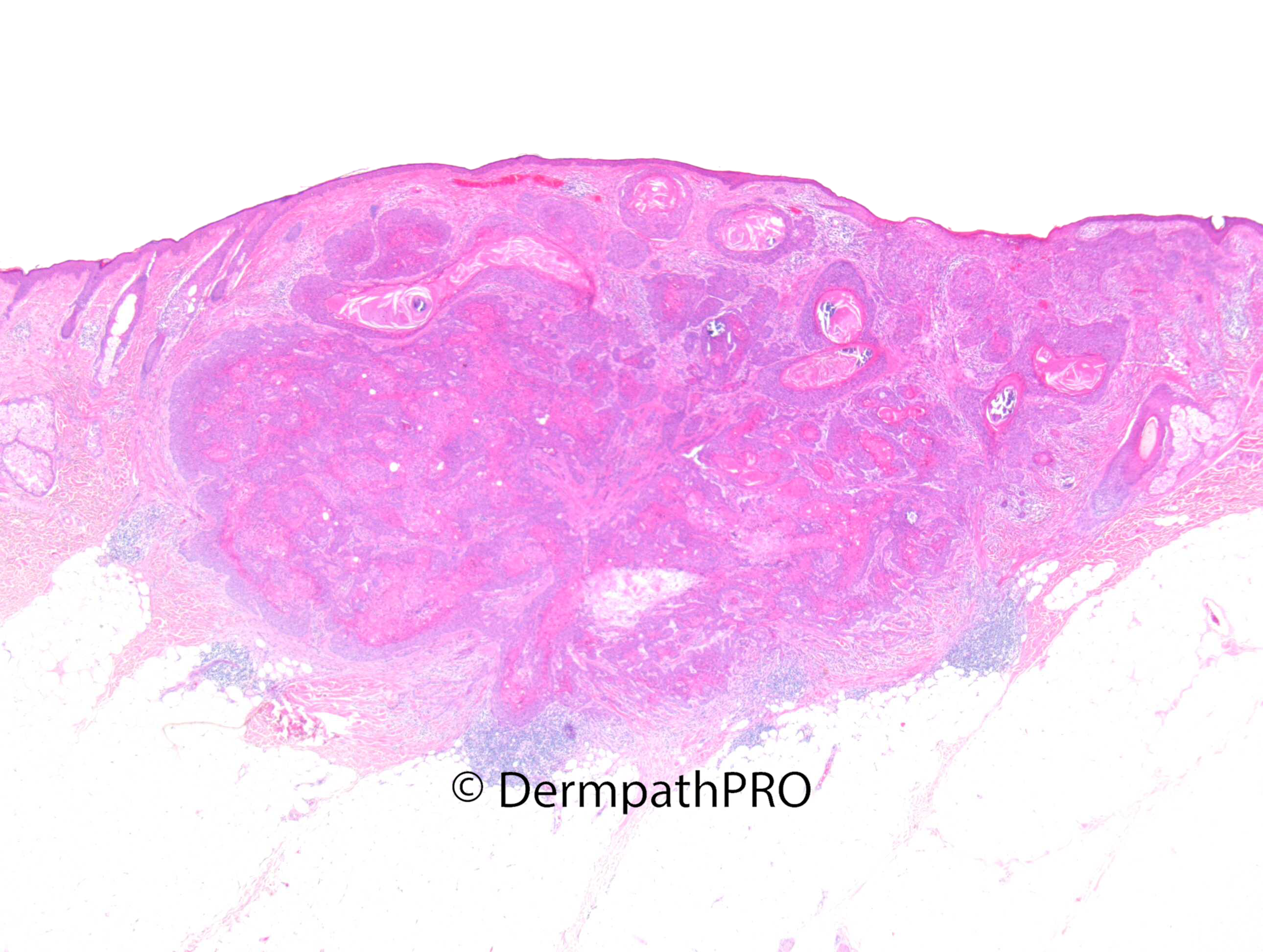
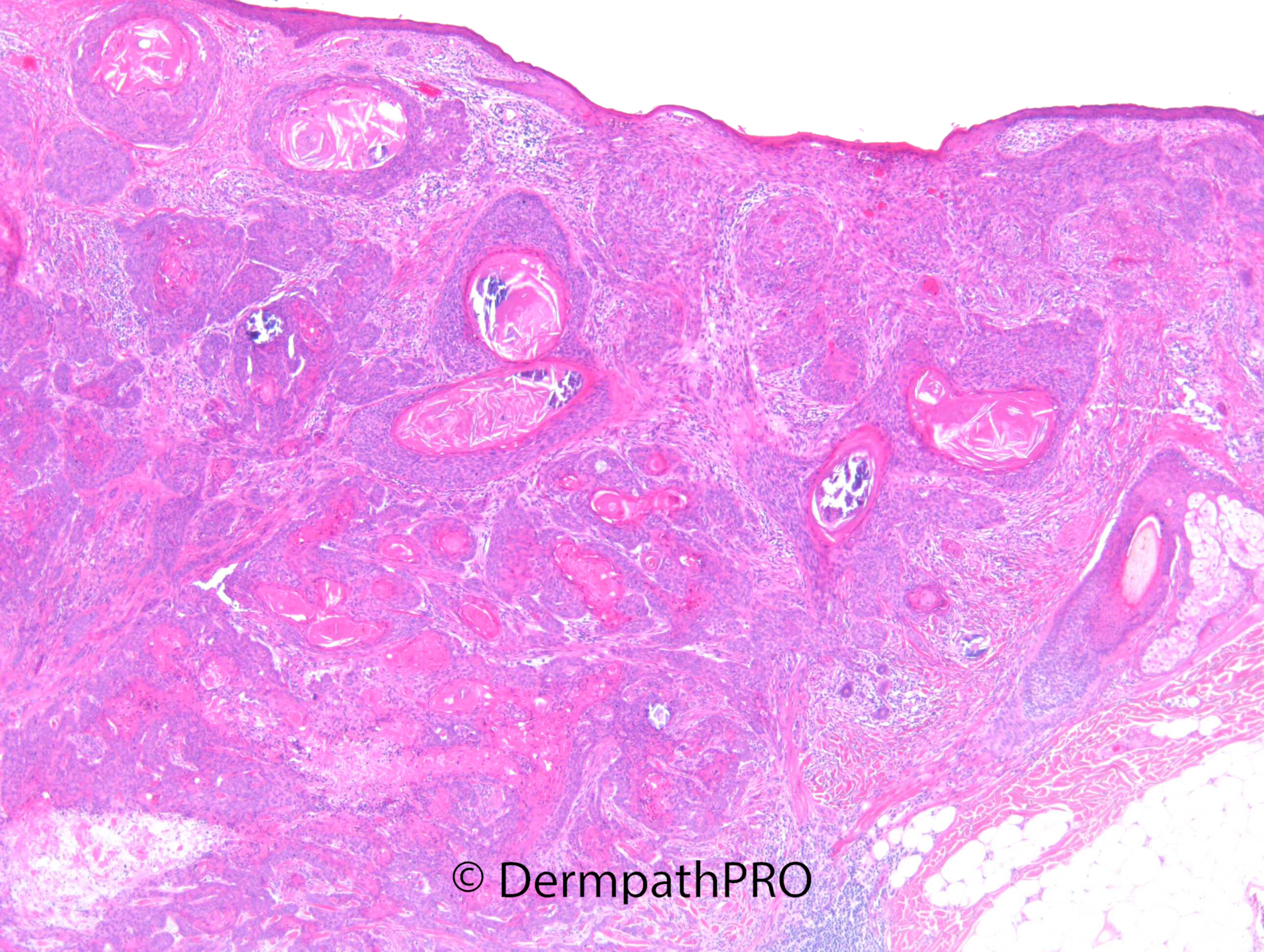
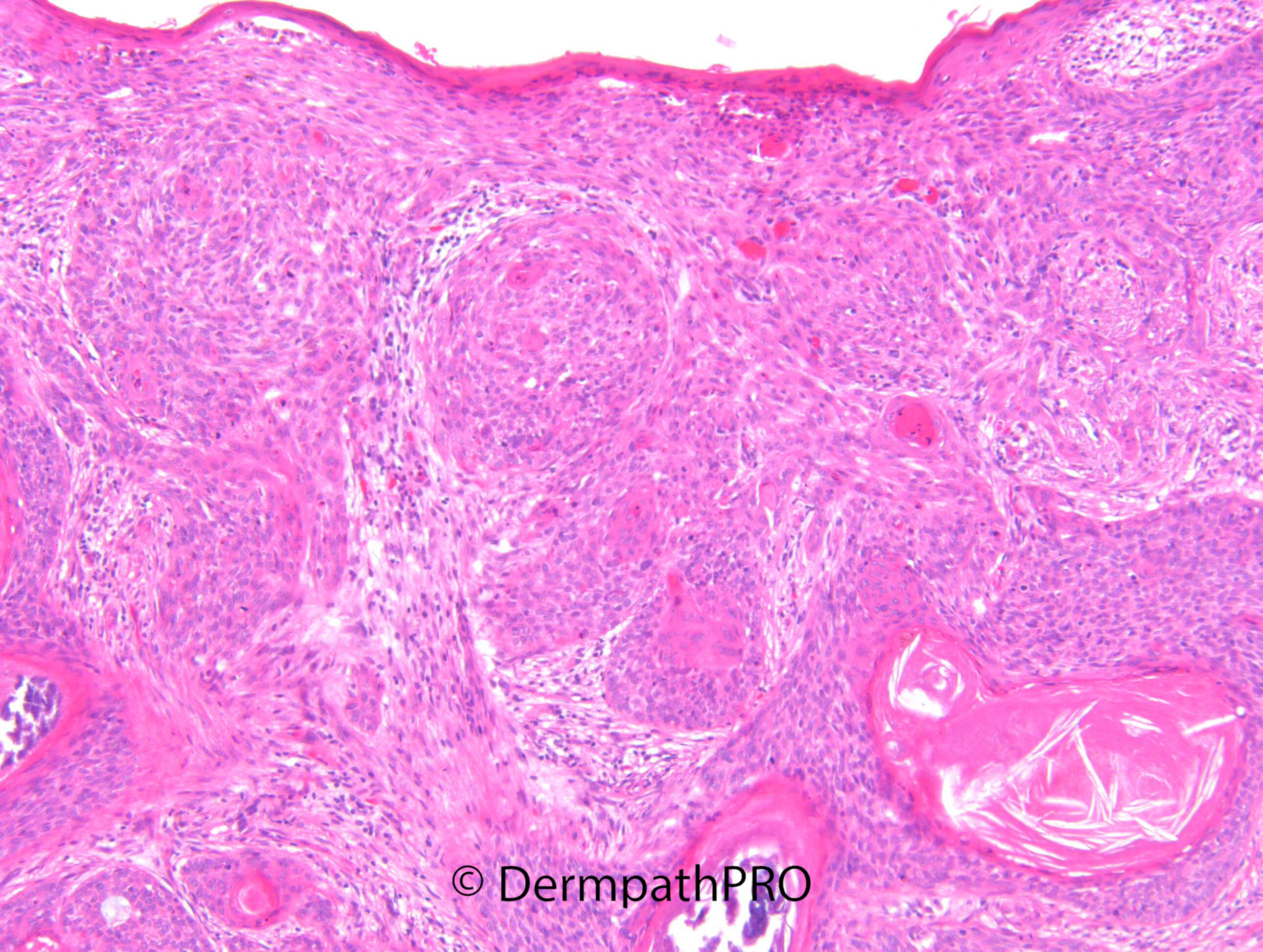
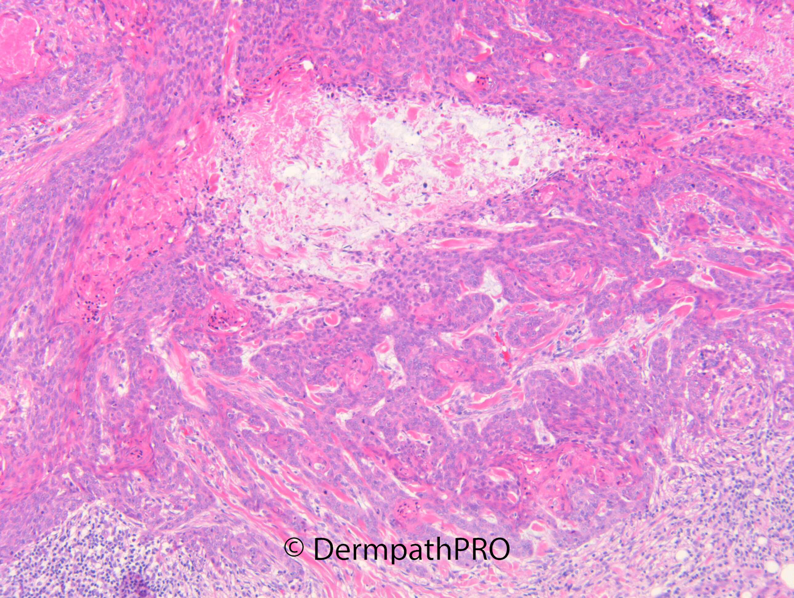
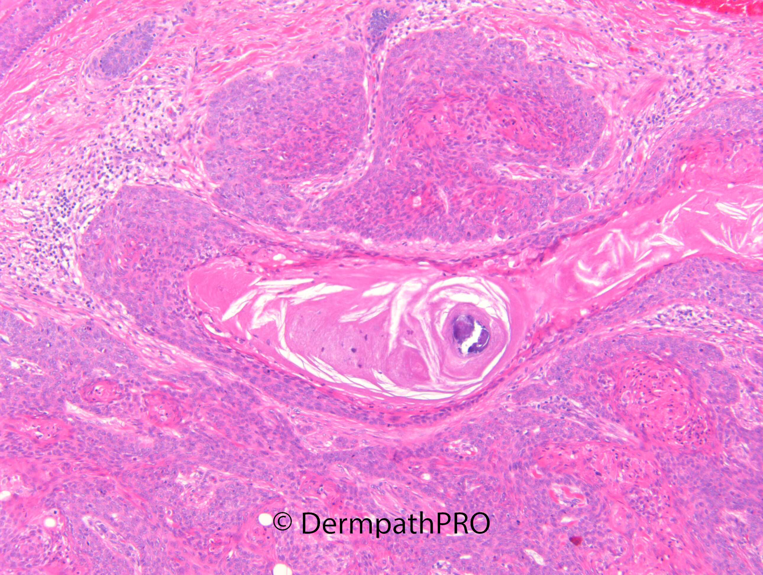
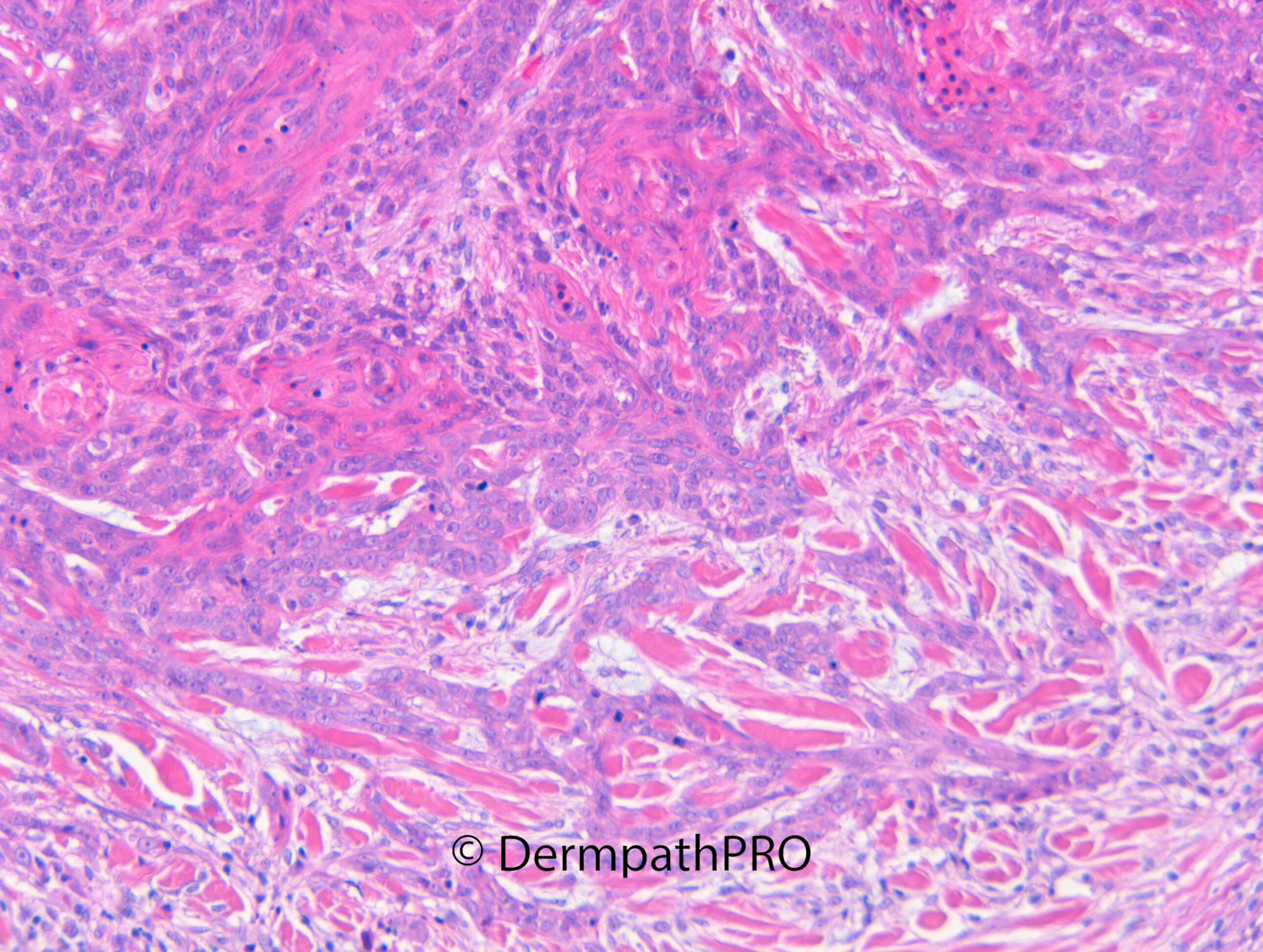
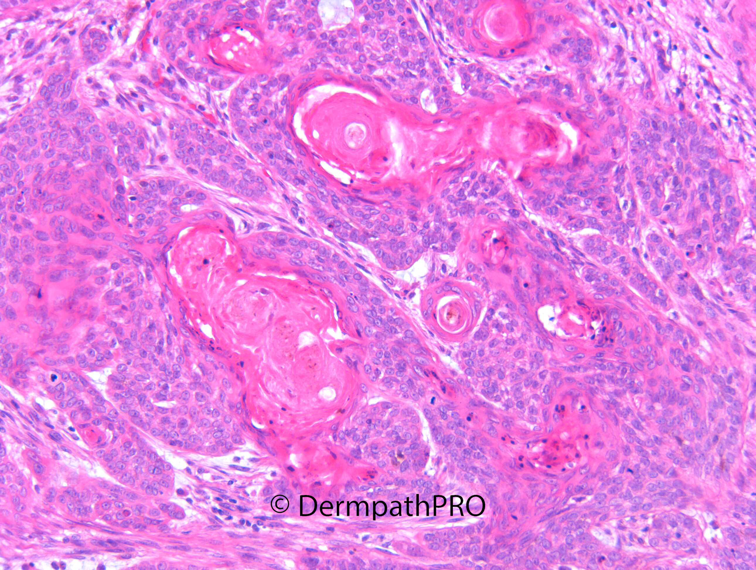
"F45. Temple. BCC.
Diagnosis: BCC, nodular keratotic and focally infiltrative (DDx Basosquamous)"


  Report Record

User Feedback


vincenzo

Posted

A difficult case. ?metatypical(basosquamous) bcc. 

Share this comment


Link to comment
share_externally

msofopoulos

Posted

On 29/05/2022 at 15:36, vincenzo said:

A difficult case. ?metatypical(basosquamous) bcc. 

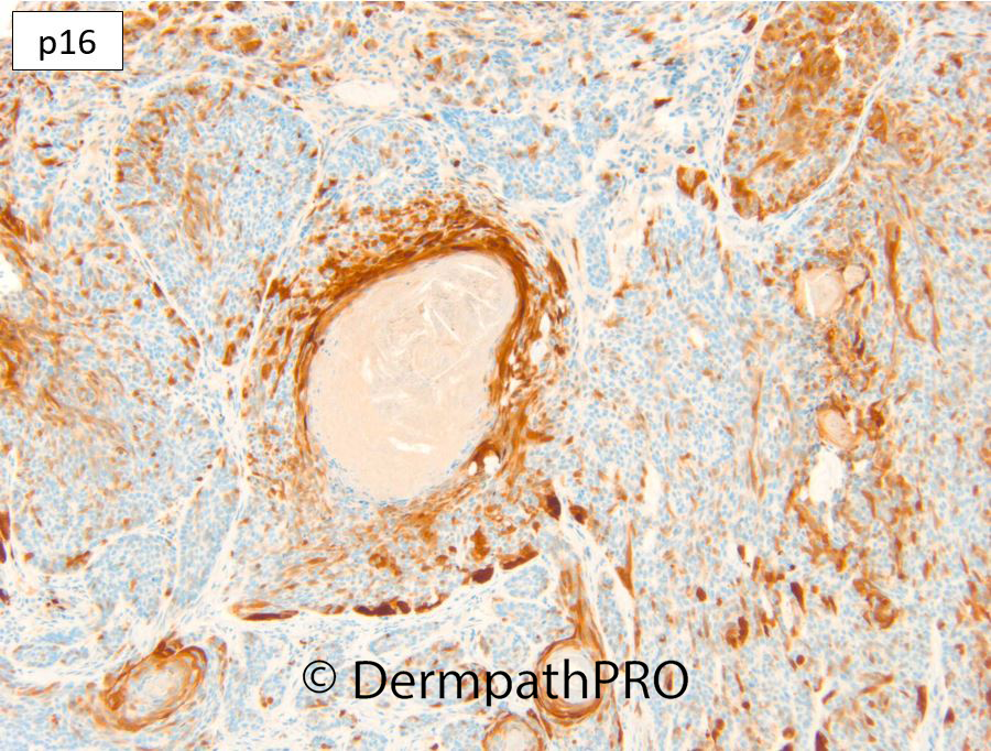
I also think that. I do not know how to interpret this  p16

Share this comment


Link to comment
share_externally

Dr. Richard Carr

Posted

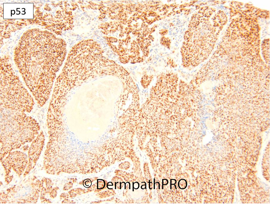
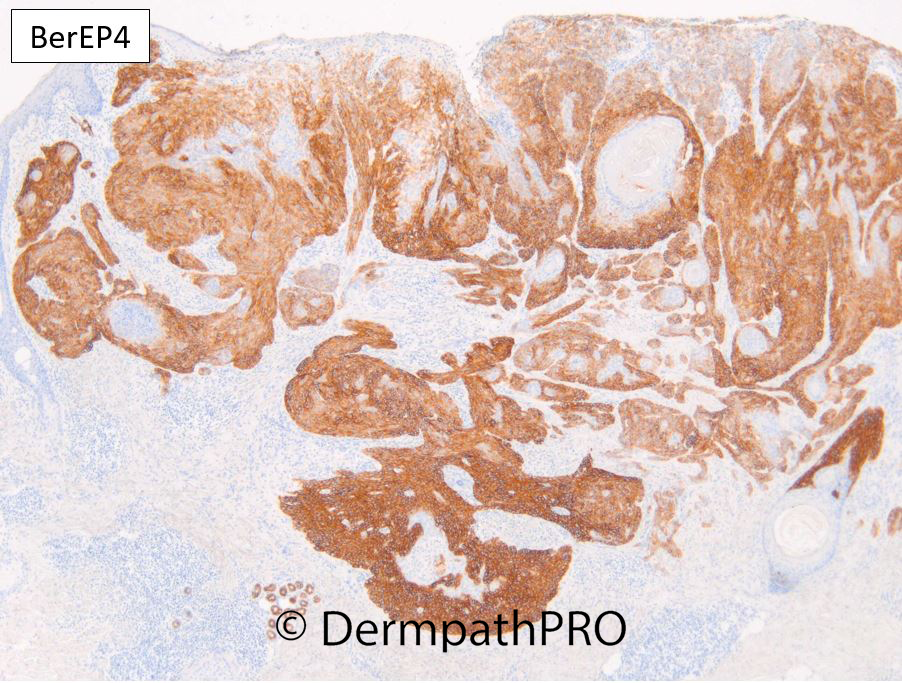
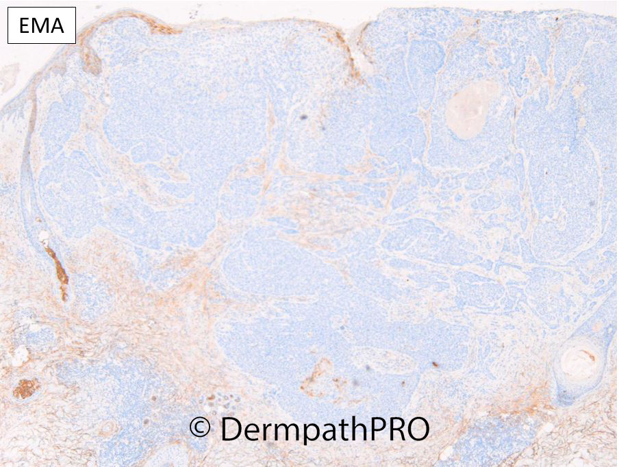
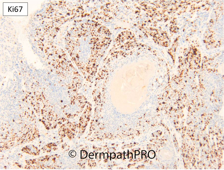
Unfortunately the diagnosis was supplied with the history (it appears). It shows the use of BerEP4 nearly always widely positive in the basaloid epithelium of BCC. Metatypical / malignant squmous component of BCC may have reduced staining for BerEP4 i.e. be more "squamous" in differentiation. EMA in my experience is ALWAYS negative in the basaloid cells of BCC but may show some staining in squamous areas. The diagnosis of "malignant squamous", "metatypical" or "basosquamous" component in BCC is subjective (I have a high threshold and would expect to see moderate to severe pleomorphism at least) and in this case I thought the squamoid atypia was mild and the BerEP4 fairly diffuse (only sparing central squamoid areas) and relative lack of EMA were more compatible with BCC nodular keratotic and focally infiltrative but not frankly basosquamous / metatypical. The latter tumours are relatively uncommon in my experience (~1-3% of BCC) but can be quite aggressive (look carefully for vascular and perineural) and in my opinion should be classified and managed as poorly differentiated cSCC. I would speculate that basosquamous BCC would have a high rate of highly aberrant p16 & p53 akin to most cSCC but this is yet to be studied and given the subjectivity of H&E and rarity of basosquamous BCC will take some time to gather systematic data. I interpretated the p16 as patchy mosaic (wild type pattern) and p53 as aberrant up-regulated (>50% to 75%) wild type which seems to fit with most BCC I'm seeing i.e. NOT highly aberrant (for p16: null, block (>90%), cytoplasmic or nuclear only; for p53: null, block (>75% moderate and strong).  Many benign tumours like SEBK, IFK, tricholemmoma etc. show similar patchy p16 although p53 is usually <50% and more often weak to moderate (congruent with proliferative/mitotic activity). A wild type p53 tends to have a congruent pattern to Ki67 (proliferative rates) in tumours both benign and many malignant but a dis-congruent p53-Ki67 is a fairly specific signature for malignancy in my experience e.g. a high and abnormally distributed Ki67 with a null p53 is a signature of malignant tumours, similarly a low Ki67 with block moderate to strong p53. In this case the p53 and Ki67 are congruent suggesting the p53 is up-regulated wild type, mainly weak to moderate and sparing differentiated areas as the Ki67 rate drops off centrally too.

Share this comment


Link to comment
share_externally


Join the conversation

You can post now and register later. If you have an account, sign in now to post with your account.

Guest
Add a comment...

×   Pasted as rich text.   Paste as plain text instead

  Only 75 emoji are allowed.

×   Your link has been automatically embedded.   Display as a link instead

×   Your previous content has been restored.   Clear editor

×   You cannot paste images directly. Upload or insert images from URL.

Loading...

×
×
  • Create New...