Jump to content
In this section we have spot diagnoses posted on a daily basis since June 2010, now over 4000! You can review the archived cases and read the suggested diagnoses by users and the final comment by the contributors.
Case are uploaded each week day by 10 am UK time with the correct diagnosis will generally be posted at 8 pm UK time. Why not view the most recent spot diagnosis and proffer a diagnosis?

Case Number : Case 4124 - 10 Nov 2022 Posted By: Saleem Taibjee

Please read the clinical history and view the images by clicking on them before you proffer your diagnosis.
Submitted Date :
   (0 reviews)

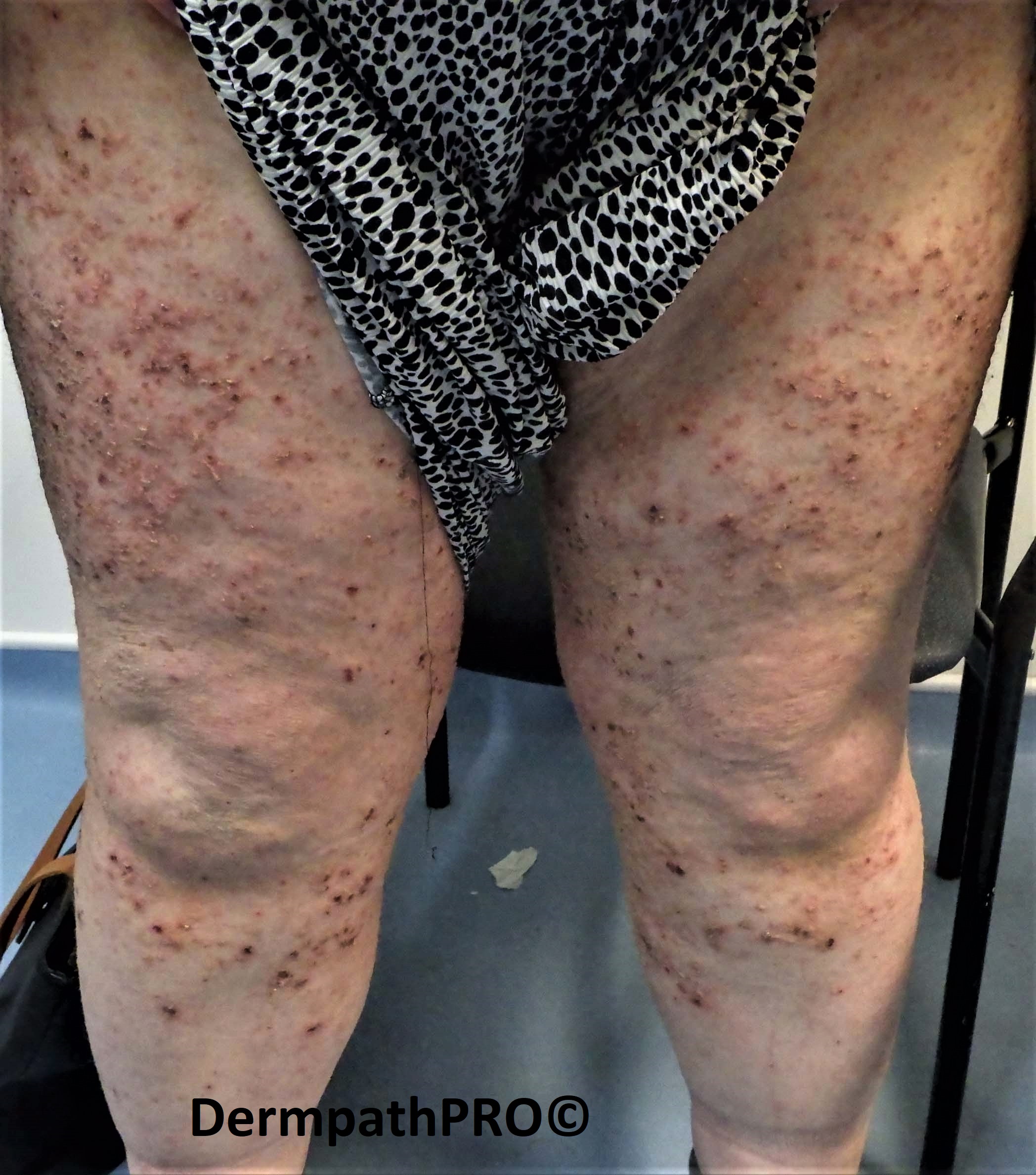
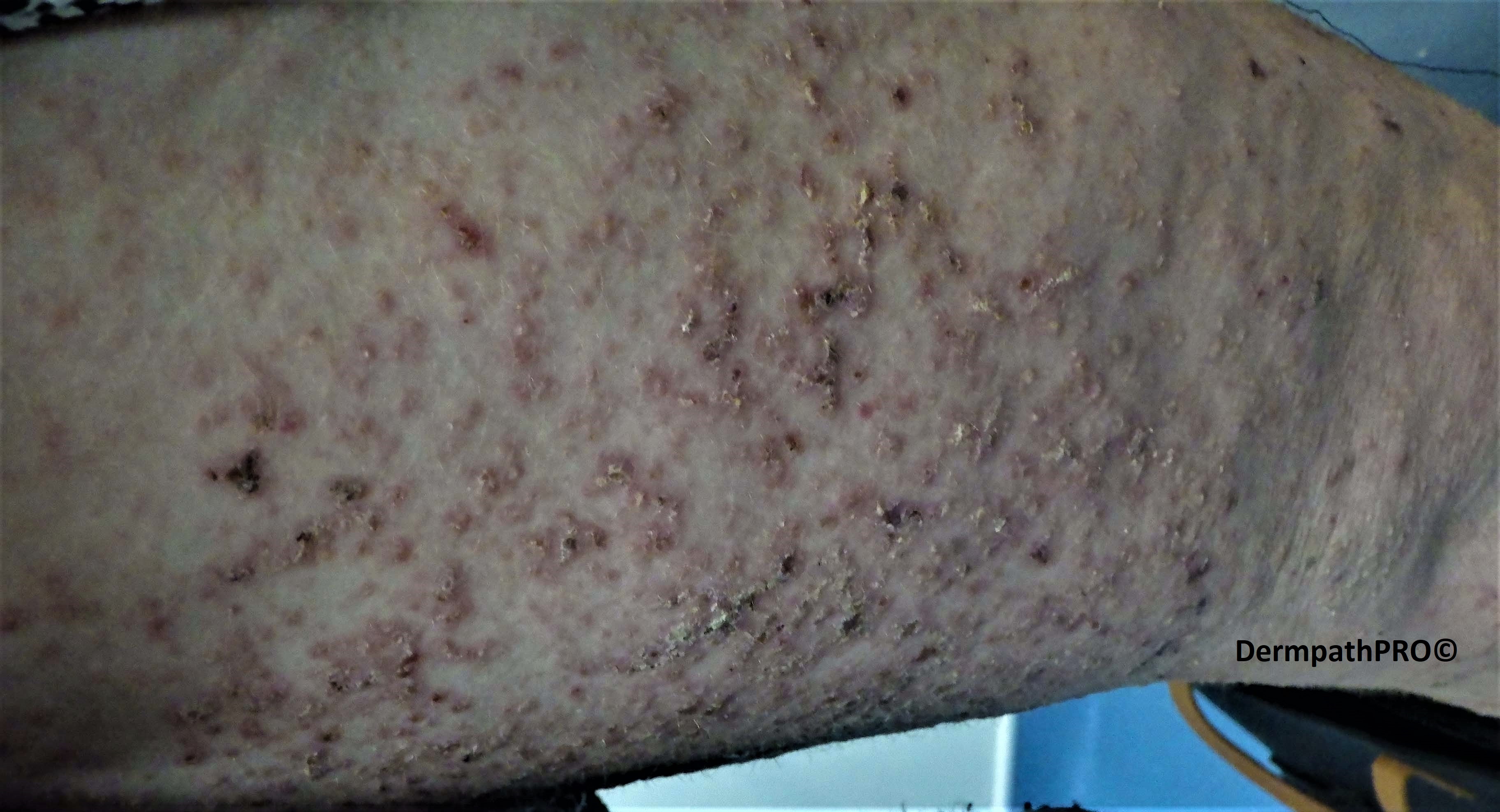
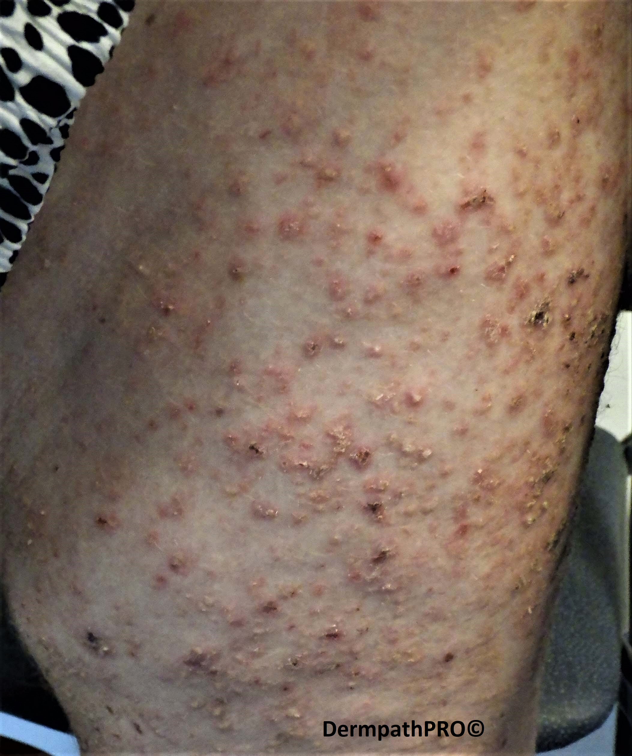
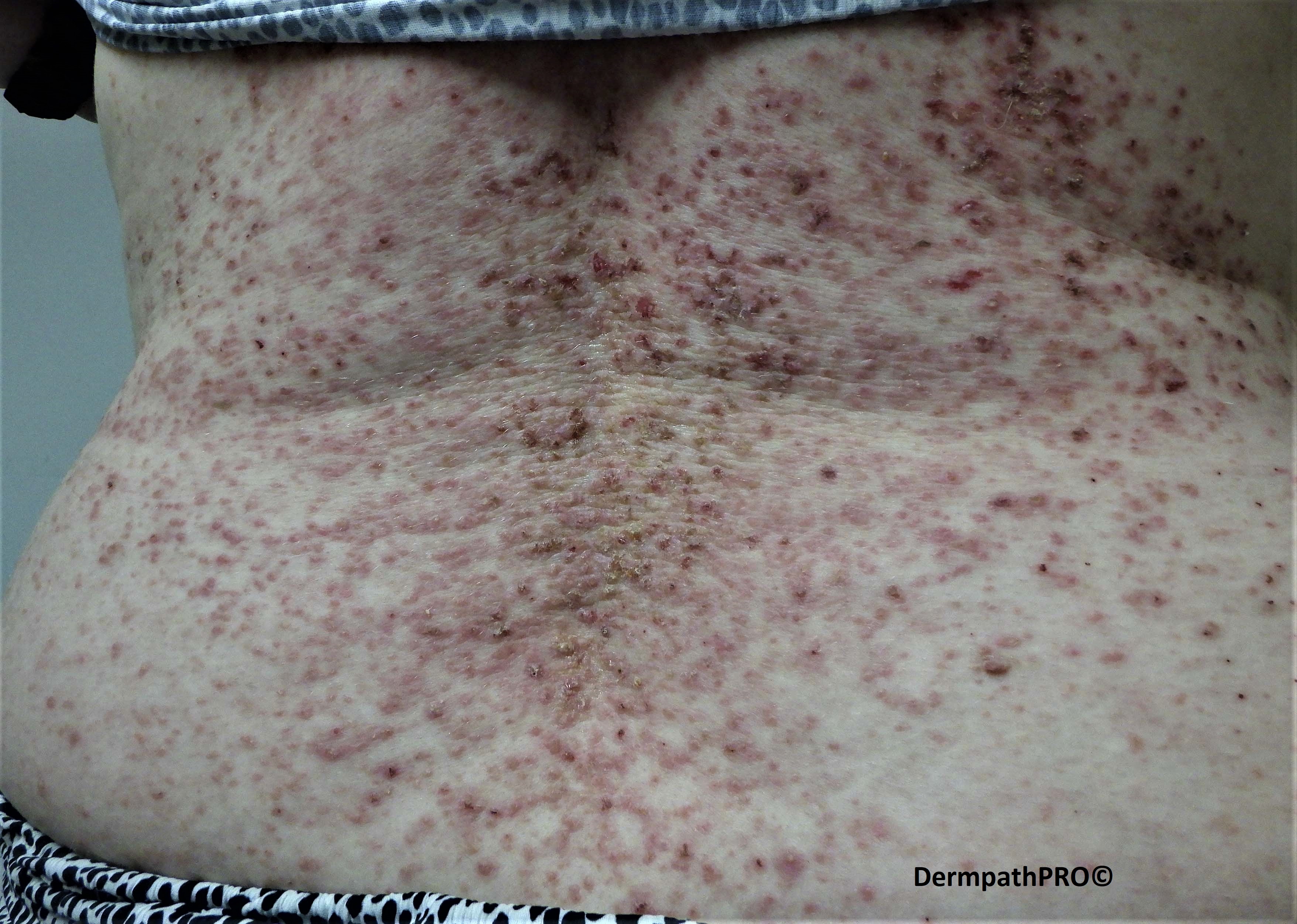
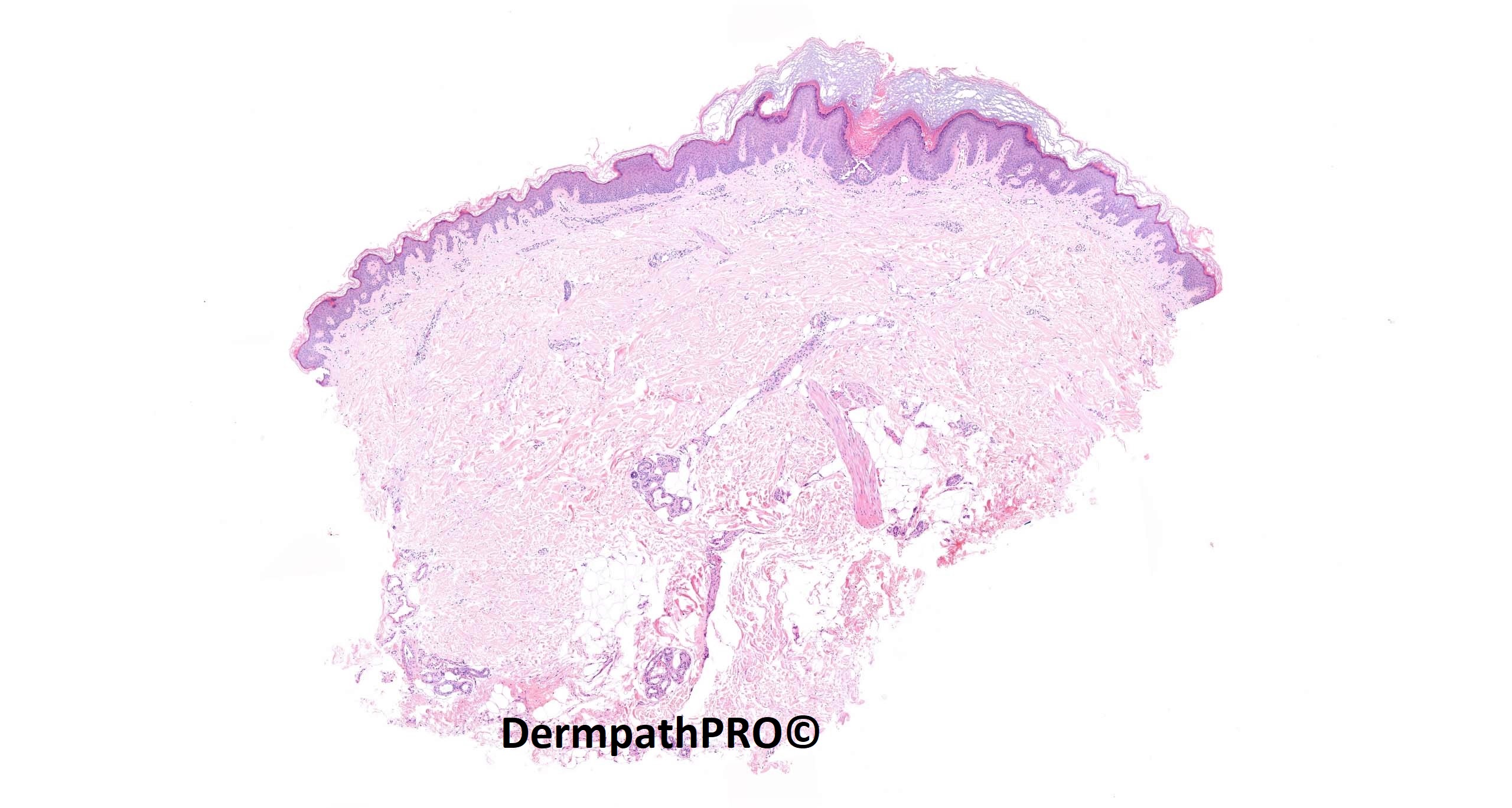
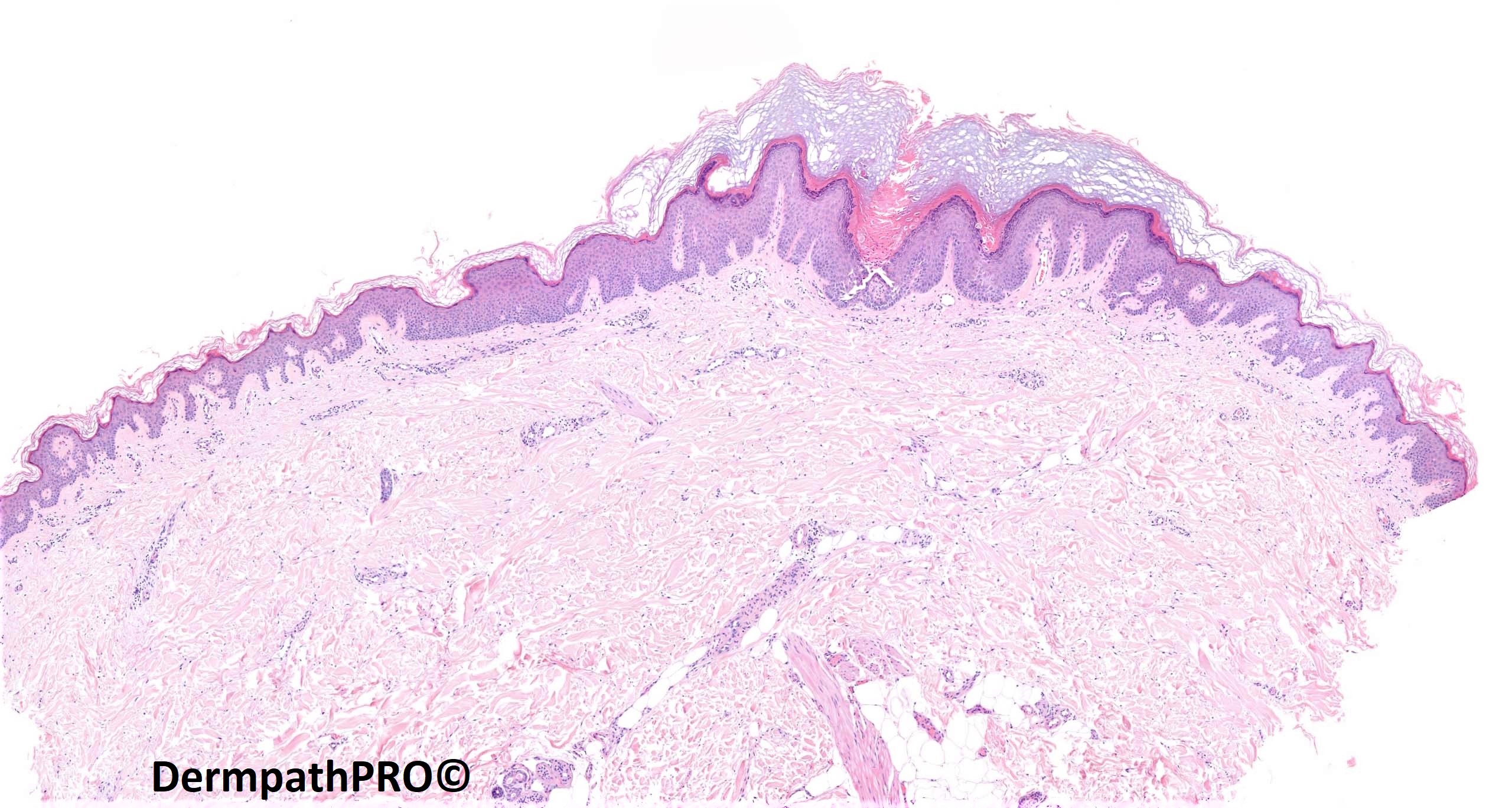
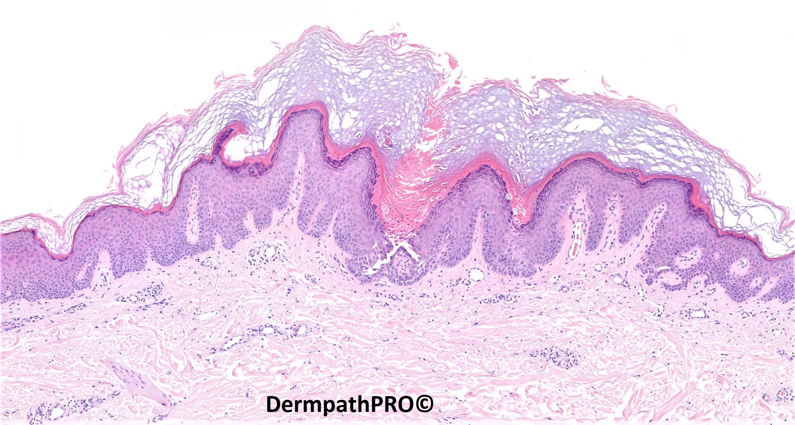
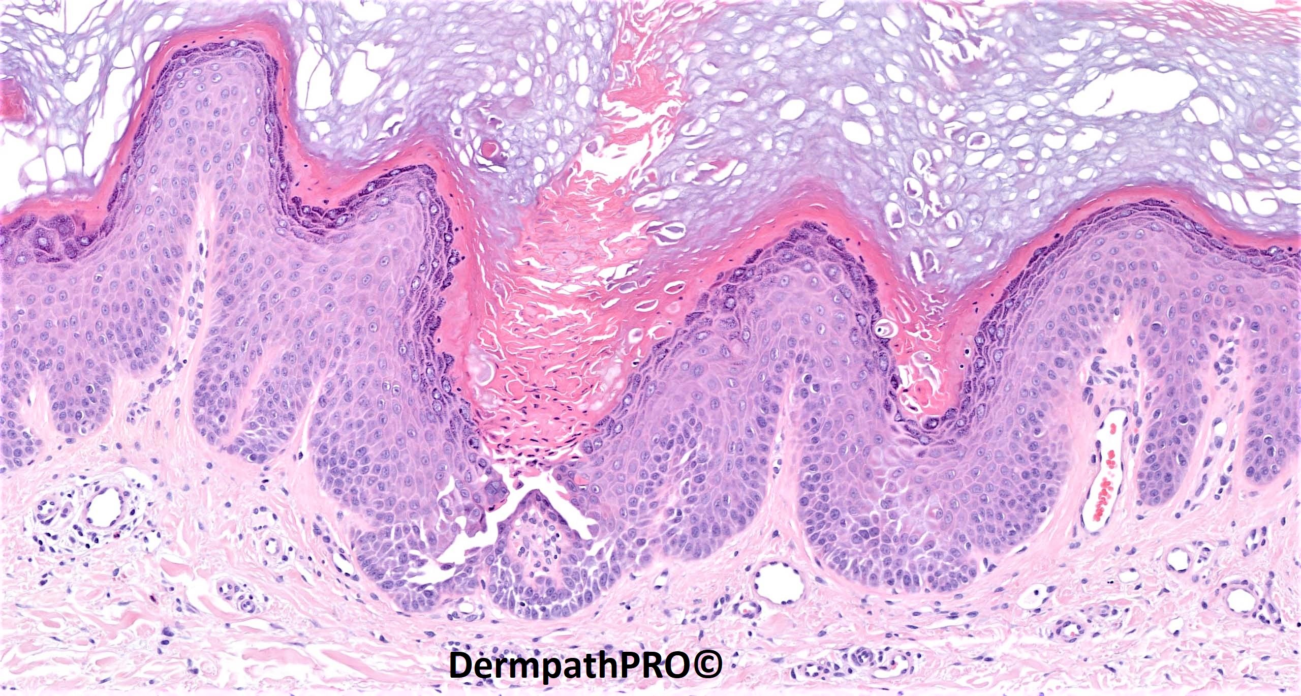
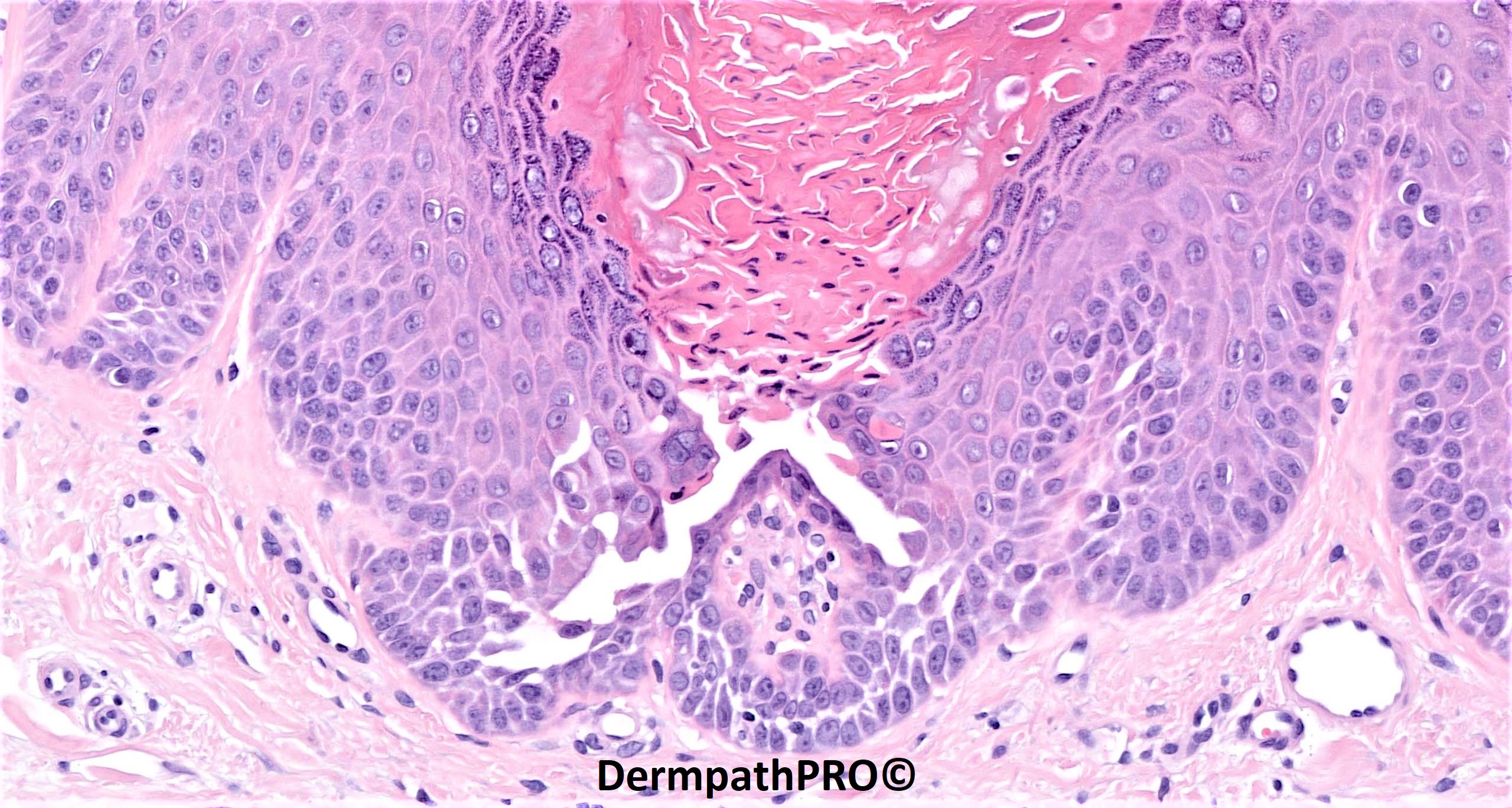
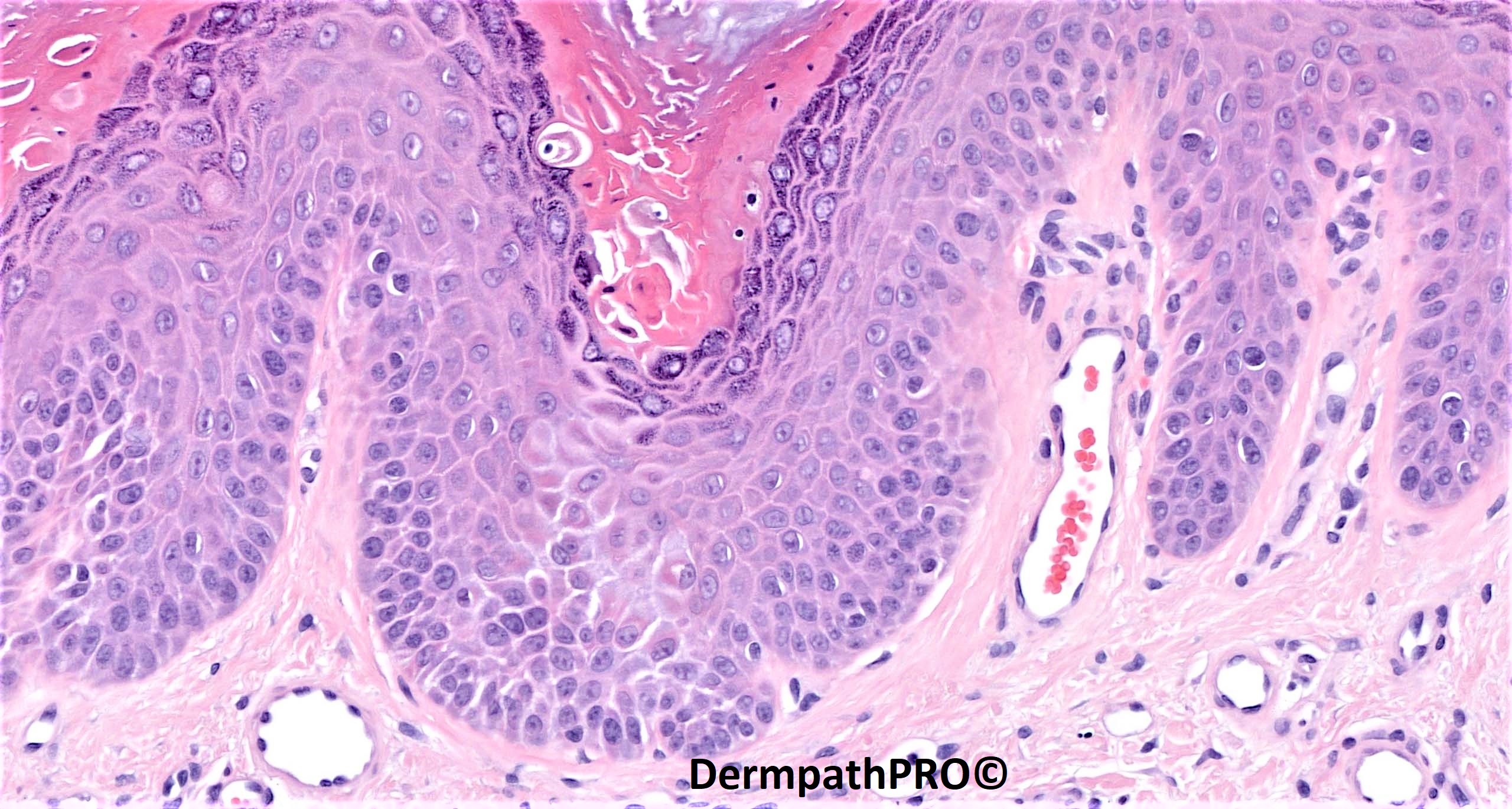
75F, recent flare of extensive rash.


  Report Record

User Feedback


'Dirty - warty' papules histologically showing acantholytic dyskeratosis diagnostic of Darier disease. Exacerbation could be due to summer. 


Share this comment


Link to comment
share_externally

msofopoulos

Posted

Darier?, I think i see grains. Did she had v shaped nailtips?

Share this comment


Link to comment
share_externally

Saman Fatah

Posted

Thanks Saleem for sharing clinical photographs and superb-quality photomicrographs.

It is Darier’s, the photos shows rather large numbers of erosions (some monomorphic) especially on back. In context of an acute widespread flare up, one has to consider super-added infective causes (bacterial/viral (HSV). 

Iam not sure there is a possible multi-nucleate keratinocyte above the acantholysis in the penultimate photo or early viral related cytopathic changes in the last photo or me imagining/hallucinating it! will wait for your constructive comments to enlighten us in due course. 


Share this comment


Link to comment
share_externally

Eman El-Nabarawy

Posted

On 16/11/2022 at 09:03, Saman Fatah said:

Thanks Saleem for sharing clinical photographs and superb-quality photomicrographs.

It is Darier’s, the photos shows rather large numbers of erosions (some monomorphic) especially on back. In context of an acute widespread flare up, one has to consider super-added infective causes (bacterial/viral (HSV). 

Iam not sure there is a possible multi-nucleate keratinocyte above the acantholysis in the penultimate photo or early viral related cytopathic changes in the last photo or me imagining/hallucinating it! will wait for your constructive comments to enlighten us in due course. 


Agree..

Share this comment


Link to comment
share_externally

Saleem Taibjee

Posted

Apologies for the late 'answer' on this one.

Yes, this is a recent case of Darier disease with typical acantholytic dyskeratosis. I wanted to include it because of the nice clinical and histological comparison. As alluded to above, it is worth considering added infection (bacterial or herpes) in patients flaring with this condition, although in this particular instance I think it was just an unexplained flare of the disease itself.

BW

Saleem

Share this comment


Link to comment
share_externally


Join the conversation

You can post now and register later. If you have an account, sign in now to post with your account.

Guest
Add a comment...

×   Pasted as rich text.   Paste as plain text instead

  Only 75 emoji are allowed.

×   Your link has been automatically embedded.   Display as a link instead

×   Your previous content has been restored.   Clear editor

×   You cannot paste images directly. Upload or insert images from URL.

Loading...

×
×
  • Create New...