Jump to content
In this section we have spot diagnoses posted on a daily basis since June 2010, now over 4000! You can review the archived cases and read the suggested diagnoses by users and the final comment by the contributors.
Case are uploaded each week day by 10 am UK time with the correct diagnosis will generally be posted at 8 pm UK time. Why not view the most recent spot diagnosis and proffer a diagnosis?

Case Number : Case 4099 - 06 Oct 2022 Posted By: Saleem Taibjee

Please read the clinical history and view the images by clicking on them before you proffer your diagnosis.
Submitted Date :
   (0 reviews)

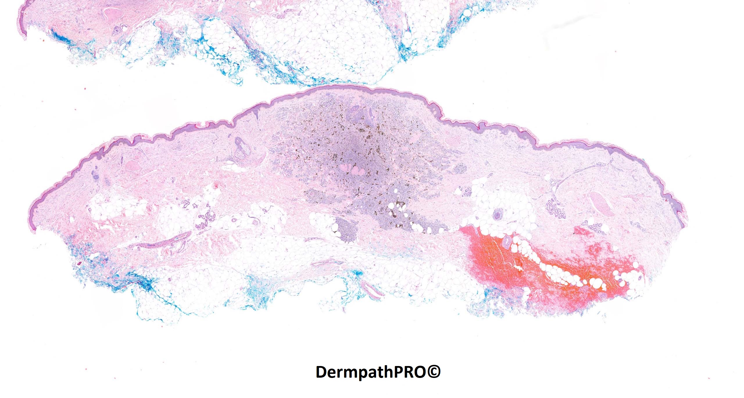
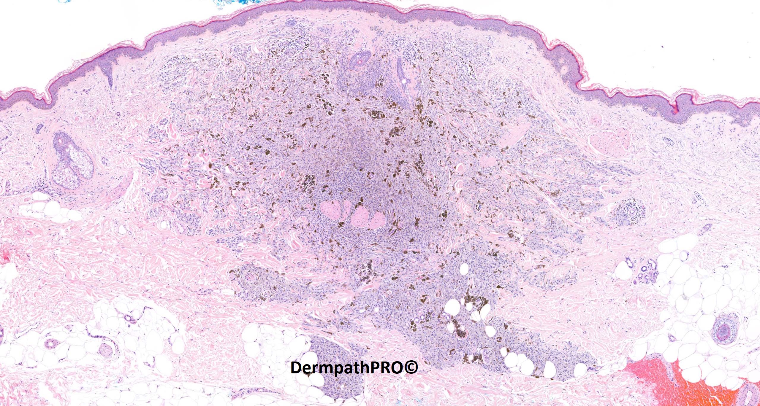
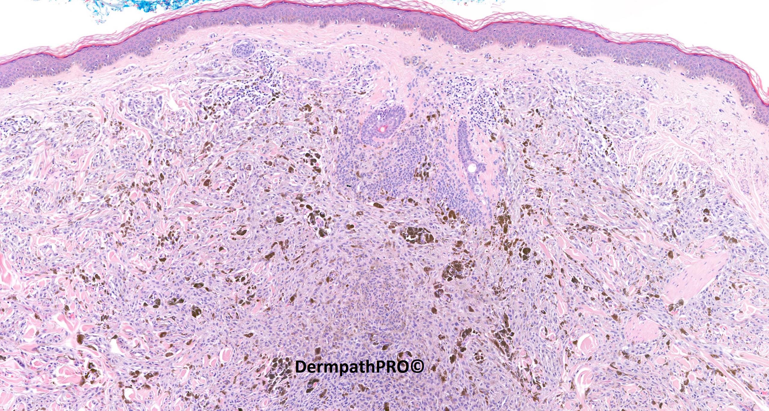
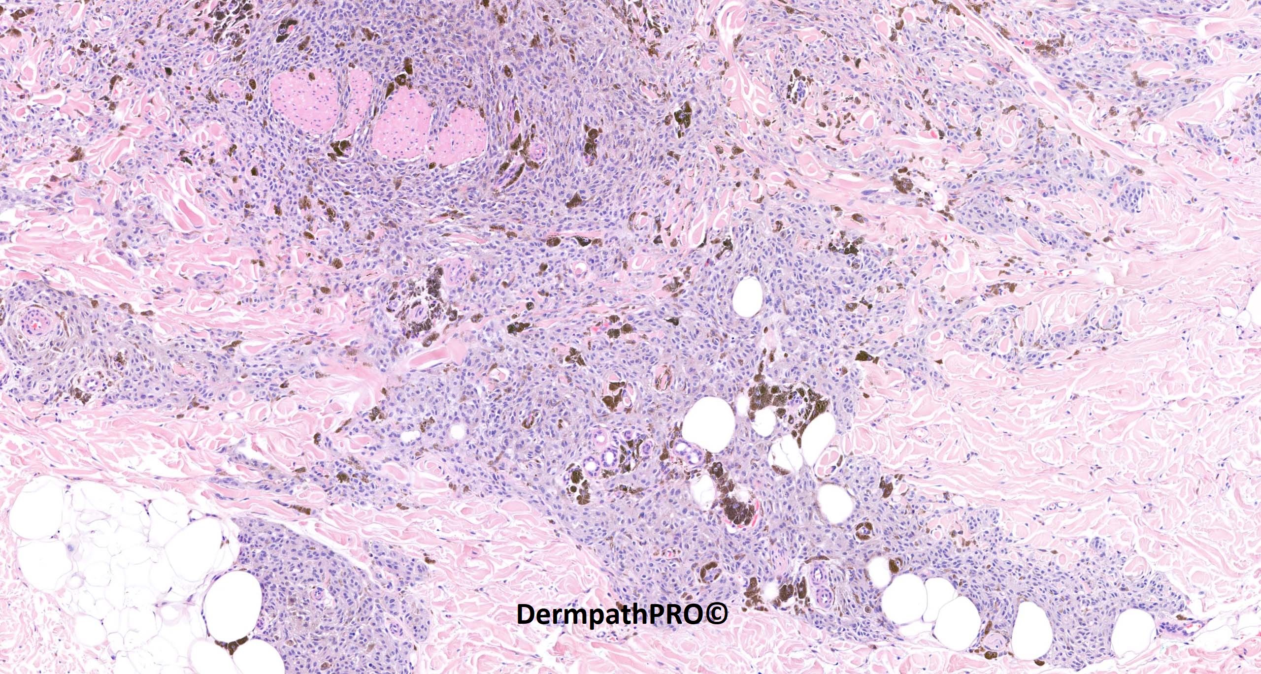
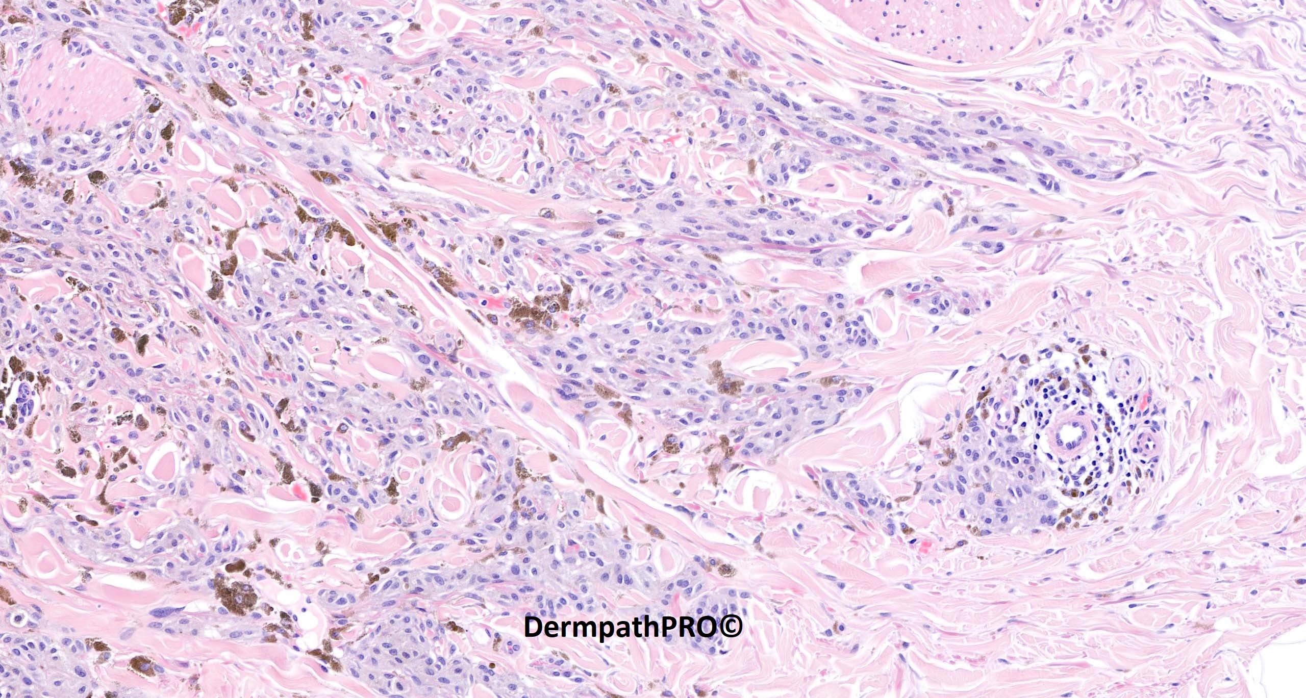
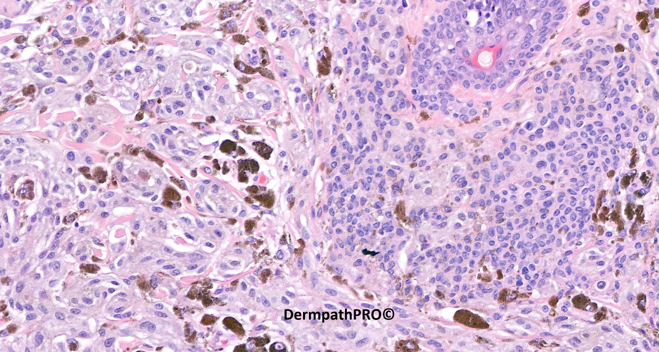
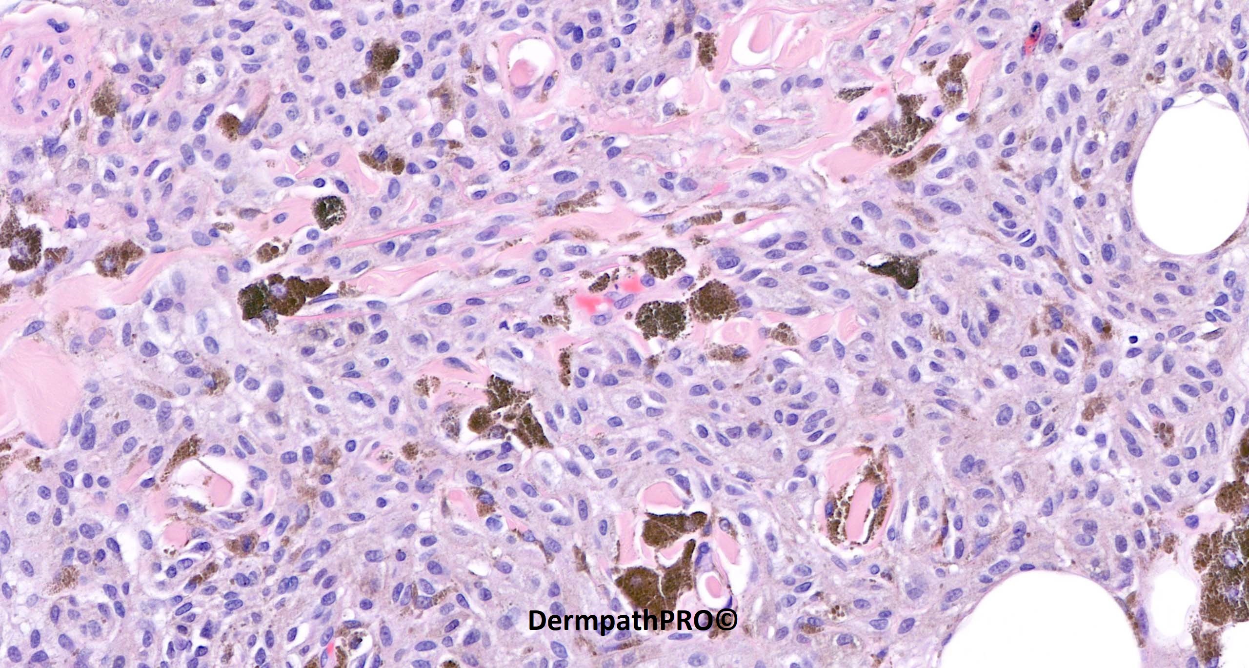
62F excision right upper arm ?blue naevus ?MM


  • Like 1
  Report Record

User Feedback


msofopoulos

Posted

It could be a DPN. I think most of them display (or evolve from) a common nevus (with a diverse b-catenin staining pattern membrane and cytoplasm vs membrane only)

Share this comment


Link to comment
share_externally

Yes. A DPN cmbined with common intradermal nevus. 

Share this comment


Link to comment
share_externally

21 hours ago, msofopoulos said:

It could be a DPN. I think most of them display (or evolve from) a common nevus (with a diverse b-catenin staining pattern membrane and cytoplasm vs membrane only)

Talked to Dr. Brenn about the terminology, he wants to dispose of the term combined nevus in the context of DPN. He believes these lesions start out as banal nevus with a second hit (activating mutations in beta-catenin, conceptually more of a specific flavor of clonal nevus) rather than collision/combined. A "pure" DPN might have acquired the second hit very early and the precedent banal component is no longer visible. Postulations supported here:Combined activation of MAP kinase pathway and β-catenin signaling cause deep penetrating nevi | Nature Communications

Share this comment


Link to comment
share_externally

msofopoulos

Posted

2 hours ago, Shawn said:

he wants to dispose of the term combined nevus in the context of DPN

After this paper from I think of them as biphenotypic and not as combined nevi, but we are somewhat forced to use the WHO nomenclature (at least here in Greece). I think that in the next blue book we will see more of the term melanocytoma.

Share this comment


Link to comment
share_externally

Saleem Taibjee

Posted

Yes, this is a deep penetrating naevus. I wonder if many of these lesions were historically (and perhaps even currently) diagnosed as blue naevus clinically and histologically. But, of course, the cytology is quite different. The beta-catenin immunostain is shown below, and nicely shows the differential pattern of staining between the DPN cells (with more intense nuclear and cytoplasmic staining) and conventional smaller naevocellular melanocytes as depicted in the 5th image above (with weaker staining sparing the nucleus). Not shown here, but S100 was strong which also tends to argue against blue naevus.

The comments relating to classification above are interesting and pertinent.

My perspective is that many DPN lesions have clearly arisen due to a second hit in a pre-existing naevus, and in that sense combined naevus is perhaps an outdated term. But I suppose it doesn't matter as long as we can recognise such lesions, and distinguish from melanoma arising in a naevus, and from either blue or Spitz spectrum.

In this particular case the cytology is extremely bland, with no mitoses, and I would venture Deep Penetrating Naevus. But I think if any significant cytological atypia or mitotic activity, Deep Penetrating Melanocytoma is a more appropriate term, and in the more worrying examples ideally such lesions should be subjected to more detailed study such as looking for a TERT promoter mutation (and other genes such as TP53, CDKN2A, PTEN, etc), array CGH, or FISH, to help gauge risk and determine management.

BW, Saleem

image.jpeg

image.jpeg


Share this comment


Link to comment
share_externally


Join the conversation

You can post now and register later. If you have an account, sign in now to post with your account.

Guest
Add a comment...

×   Pasted as rich text.   Paste as plain text instead

  Only 75 emoji are allowed.

×   Your link has been automatically embedded.   Display as a link instead

×   Your previous content has been restored.   Clear editor

×   You cannot paste images directly. Upload or insert images from URL.

Loading...

×
×
  • Create New...