Jump to content
In this section we have spot diagnoses posted on a daily basis since June 2010, now over 4000! You can review the archived cases and read the suggested diagnoses by users and the final comment by the contributors.
Case are uploaded each week day by 10 am UK time with the correct diagnosis will generally be posted at 8 pm UK time. Why not view the most recent spot diagnosis and proffer a diagnosis?

Case Number : Case 4109 - 20 Oct 2022 Posted By: Saleem Taibjee

Please read the clinical history and view the images by clicking on them before you proffer your diagnosis.
Submitted Date :
   (0 reviews)

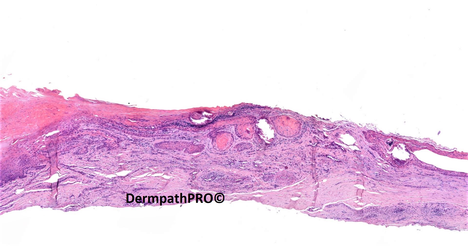
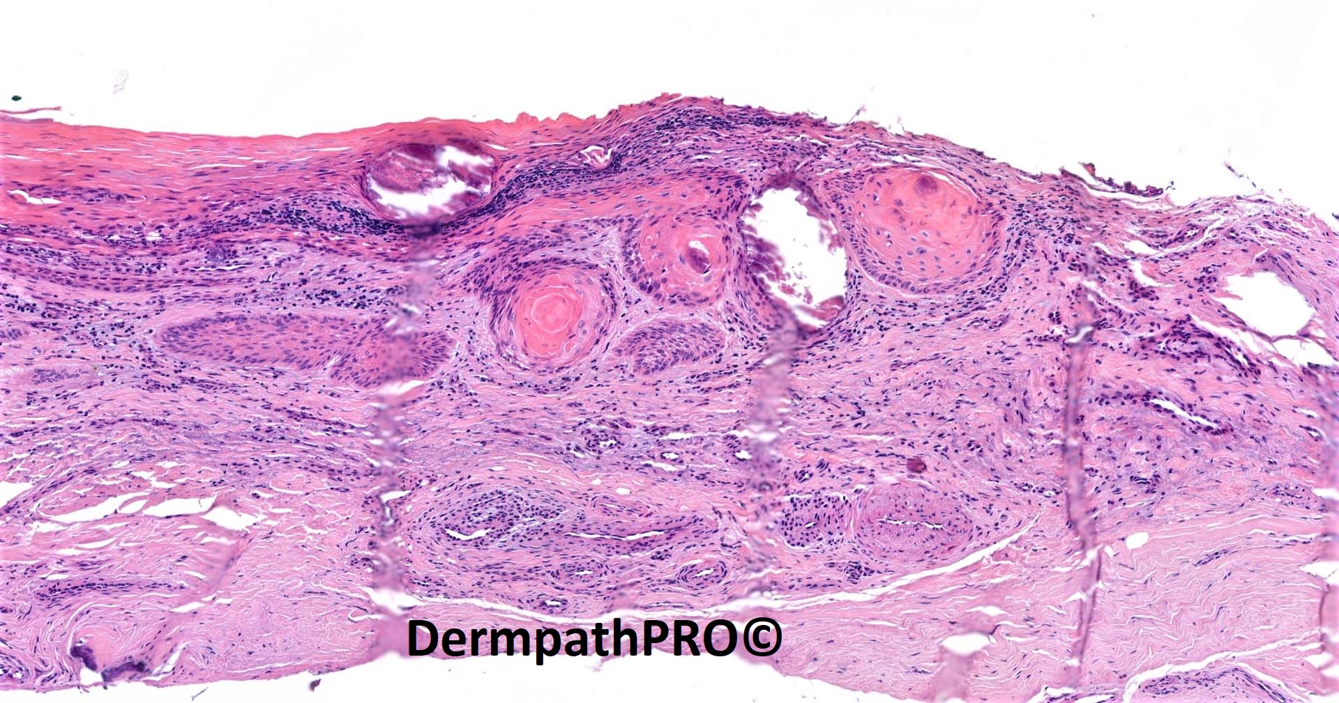
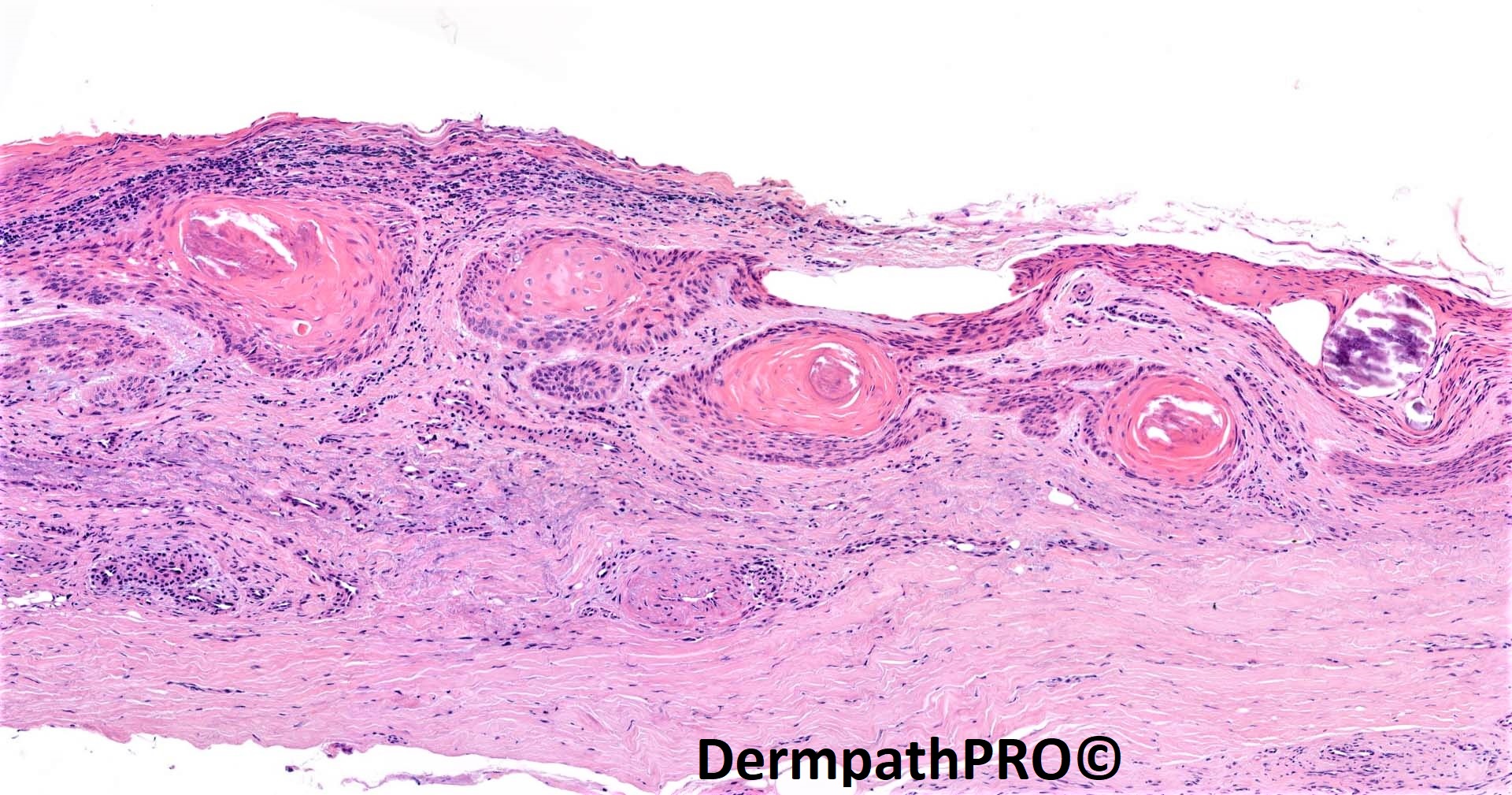
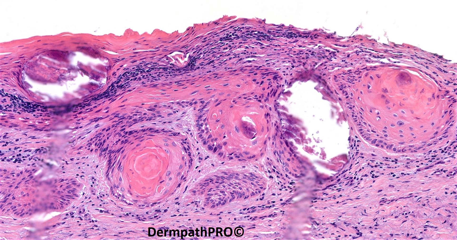
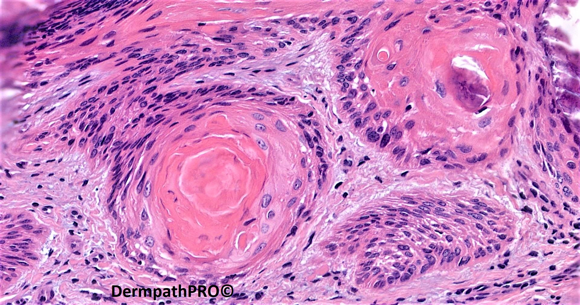
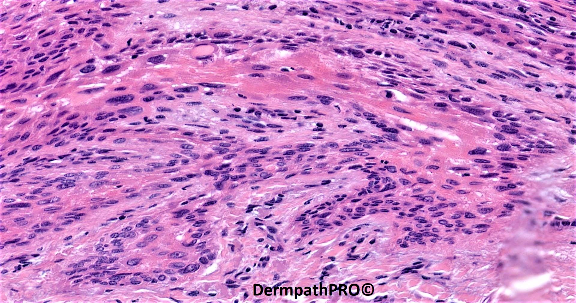
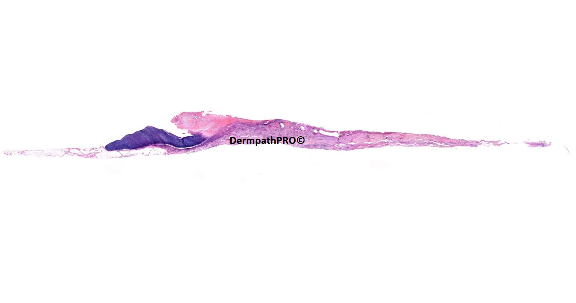
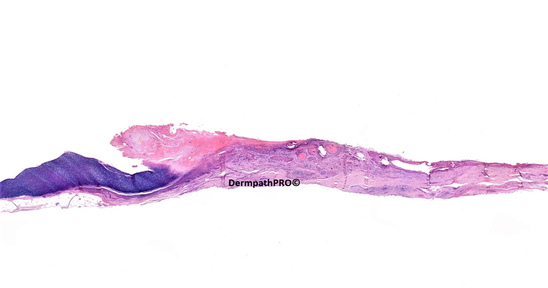
70M, full thickness longitudinal excision of lateral third of nail bed, left ring finger, ‘subungual fibrous papule’

  • Like 1
  • Thanks 1
  Report Record

User Feedback


I think this is a Onychopapilloma (OP) associated with subepidermal onycholemmal cysts. The calcifications can be seen in these cysts and they are not uncommon in OP

Share this comment


Link to comment
share_externally

Saleem Taibjee

Posted

Not so many responses this time, perhaps reflecting how we all struggle with nail specimens.

So well done to Igor....

I was very grateful to Prof Eckhart Haneke for his expert opinion on this case, as follows:

"In my special onychopathology practice, this is not so uncommon. I have no doubt that it is an onychopapilloma with some secondary infection explaining the neutrophils in the distal keratotic tip of the lesion. The epithelial buds and cyst formation with a tendency to calcification are very characteristic. Some nuclear atypia is normal for onychopapillomas, particularly binucleate cells and an occasional apoptotic cell.

This is essentially a benign lesion. I would treat it as such. However, as two consulting histopathologists are not sure about the benignity I would recommend to just follow the patient once yearly. Recurrences are relatively common and not necessarily a sign of a malignant lesion."

McKee’s textbook also states: Pathology indicates papillomatous acanthosis of the distal matrix spreading to the nail bed, longitudinal canaliform dystrophy of the ventral nail plate, matrix-like metaplasia of the nail bed and distal subungual hyperkeratosis.

BW
Saleem

Share this comment


Link to comment
share_externally

Sorry, I'm late. Very interesting case, especially for a general pathologist like me. I'm learning, in this case, beacause my thought was an early SCC. Thanks, Saleem. 

Share this comment


Link to comment
share_externally


Join the conversation

You can post now and register later. If you have an account, sign in now to post with your account.

Guest
Add a comment...

×   Pasted as rich text.   Paste as plain text instead

  Only 75 emoji are allowed.

×   Your link has been automatically embedded.   Display as a link instead

×   Your previous content has been restored.   Clear editor

×   You cannot paste images directly. Upload or insert images from URL.

Loading...

×
×
  • Create New...