Jump to content
In this section we have spot diagnoses posted on a daily basis since June 2010, now over 4000! You can review the archived cases and read the suggested diagnoses by users and the final comment by the contributors.
Case are uploaded each week day by 10 am UK time with the correct diagnosis will generally be posted at 8 pm UK time. Why not view the most recent spot diagnosis and proffer a diagnosis?

Case Number : Case 4084 - 15 September 2022 Posted By: Saleem Taibjee

Please read the clinical history and view the images by clicking on them before you proffer your diagnosis.
Submitted Date :
   (0 reviews)

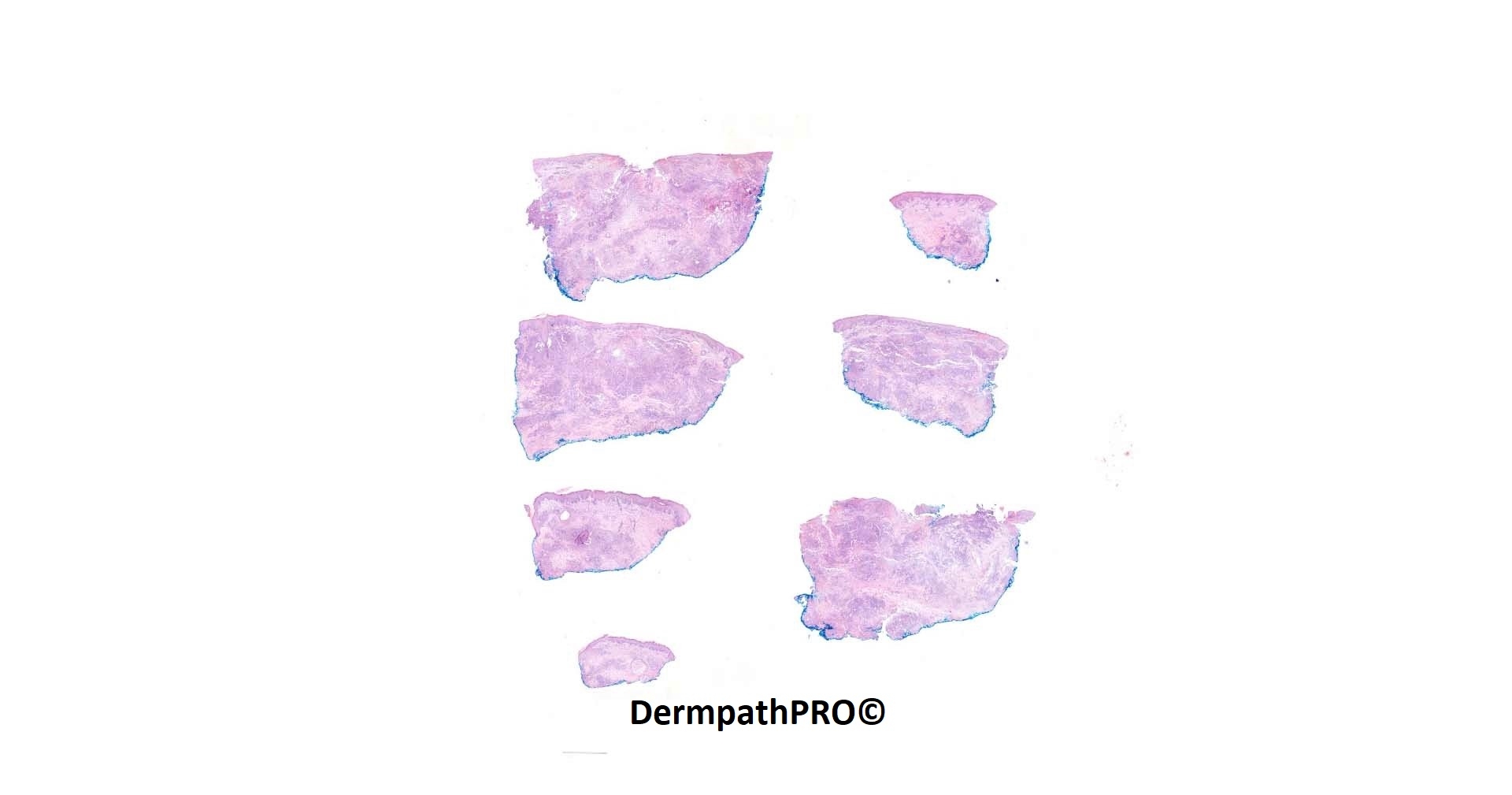
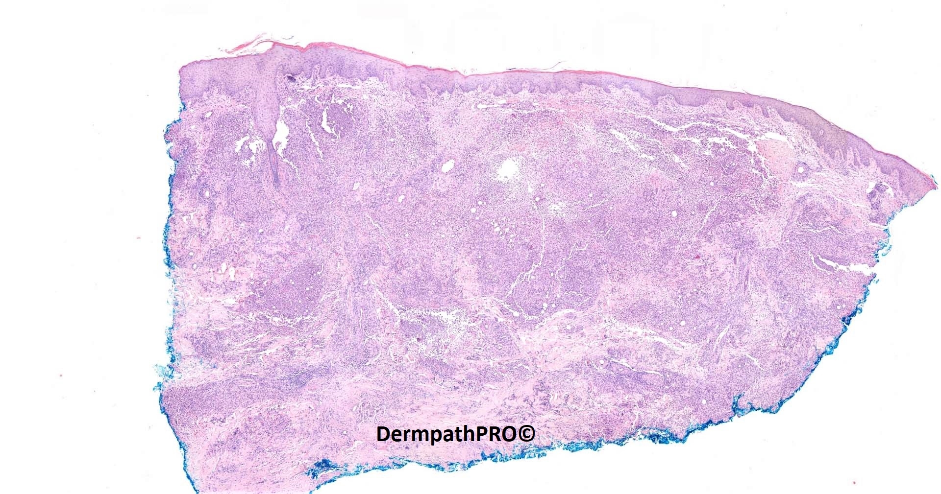
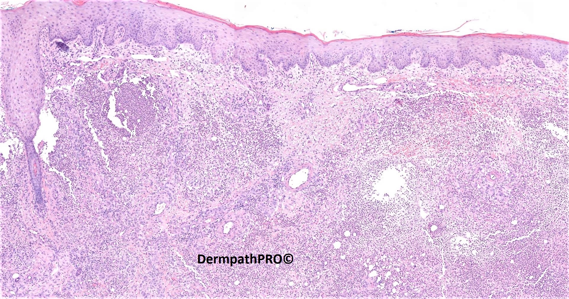
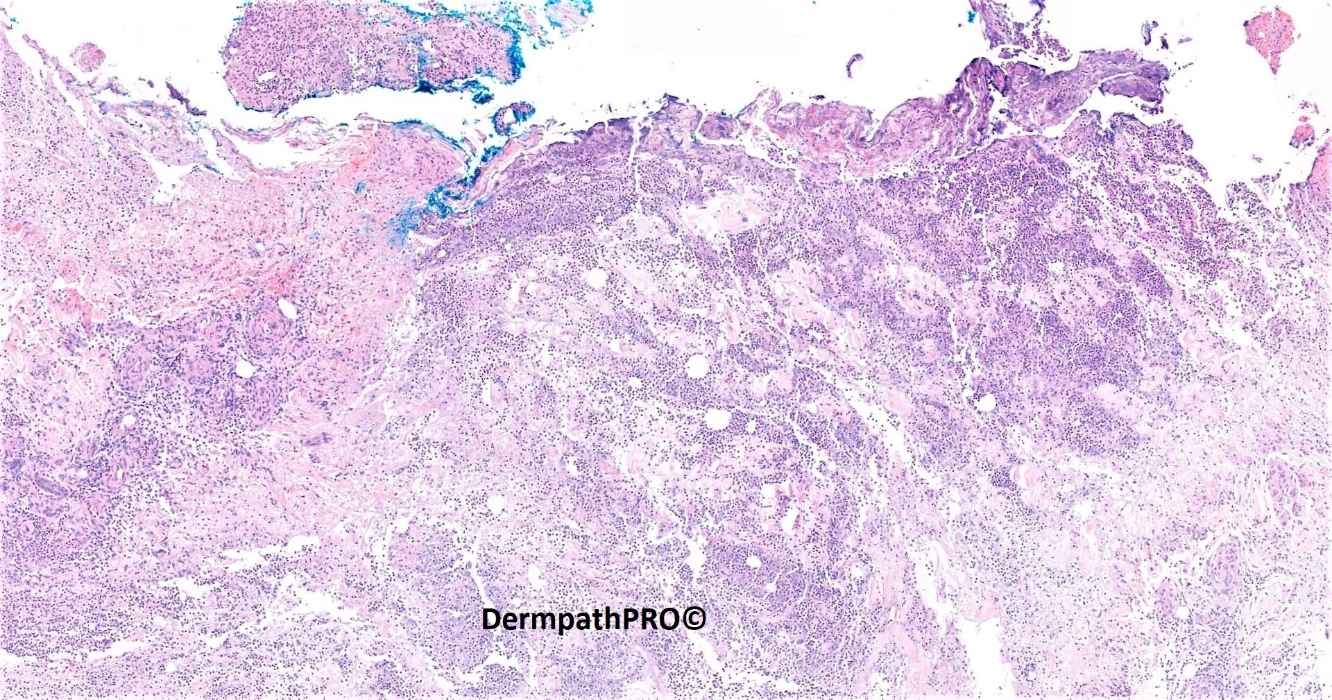
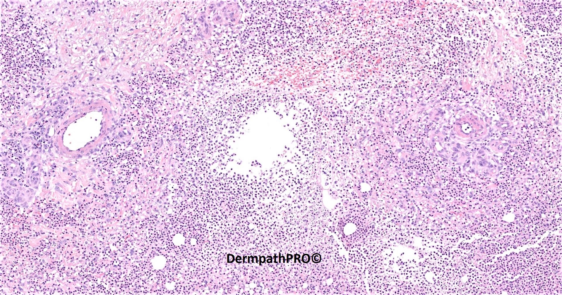
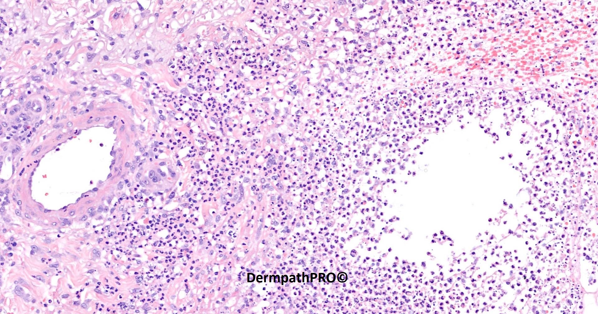
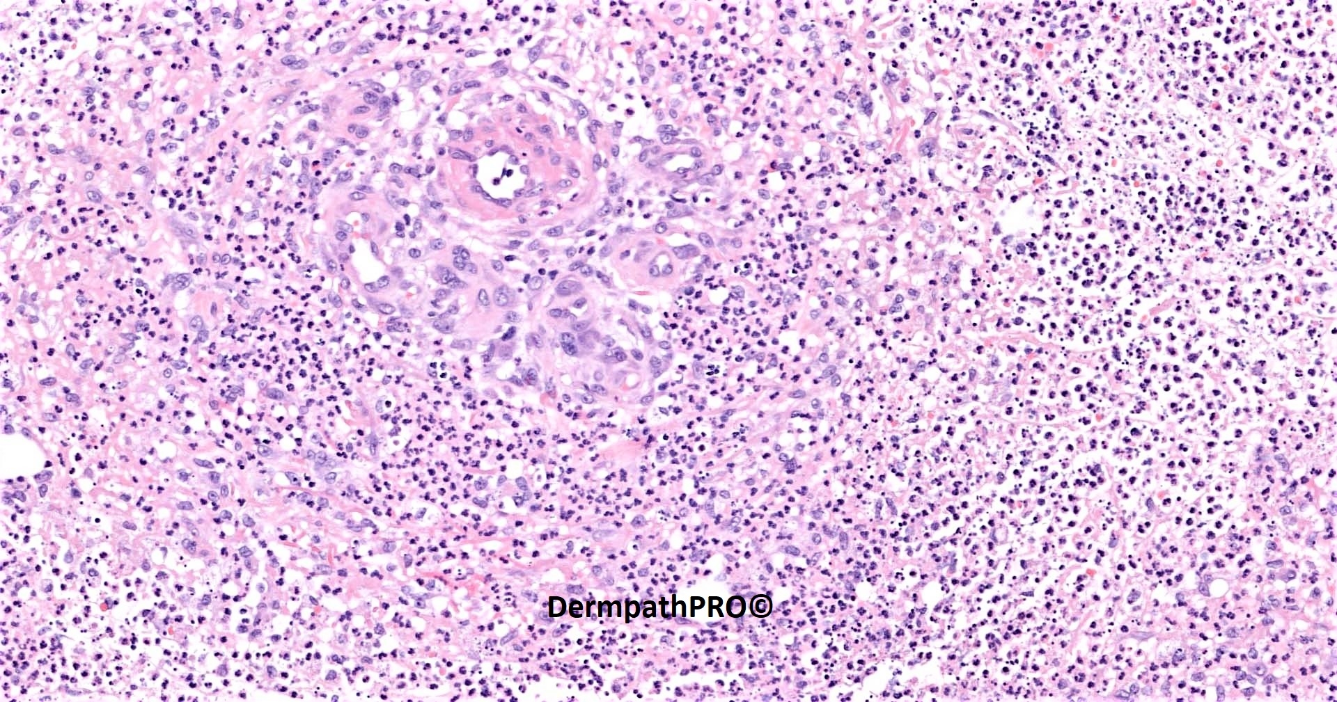
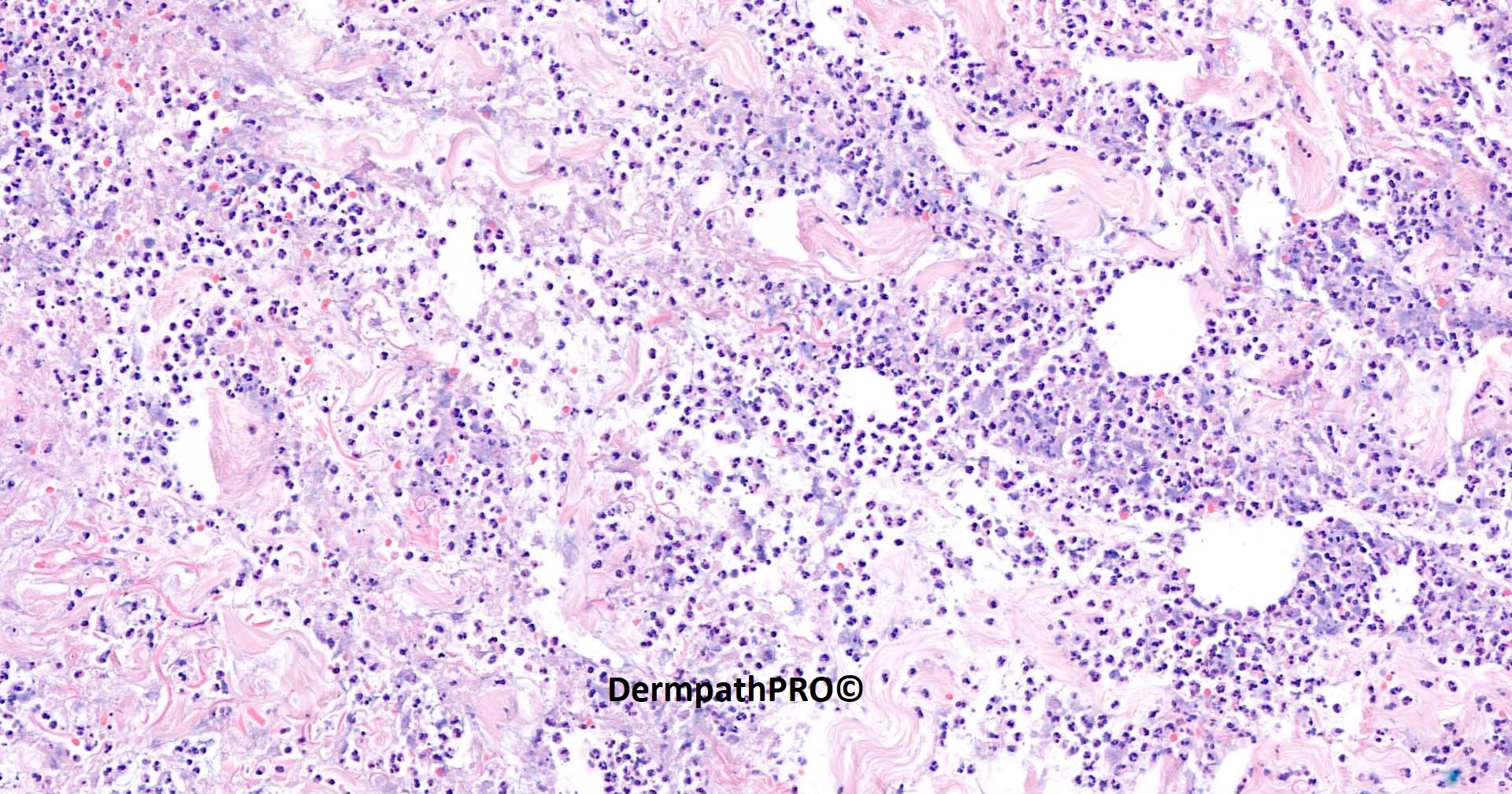
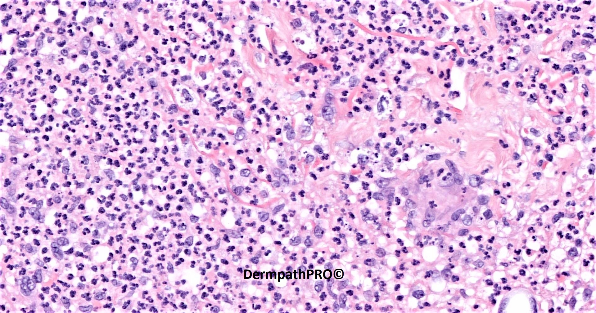
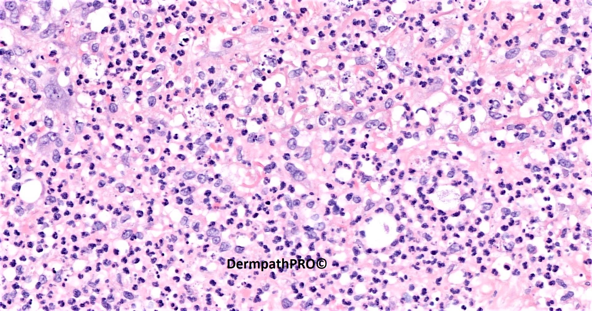
82M biopsy right calf: crusted lesions on right lower leg ?infected eczema ?malignancy


  Report Record

User Feedback


Neutrophilic dermatosis, could be pyoderma gangrenosum in the setting of hematological malignancy, rheumatoid arthritis or inflammatory bowel disease.

Share this comment


Link to comment
share_externally

Meenakshi Batrani

Posted

Need to exclude fungal, some unusual bacterial and atypical mycobacterial infection. 

Share this comment


Link to comment
share_externally

Saman Fatah

Posted

Infection especially rapidly growing NTM species (Non-Tubeculous Mycobacteria) like M.Chelonae and others. In the last image the white circular spaces on right lower corner contains possible organisms.

Ultimately special stains and tissue culture is required to confirm. 

Was the patient immunocompromised or on Prednisolone?

Share this comment


Link to comment
share_externally

Eman El-Nabarawy

Posted

Of course infection needs to be ruled out. But I d like to add anaplastic large T cell lymphoma as a mimicker of inflammatory conditions to the DD.

Share this comment


Link to comment
share_externally

Saleem Taibjee

Posted

Congratulations to Saman. Spot on... this was a recent case of confirmed Mycobacterium chelonae. The patient was indeed on methotrexate for severe eczema, and thus immunocompromised. He is a keen gardener, so presumably acquired the infection in that environment. The circular spaces which Saman points out are indeed where to look for the organisms. The ZN stain is shown below. There were numerous organisms suggestive of a fast-grower.

The key message here is that atypical mycobacterial infections do not always show much in the way of granulomatous inflammation, and as in this case, can comprise mostly neutrophils/abscess-like picture. So remember to include a ZN stain also. I have seen cases where Wade-Fite is positive, but ZN is negative. However, the Wade-Fite technique is more difficult, and in fact our own lab has now stopped providing Wade-Fite, but not an issue for this particular case.

image.jpegimage.jpeg


Share this comment


Link to comment
share_externally


Join the conversation

You can post now and register later. If you have an account, sign in now to post with your account.

Guest
Add a comment...

×   Pasted as rich text.   Paste as plain text instead

  Only 75 emoji are allowed.

×   Your link has been automatically embedded.   Display as a link instead

×   Your previous content has been restored.   Clear editor

×   You cannot paste images directly. Upload or insert images from URL.

Loading...

×
×
  • Create New...