Jump to content
In this section we have spot diagnoses posted on a daily basis since June 2010, now over 4000! You can review the archived cases and read the suggested diagnoses by users and the final comment by the contributors.
Case are uploaded each week day by 10 am UK time with the correct diagnosis will generally be posted at 8 pm UK time. Why not view the most recent spot diagnosis and proffer a diagnosis?

Case Number : Case 4629 - 05 August 2025 Posted By: Admin_Dermpath

Please read the clinical history and view the images by clicking on them before you proffer your diagnosis.
Submitted Date :
   (0 reviews)

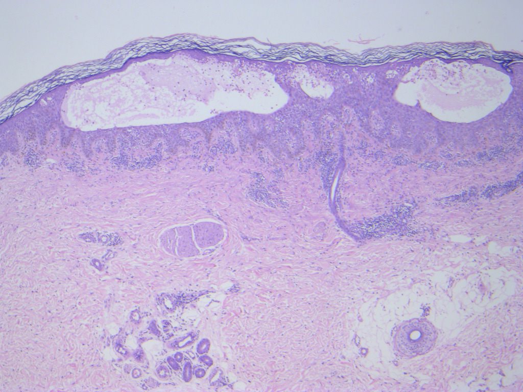
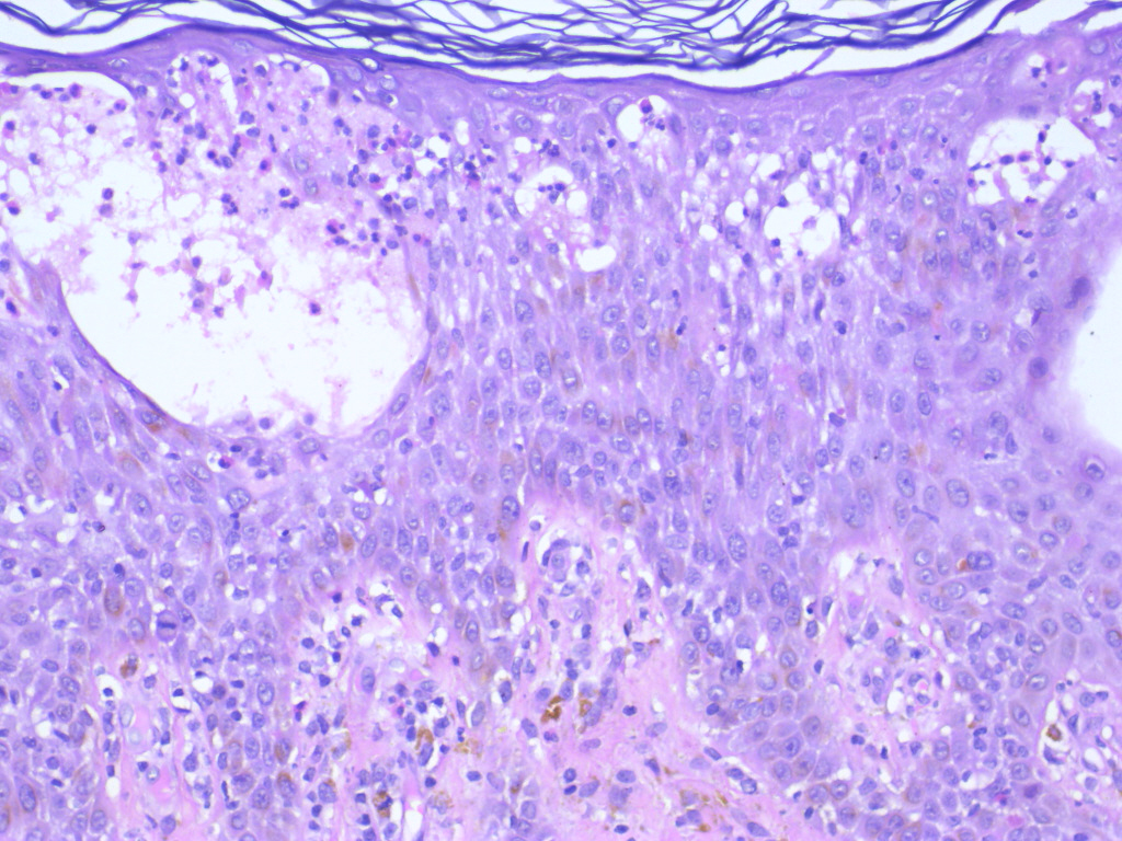
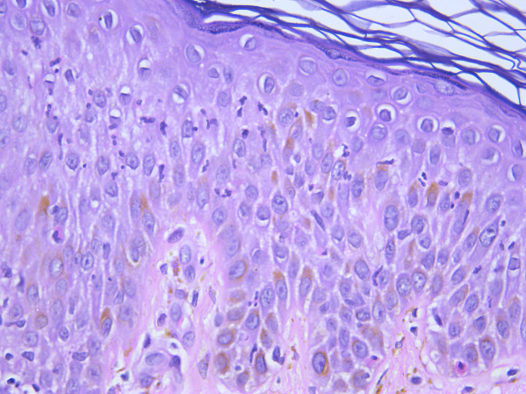
60 year female, presented with eroded vesicular lesions on itchy erythematous skin since 8-9 years.No oral lesions. Responds to Dapsone. Clinical considerations were dermatitis herpetiformis and bullous pemphigoid.


  Report Record

User Feedback


Dr. Shoba Ramesh

Posted

prebullous pemphigoid 

Share this comment


Link to comment
share_externally

Richard Logan

Posted

I presume the description "since 8-9 years" really means "for" 8-9 years, rather then "since the age of 8-9 years", which of course raises other possible diagnoses, however unlikely.

There are intra-epidermal vesicles containing eosinophils, but no suggestion of a sub-epidermal split.  I don't think the intra-epidermal position of the vesicles is due to re-epithelialisation after healed blistering.  Such eosinophil-containing vesicles can be seen in the pre-bullous phase of pemphigoid but it would be unusual for this phase to persist for 8-9 years without "maturing" into overt bullous pemphigoid.  Obviously immunofluorescence would clinch the diagnosis.  If negative, then alternative diagnoses would have to be considered such as an "eczematous" drug reaction, insect bites etc.

Share this comment


Link to comment
share_externally

msofopoulos

Posted

I would wouldnt rule out BP and Duhring but how about prurigo pigmentosa (nagashima)? For BP and Duhring the split is not in the right place (subepidermal). Would like to see DIF as the first two have pretty distinct patterns and the third is negative (i think)

Share this comment


Link to comment
share_externally

Meenakshi Batrani

Posted

DIF showed intercellular positivity with IgG and C3. Thus, the diagnosis is pemphigus herpetiformis which usually shows eosinophilic and/or neutrophilic spongiosis with intraepidermal vesicles / pustules, while acantholysis may be inconspicuous in many cases.

Share this comment


Link to comment
share_externally

Meenakshi Batrani

Posted

On 18/08/2025 at 14:50, Richard Logan said:

I presume the description "since 8-9 years" really means "for" 8-9 years, rather then "since the age of 8-9 years", which of course raises other possible diagnoses, however unlikely.

There are intra-epidermal vesicles containing eosinophils, but no suggestion of a sub-epidermal split.  I don't think the intra-epidermal position of the vesicles is due to re-epithelialisation after healed blistering.  Such eosinophil-containing vesicles can be seen in the pre-bullous phase of pemphigoid but it would be unusual for this phase to persist for 8-9 years without "maturing" into overt bullous pemphigoid.  Obviously immunofluorescence would clinch the diagnosis.  If negative, then alternative diagnoses would have to be considered such as an "eczematous" drug reaction, insect bites etc.

Yes. It is for 8-9 years, some grammatical errors while writing the history.  

Share this comment


Link to comment
share_externally


Join the conversation

You can post now and register later. If you have an account, sign in now to post with your account.

Guest
Add a comment...

×   Pasted as rich text.   Paste as plain text instead

  Only 75 emoji are allowed.

×   Your link has been automatically embedded.   Display as a link instead

×   Your previous content has been restored.   Clear editor

×   You cannot paste images directly. Upload or insert images from URL.

Loading...

×
×
  • Create New...