Jump to content
In this section we have spot diagnoses posted on a daily basis since June 2010, now over 4000! You can review the archived cases and read the suggested diagnoses by users and the final comment by the contributors.
Case are uploaded each week day by 10 am UK time with the correct diagnosis will generally be posted at 8 pm UK time. Why not view the most recent spot diagnosis and proffer a diagnosis?

Case Number : CASE 4613 - 29 March 2025 Posted By: Admin_Dermpath

Please read the clinical history and view the images by clicking on them before you proffer your diagnosis.
Submitted Date :
   (0 reviews)

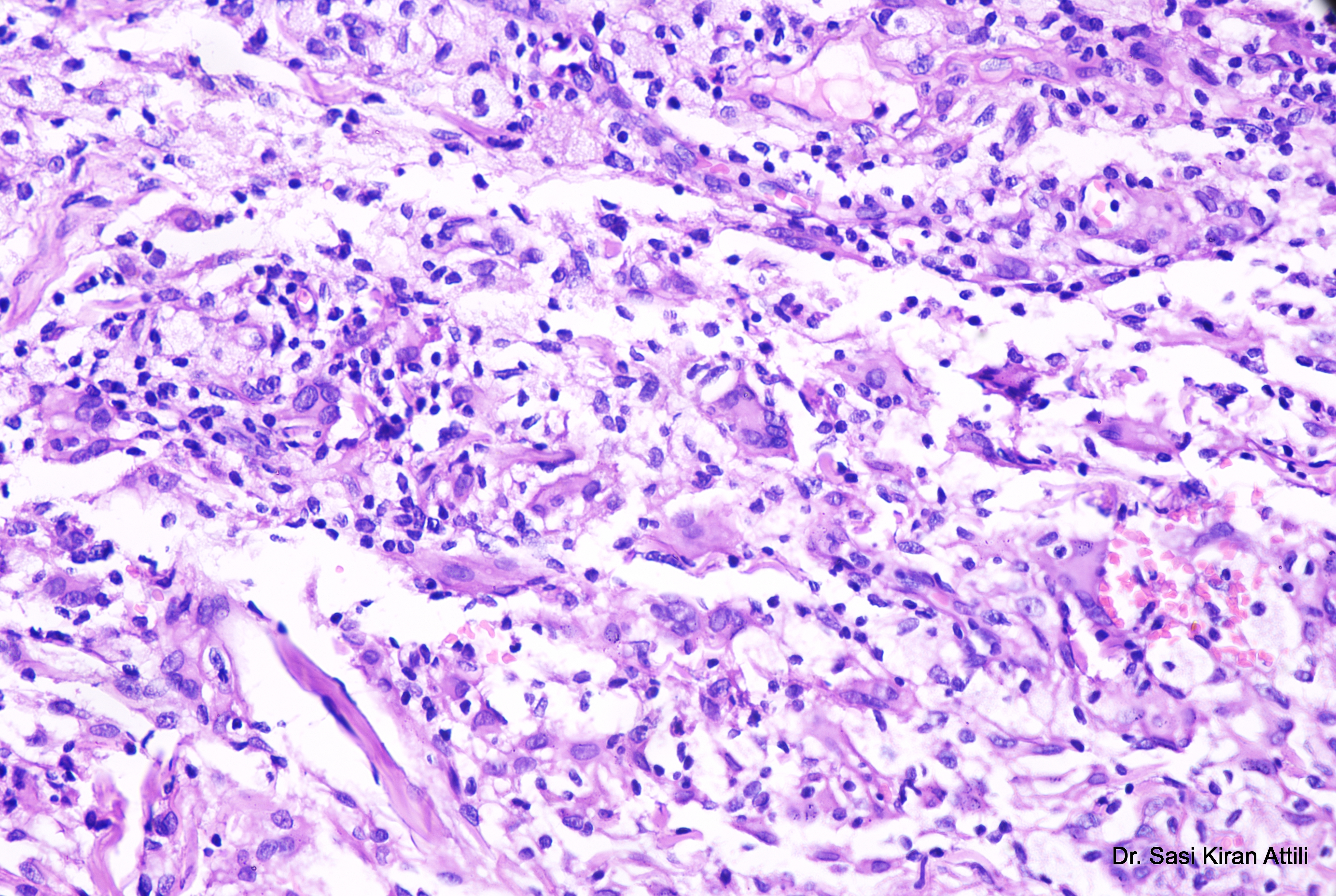
A 21-year-old male presented with a 2-year history of multiple brownish skin lesions over the face, neck, both axillae, both upper limbs, both popliteal fossae, and both flanks, with aggregation on the flanks over the last 4 months. Examination revealed multiple confluent necrotic macules and papules over the face, auricular area, axillae, inguinal regions, knees, and flanks, with ill-defined yellowish macules on the upper and lower lips and buccal mucosae. Genital mucosa was normal. Cerebral


  Report Record

User Feedback


Dr. Shoba Ramesh

Posted (edited)

histiocytes, giant cells and edema and extravasation of RBC, papulonecrotic TB. 

Edited by Dr. Shoba Ramesh
better understanding of cell infiltates and CPC

Share this comment


Link to comment
share_externally

Richard Logan

Posted

We have to tie up the clinical description of necrotic macules and papules with the granulomatous histology including many giant cells.  I am inclined towards an infective aetiology, along the lines of papulo-necrotic tuberculid, or perhaps leprosy.  Certainly special stains are called for here.

Share this comment


Link to comment
share_externally

vincenzo

Posted

Difficult. ?Necrobiotic xanthogranuloma. 

Share this comment


Link to comment
share_externally

Richard Logan

Posted

1 hour ago, vincenzo said:

Difficult. ?Necrobiotic xanthogranuloma. 

I think it would be most unusual for NXG to cause numerous brownish lesions scattered so widely.  NXG usually looks yellow, with at most a few lesions, mainly around the eyes.

Share this comment


Link to comment
share_externally

Sasi Attili

Posted

This was diagnosed as Non-Langerhan Cell Histiocytosis. CD68+ve. S100 and Cd1a -ve. 

Share this comment


Link to comment
share_externally


Join the conversation

You can post now and register later. If you have an account, sign in now to post with your account.

Guest
Add a comment...

×   Pasted as rich text.   Paste as plain text instead

  Only 75 emoji are allowed.

×   Your link has been automatically embedded.   Display as a link instead

×   Your previous content has been restored.   Clear editor

×   You cannot paste images directly. Upload or insert images from URL.

Loading...

×
×
  • Create New...