Jump to content
In this section we have spot diagnoses posted on a daily basis since June 2010, now over 4000! You can review the archived cases and read the suggested diagnoses by users and the final comment by the contributors.
Case are uploaded each week day by 10 am UK time with the correct diagnosis will generally be posted at 8 pm UK time. Why not view the most recent spot diagnosis and proffer a diagnosis?

Case Number : Case 1468 -09 February Posted By: Guest

Please read the clinical history and view the images by clicking on them before you proffer your diagnosis.
Submitted Date :
   (0 reviews)

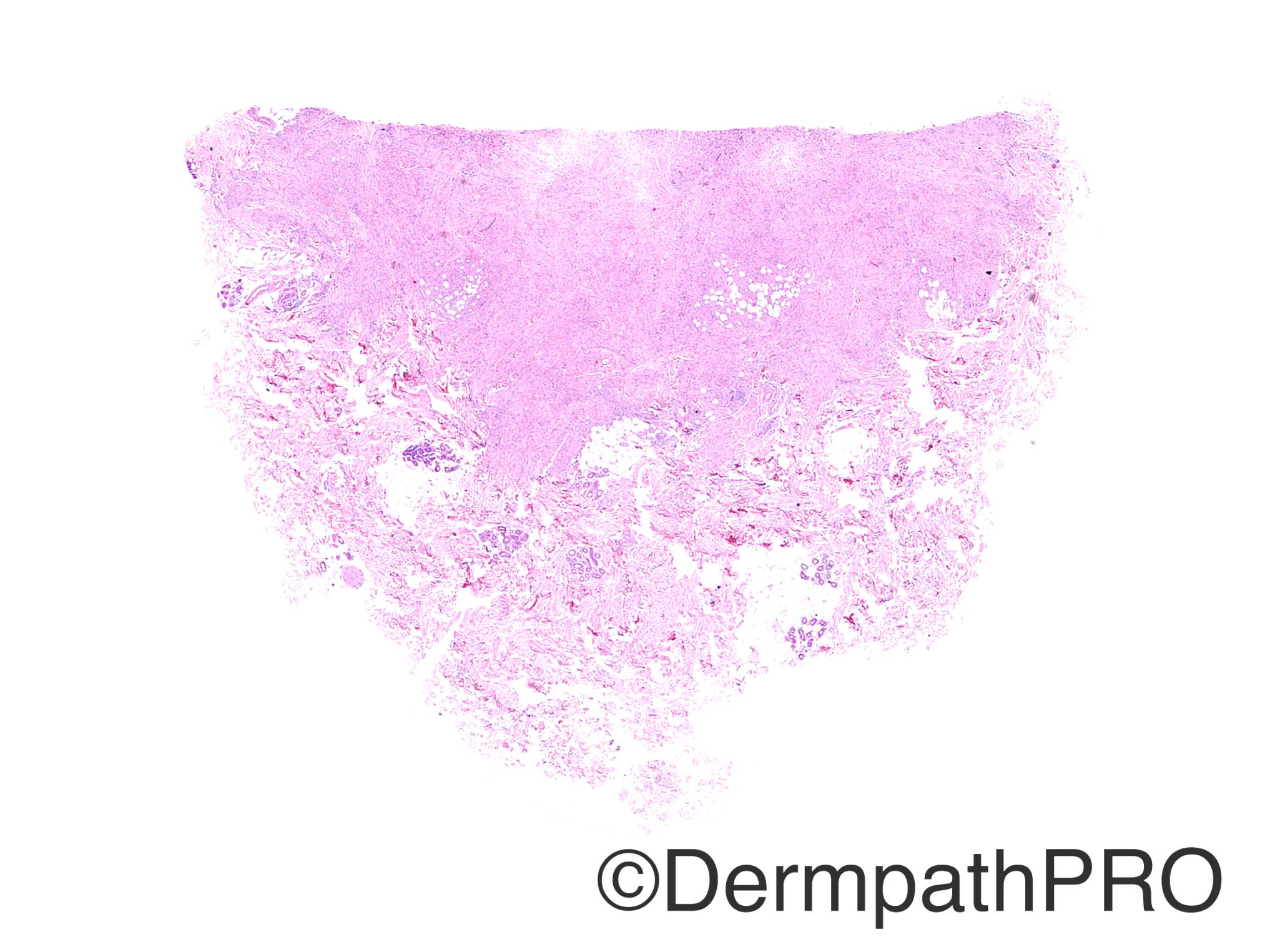
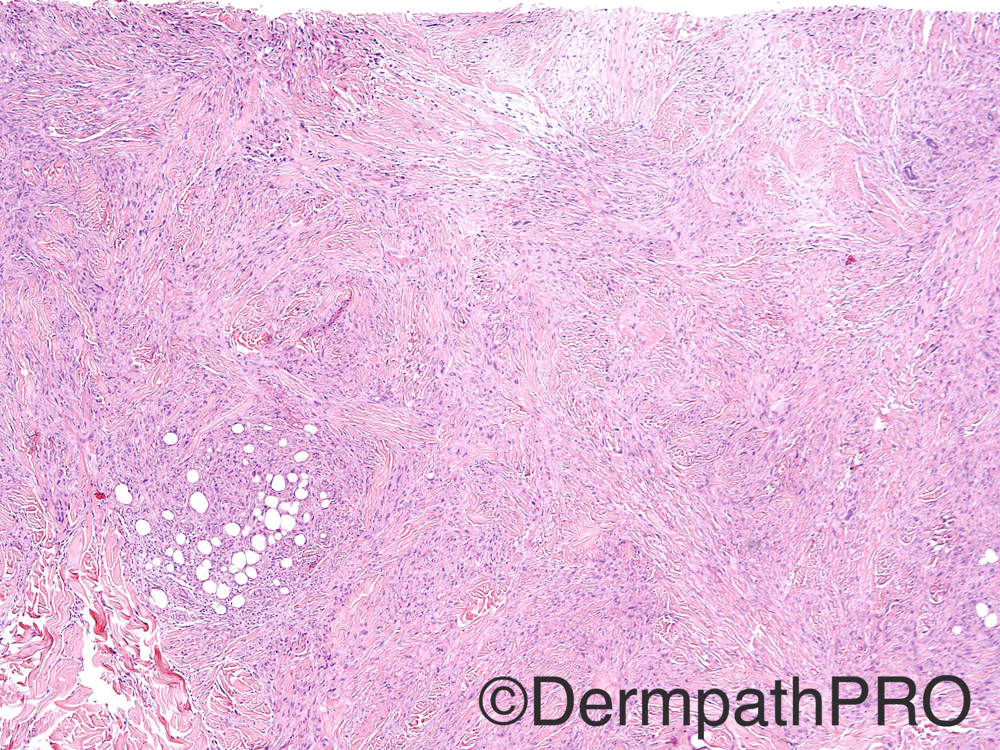
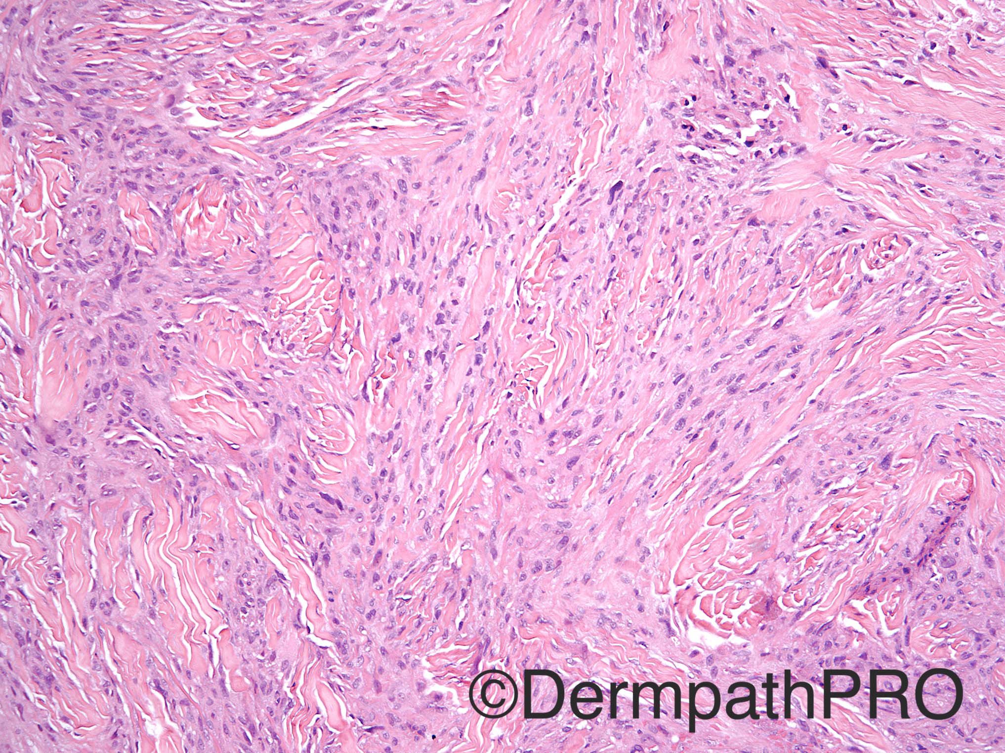
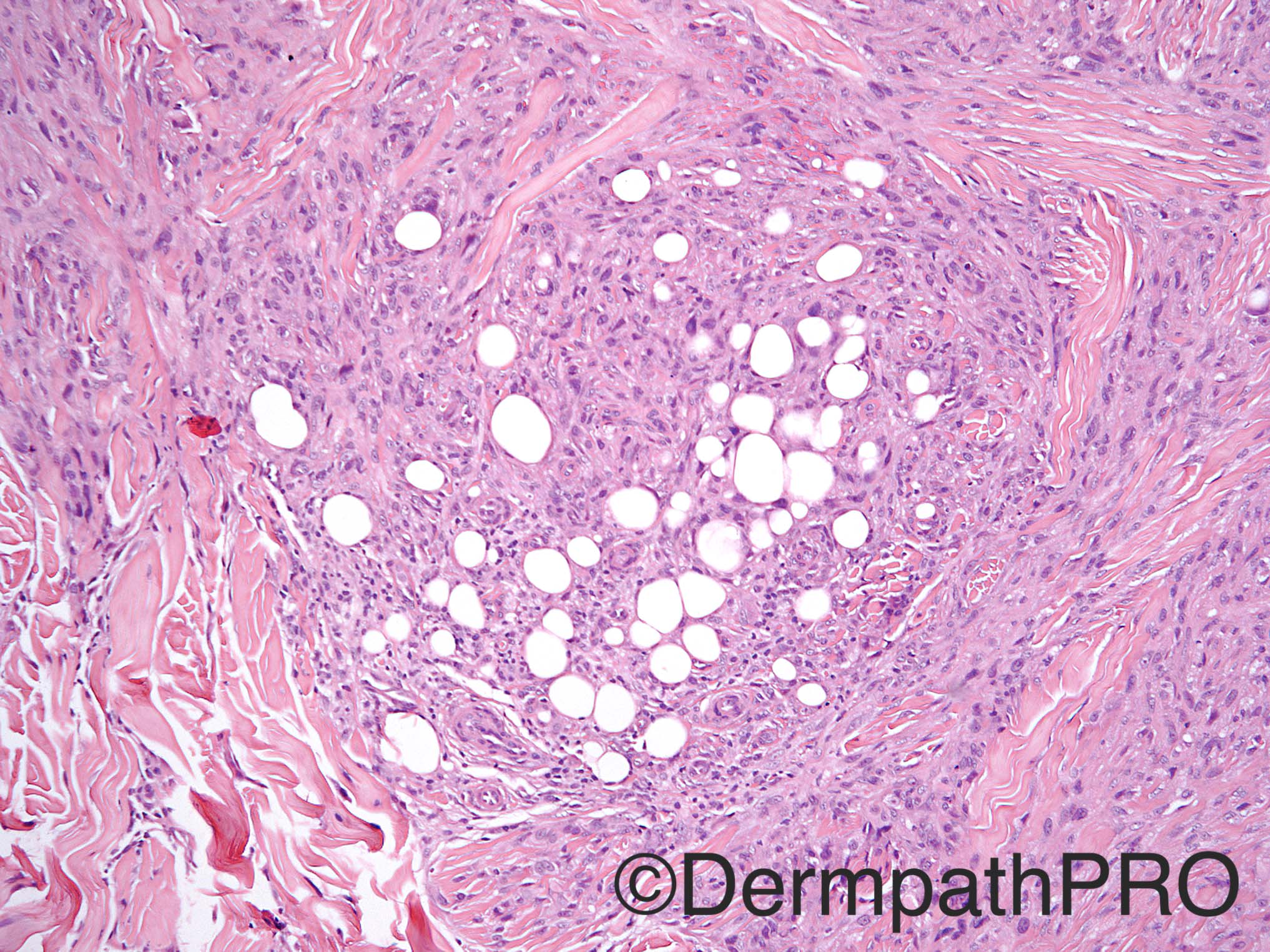
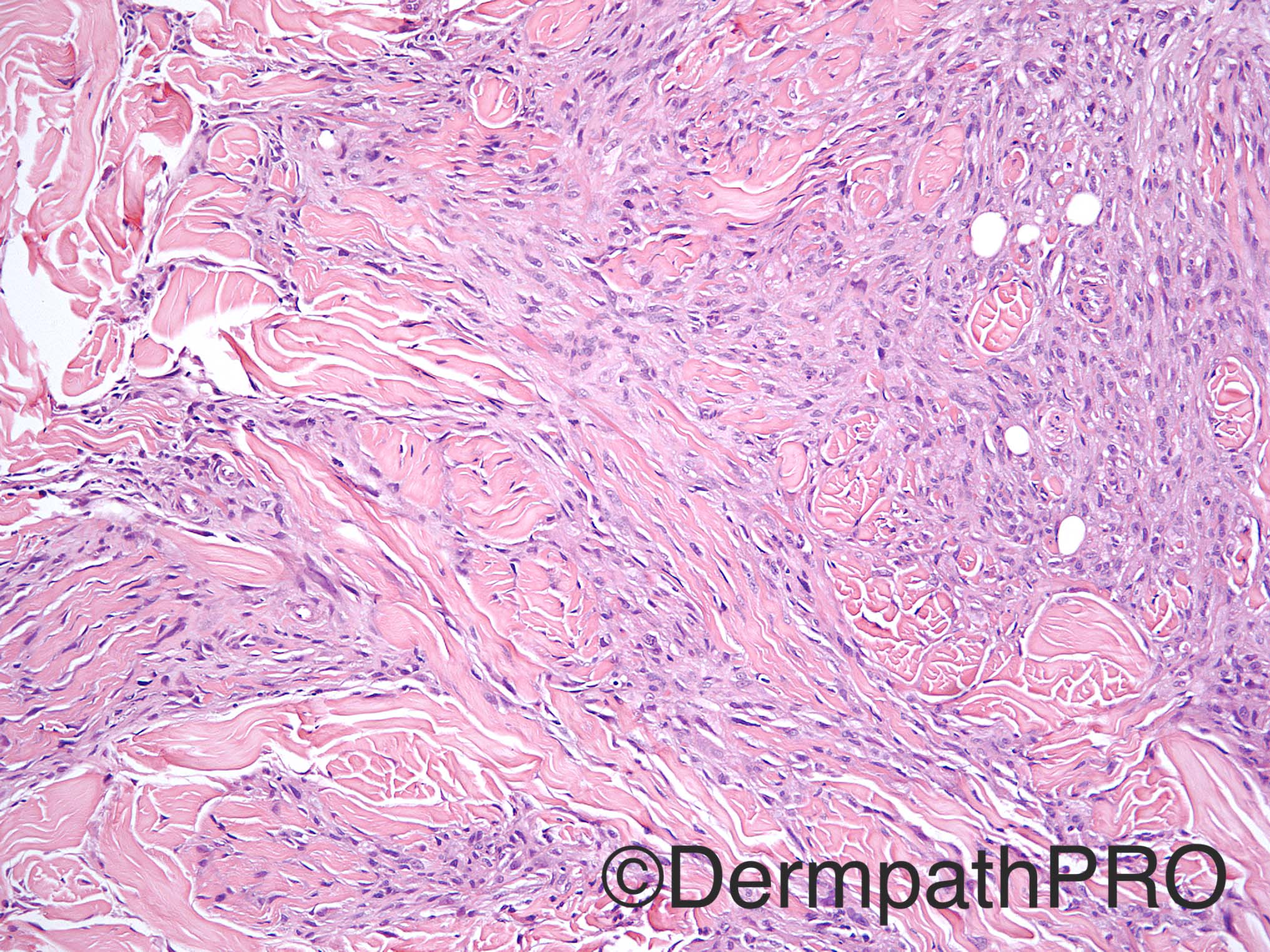
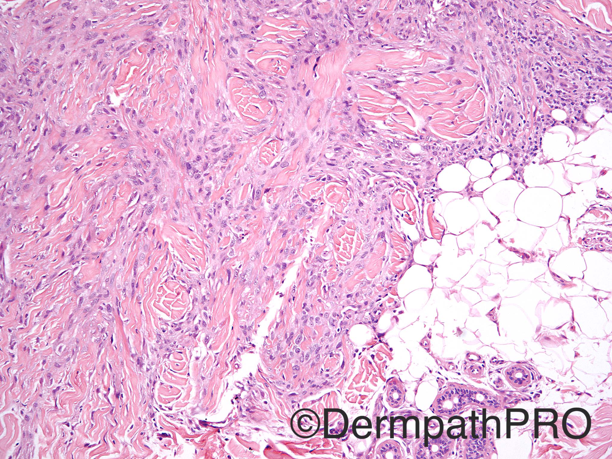
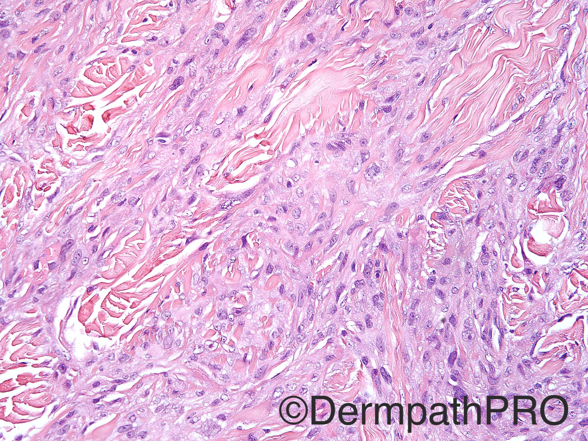
Case History: 38 year old man with numerous nodules on the skin, including the scalp. Biopsy from left shoulder.

Case posted by Dr Uma Sundram


  • Like 1
  Report Record

User Feedback


biswanath behera

Posted

Cords and fascicles of spindle shaped cells forming storiform pattern, collagen trapping, involvement of subcutis, pleomorphism but no mitotic figures.,


? Eruptive Dermatofibroma, the present nodule showing histopathological features suggestive of cellular Dermatofibroma

Share this comment


Link to comment
share_externally

Dr. Mona Abdel-Halim

Posted

I have the same impression of dermatofibroma, in correlation with the clinical pucture, thinking eruptive DF.. Haven't seen it before!!

Share this comment


Link to comment
share_externally

Raul Perret

Posted

This is indeed a challenging case, when i saw the first picture my first thought was cellular dermatofibroma. But after continuing with the images I was less sure of this idea. I see a dermal proliferation with irregular borders  disposed mainly in fascicles of different size and orientation, cytologically the cells seem to have diffuse atypia with irregular nuclear membranes, small nucleoli and inconspicuous cell borders; some of the cells are clearly fusiform with blunted nuclear borders others are not. I dont perceive mitotic figures. Of particular interest is the adipose tissue seen on picture 5, the presence of adypocites interspersed with a fusiform proliferation would normally make me think of a primary adipocytic neoplasm but I am really not convinced here. Finally the clinical setting makes me also consider a primary cutaneous smooth muscle neoplasm. So, to sum up, I would perform a wide panel of immunos (ck, s100, desmin, smooth muscle actin, caldesmon, factor XIII, ki67) I am not sure but I think this is a smooth muscle neoplasm in the range of leyomioma/aytipical intradermal smooth muscle neoplasm (as proposed by Fletcher) 

Share this comment


Link to comment
share_externally

Nitin Khirwadkar

Posted

Thinking of an eruptive cellular dermatofibroma. These have been described in a setting of connective tissue disease. The entrapment of fat is very DFSP like. Like Raul has suggested, we require a range of IHC markers. Definitely challenging!

Share this comment


Link to comment
share_externally

Uma Sundram

Posted

V challenging case. The patient also had lung metastases. We made multiple diagnoses of cellular/ eruptive df and did a battery of immunos including s100 and desmin (both neg). At the suggestion of our soft tissue expert we did pan keratin (faint pos) and EMA (strong +). Cd34 was pos and there was loss of INI. We had initially done ck5/6 which was negative. Final DX: epithelioid sarcoma.

Share this comment


Link to comment
share_externally

Sasi Attili

Posted

Wow! difficult case. I also thought this was eruptive DF

Share this comment


Link to comment
share_externally


Join the conversation

You can post now and register later. If you have an account, sign in now to post with your account.

Guest
Add a comment...

×   Pasted as rich text.   Paste as plain text instead

  Only 75 emoji are allowed.

×   Your link has been automatically embedded.   Display as a link instead

×   Your previous content has been restored.   Clear editor

×   You cannot paste images directly. Upload or insert images from URL.

Loading...

×
×
  • Create New...