Jump to content
In this section we have spot diagnoses posted on a daily basis since June 2010, now over 4000! You can review the archived cases and read the suggested diagnoses by users and the final comment by the contributors.
Case are uploaded each week day by 10 am UK time with the correct diagnosis will generally be posted at 8 pm UK time. Why not view the most recent spot diagnosis and proffer a diagnosis?

Case Number : Case 1889 - 24 August - Dr Iskander Chaudhry (Invited) Posted By: Guest

Please read the clinical history and view the images by clicking on them before you proffer your diagnosis.
Submitted Date :
   (0 reviews)

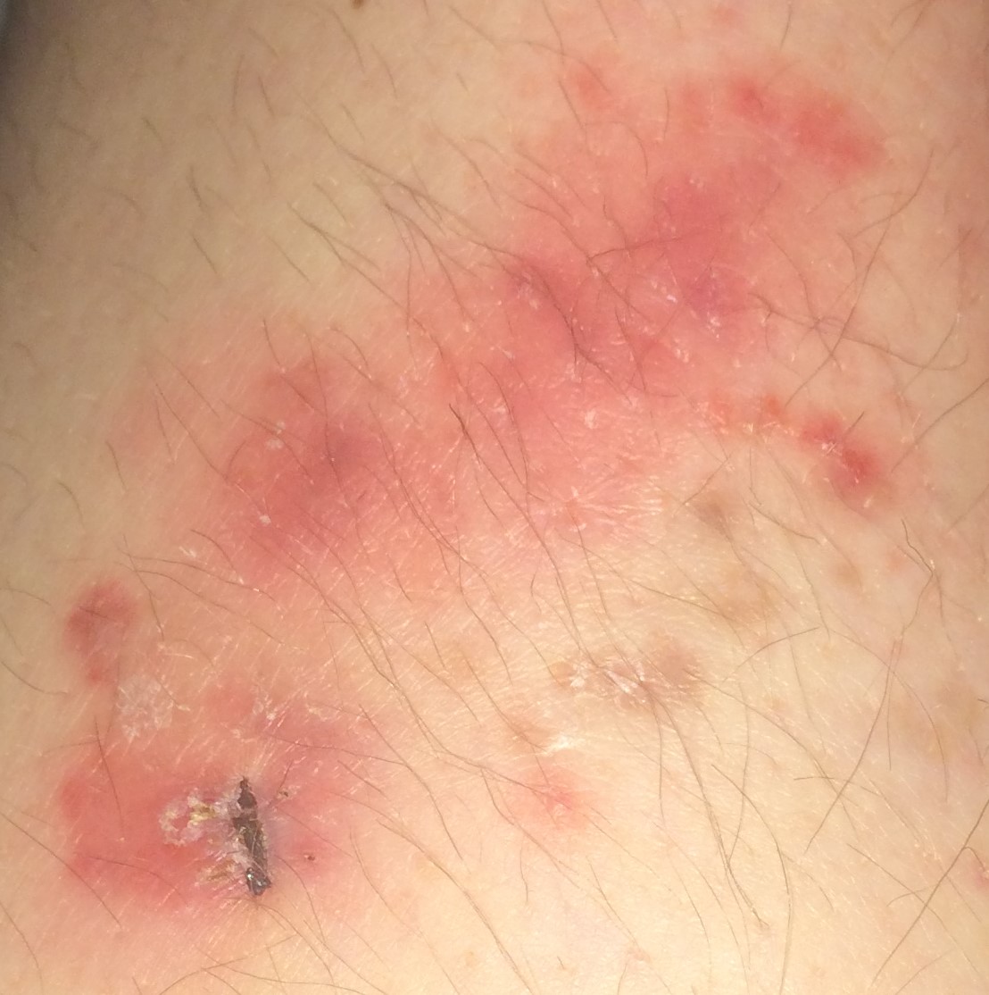
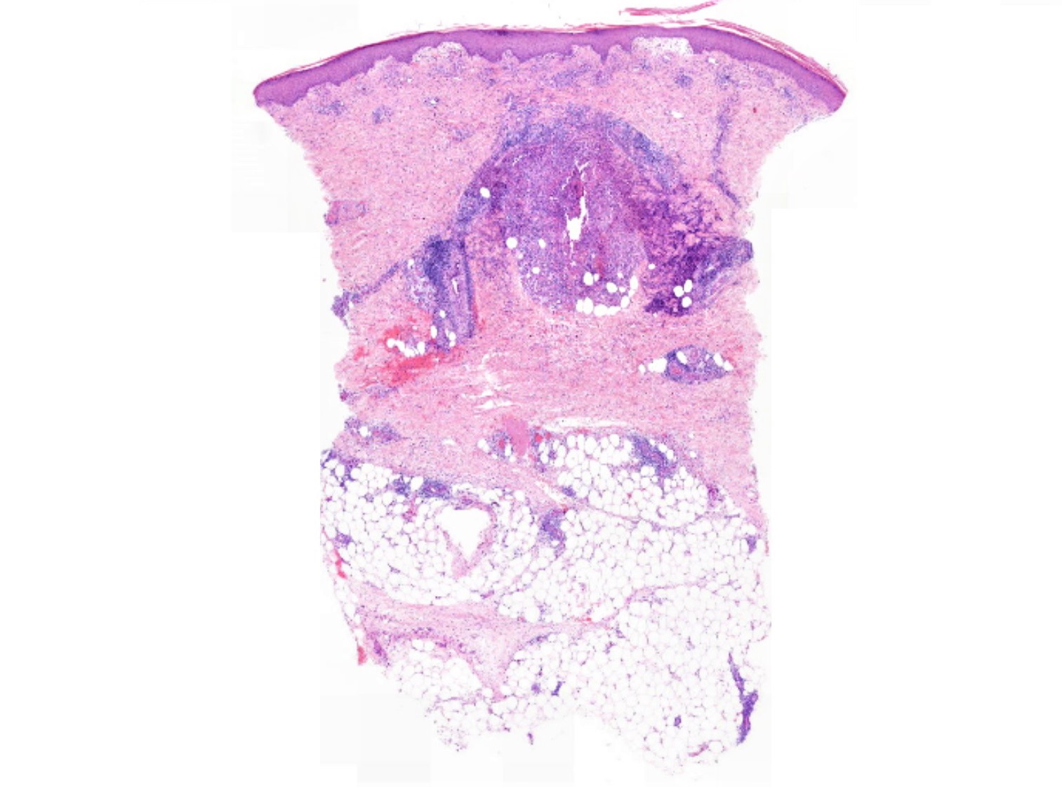
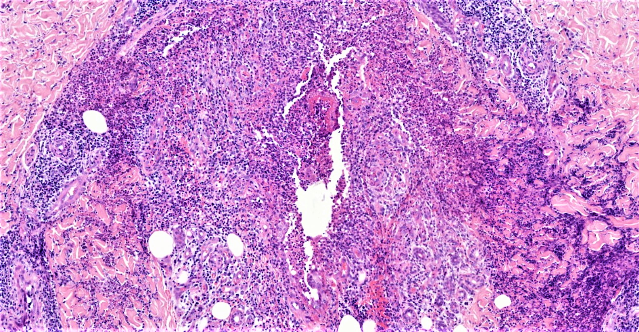
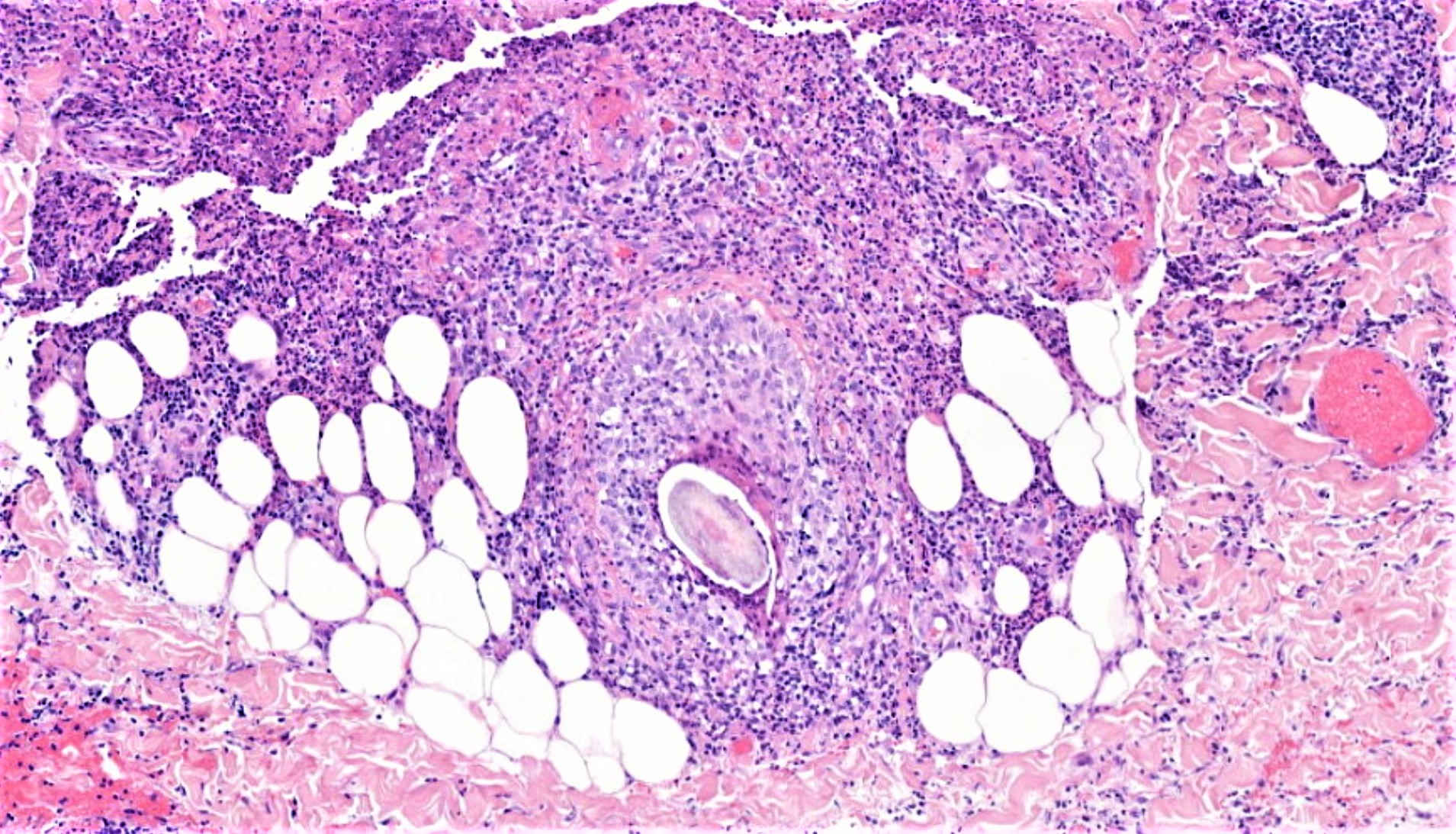
24 year old Female. Rash - left and right lower legs - comes and goes over past 13 years.

Edited by Admin_Dermpath


  Report Record

User Feedback


Raul Perret

Posted (edited)

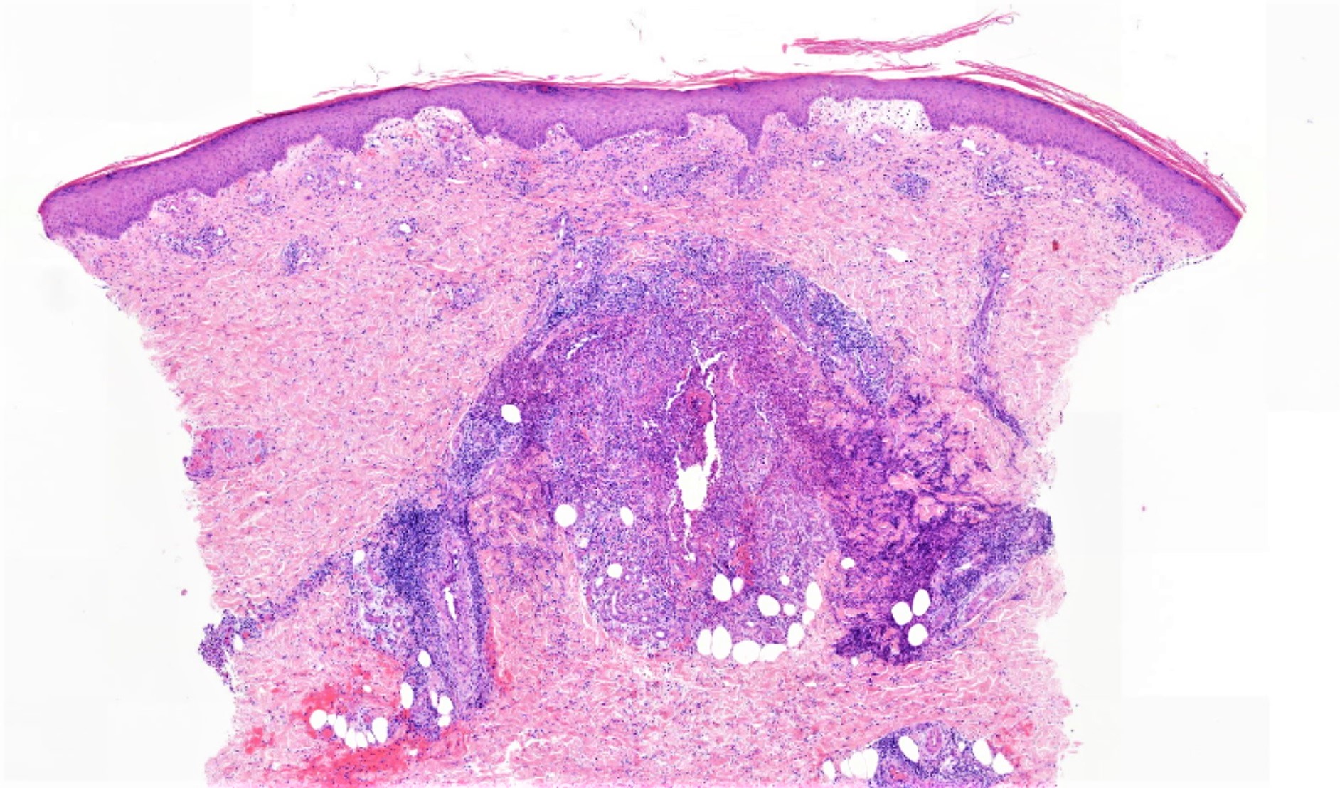
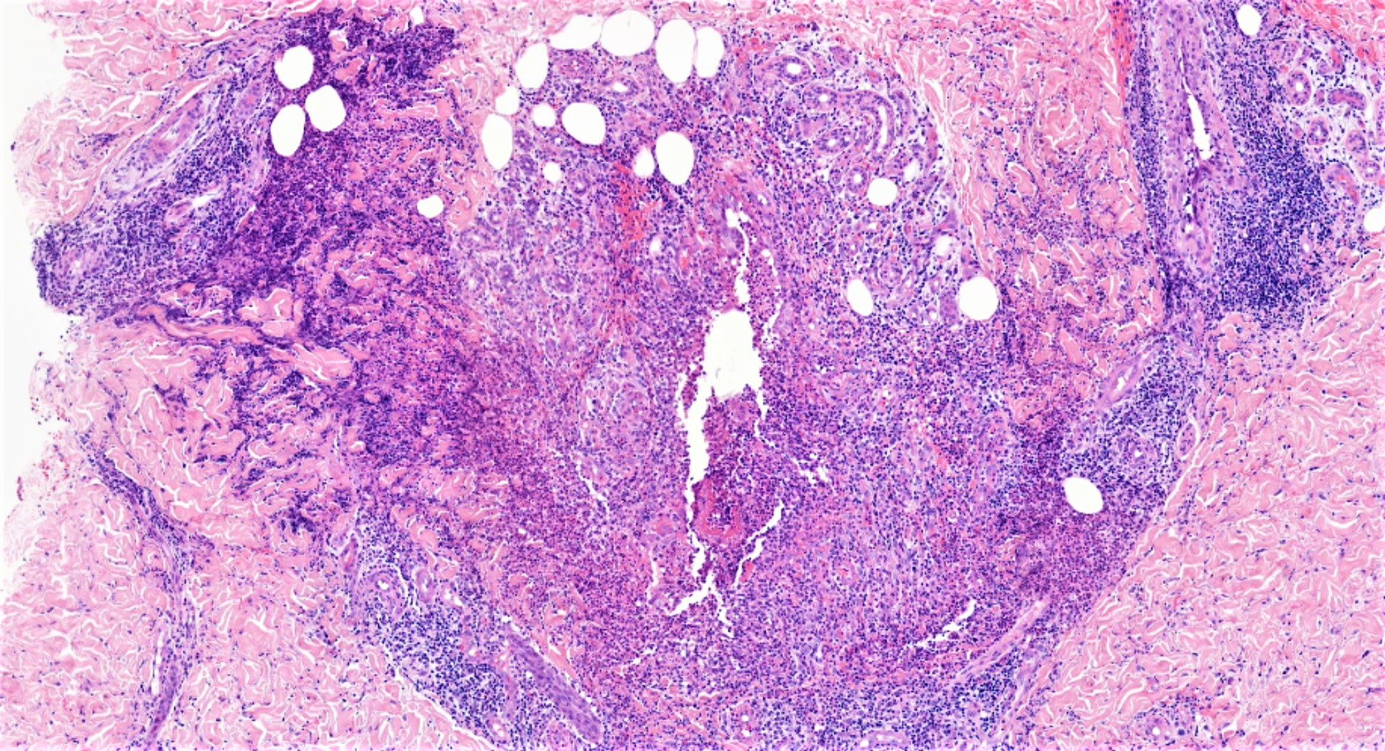
I am a bit puzzled by this case, there is mainly a lymphocytic infiltrate with excessive tropism towards blood vessels and hair follicle, destroying the later. I thought of a vasculopathic reaction process with folliculitis and prominent lymphocytes, for example Behcet's disease or lupus? but the clinical image does not perfectly fit with the typical skin manifestations of these diseases, is there any more clinical information? 


Edited by Raul Perret

Share this comment


Link to comment
share_externally

Raul Perret

Posted

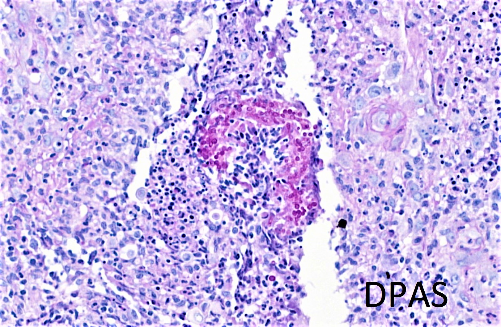
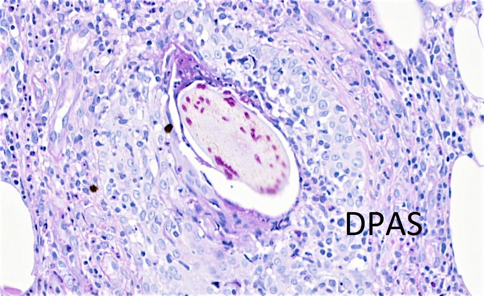
I think I would do a PAS stain too in case there are is a combination of pathologies

Share this comment


Link to comment
share_externally

Admin_Dermpath

Posted

Dear Raul

No more history than provided - lesions come and go! 

Iskander

Share this comment


Link to comment
share_externally

Cem Leblebici

Posted

Follicular Lymphomatoid Papulosis? 

Share this comment


Link to comment
share_externally

Raul Perret

Posted

Yeah I thought of that dx at the beginning but does it fit the clinical image?

Share this comment


Link to comment
share_externally

Anil Patki

Posted

Clinical features, granulomatous infiltrate in lower dermis and subcutis with panniculitis, all point to erythema induratum (Bazin's disease)

Share this comment


Link to comment
share_externally

Raul Perret

Posted

Yes this is lymphomatoid papulosis with follicular compromise I do not know what was I thinking..

Share this comment


Link to comment
share_externally

Admin_Dermpath

Posted

Dear All 

CD30 shows only scattered cells! Try again! 

Iskander

Share this comment


Link to comment
share_externally

Sasi Attili

Posted (edited)

2 hours ago, Raul Perret said:

Yes this is lymphomatoid papulosis with follicular compromise I do not know what was I thinking..

LyP localised to just the legs? Is that described?


Not sure what to call this. There are granulomatous changes around the follicular unit. Lichen scrofulosorum? Clinical doesn't quite fit though!

Edited by Sasi Attili

Share this comment


Link to comment
share_externally

Anil Patki

Posted

Recurrent insect bite reaction? But it should show eosinophils and no panniculitis!

Share this comment


Link to comment
share_externally

Raul Perret

Posted (edited)

44 minutes ago, Robledo F. Rocha said:

Considering the suppurative folliculitis with surrounding Sweet’s-like vascular reaction, sterile neutrophilic folliculitis with perifollicular vasculopathy is a possibility. Clinical history fits as well, especially if there is an associated systemic disease as Raul said above.

yeah I read about that entity Robledo, but it is supposed to be primarily neutrophils in the dermis and this looks mainly as a lymphocytic process? do you guys agree? I am clueless, I suppose immunofluorescence was not performed in this case?

Edited by Raul Perret

Share this comment


Link to comment
share_externally

Saman Fatah

Posted

It is very interesting to see clinical photos beside the photomicrographs! Admin team have to be congratulated for this new feature.

Clinically red-brown "granulomatous" plaques, some are folliculocentric with subtle superficial scale and light brown post inflammatory hyperpigmentation in older lesions. It is bilateral and intermittent, would be nice to clarify is she shaves her legs or uses other mechanical methods for hair removal?

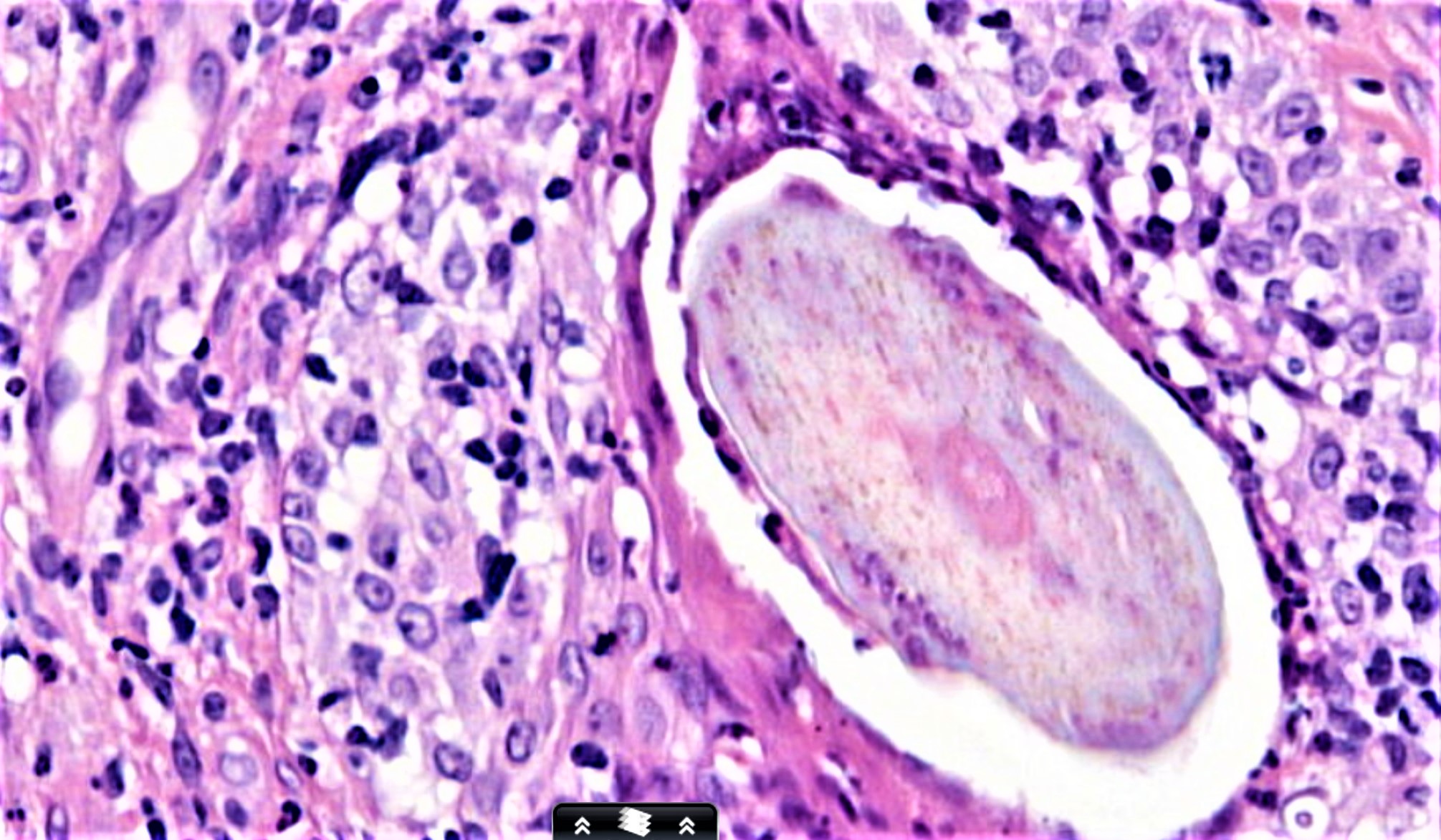
The main feature is a granulomatous infiltrate with large numbers of lymphocytes centred around what appear to be a hair shaft in the middle of dermis. It appears like a reactive process to a foreign body (hair shaft granulomas) but no obvious giant cells. I thought about possibility of Majocchi granulomas though bilaterality will be slightly unusual (but still possible). Other DDx cutaneous mycotic infection with "pseudolymphoatous" infiltrate but the histology is not typical of this. Lastly one should bear in mind externally introduced material for longstanding lesions in easily accessible sites if clinical context is appropriate and other causes excluded.


Share this comment


Link to comment
share_externally

Admin_Dermpath

Posted

Dear All 


Dr Chaudhry has added the DPAS stains. We can add clinical images if provided by the contributors with appropriate consent! Final diagnosis  ....


DermpathPRO Admin. 

Share this comment


Link to comment
share_externally

Robledo F. Rocha

Posted

Agree with Majocchi’s granuloma, but, wow!, more than one decade, how long time!

Share this comment


Link to comment
share_externally

Raul Perret

Posted

This is so interesting, the last picture was suspicious showing the hair shaft with some «tubular» structures thats why I added the comment of the PAS stain. I just thought it was highly unlikely due to the clinical. So nice to have cases like this one and keep learning everyday, thank you colleagues and the editorial team

Share this comment


Link to comment
share_externally

Sasi Attili

Posted

On 8/25/2017 at 11:15, Raul Perret said:

This is so interesting, the last picture was suspicious showing the hair shaft with some «tubular» structures thats why I added the comment of the PAS stain. I just thought it was highly unlikely due to the clinical. So nice to have cases like this one and keep learning everyday, thank you colleagues and the editorial team


Absolutely. I wondered about fungus for a fleeting second, but dismissed it based on the clinical!

Share this comment


Link to comment
share_externally


Join the conversation

You can post now and register later. If you have an account, sign in now to post with your account.

Guest
Add a comment...

×   Pasted as rich text.   Paste as plain text instead

  Only 75 emoji are allowed.

×   Your link has been automatically embedded.   Display as a link instead

×   Your previous content has been restored.   Clear editor

×   You cannot paste images directly. Upload or insert images from URL.

Loading...

×
×
  • Create New...