Jump to content
In this section we have spot diagnoses posted on a daily basis since June 2010, now over 4000! You can review the archived cases and read the suggested diagnoses by users and the final comment by the contributors.
Case are uploaded each week day by 10 am UK time with the correct diagnosis will generally be posted at 8 pm UK time. Why not view the most recent spot diagnosis and proffer a diagnosis?

Case Number : Case 1890 - 25 August Dr Carr Posted By: Guest

Please read the clinical history and view the images by clicking on them before you proffer your diagnosis.
Submitted Date :
   (0 reviews)

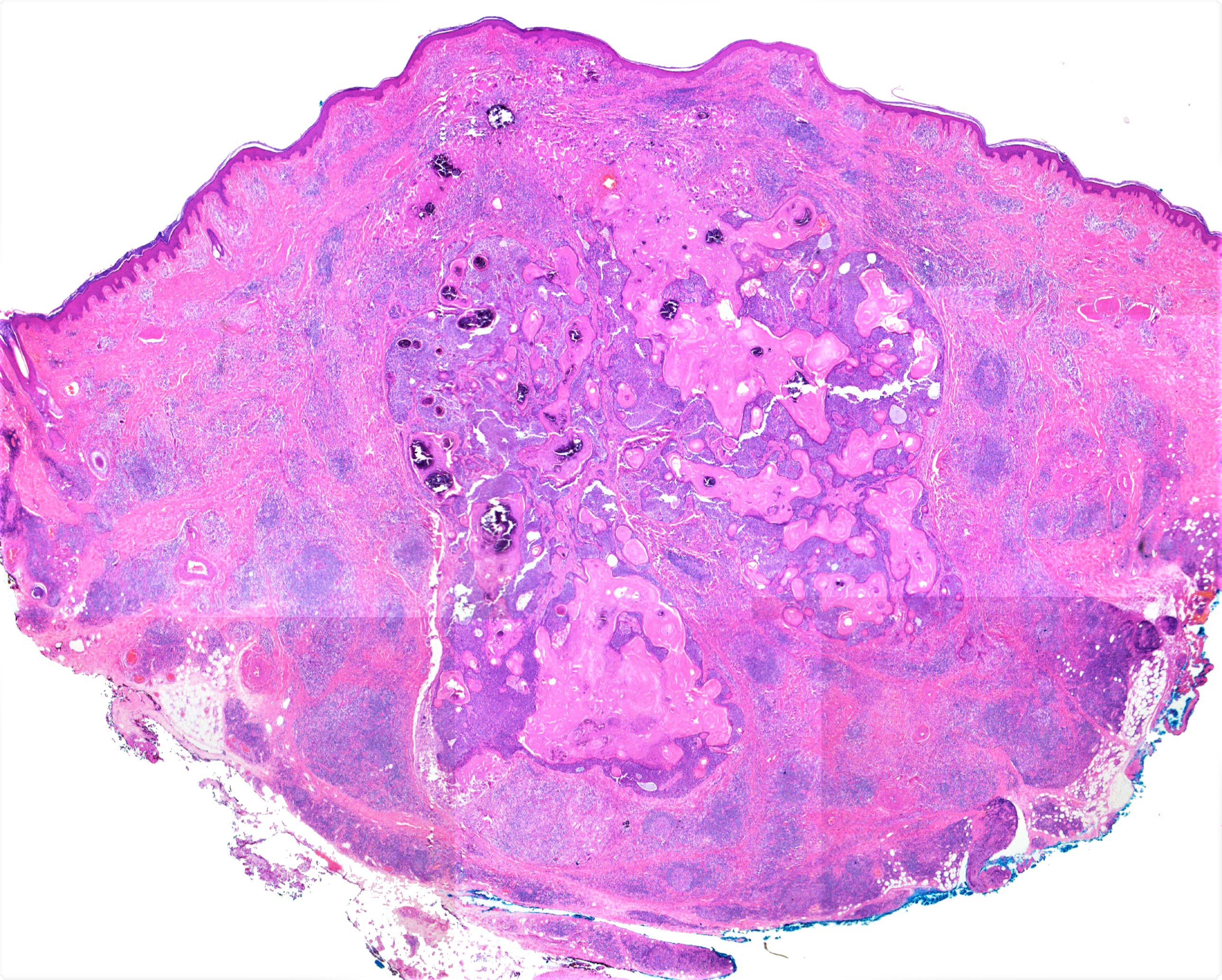
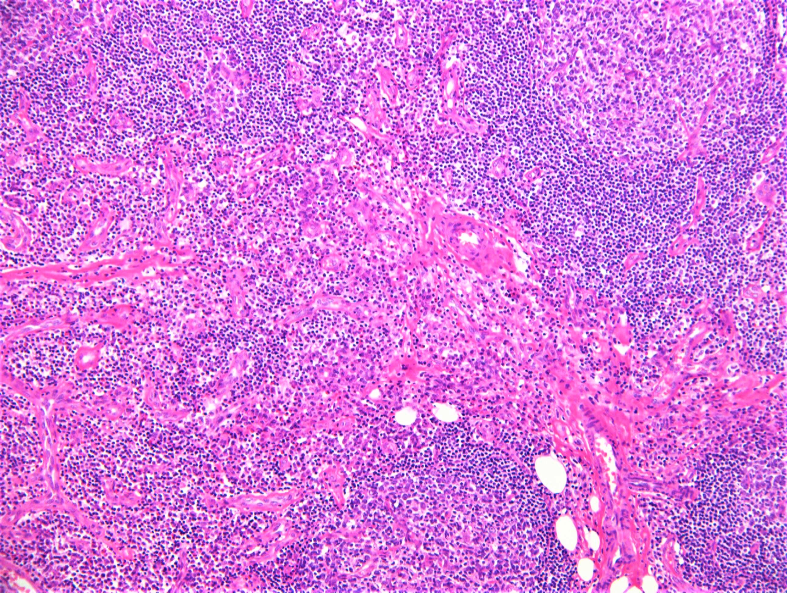
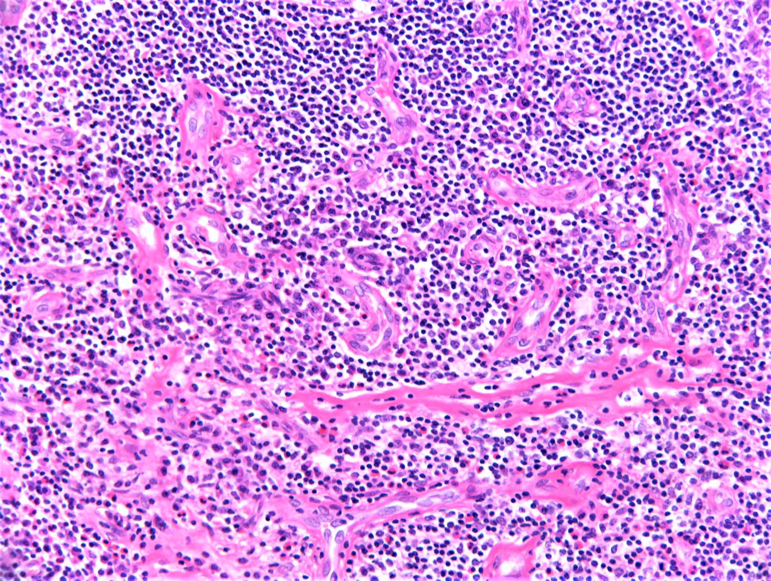
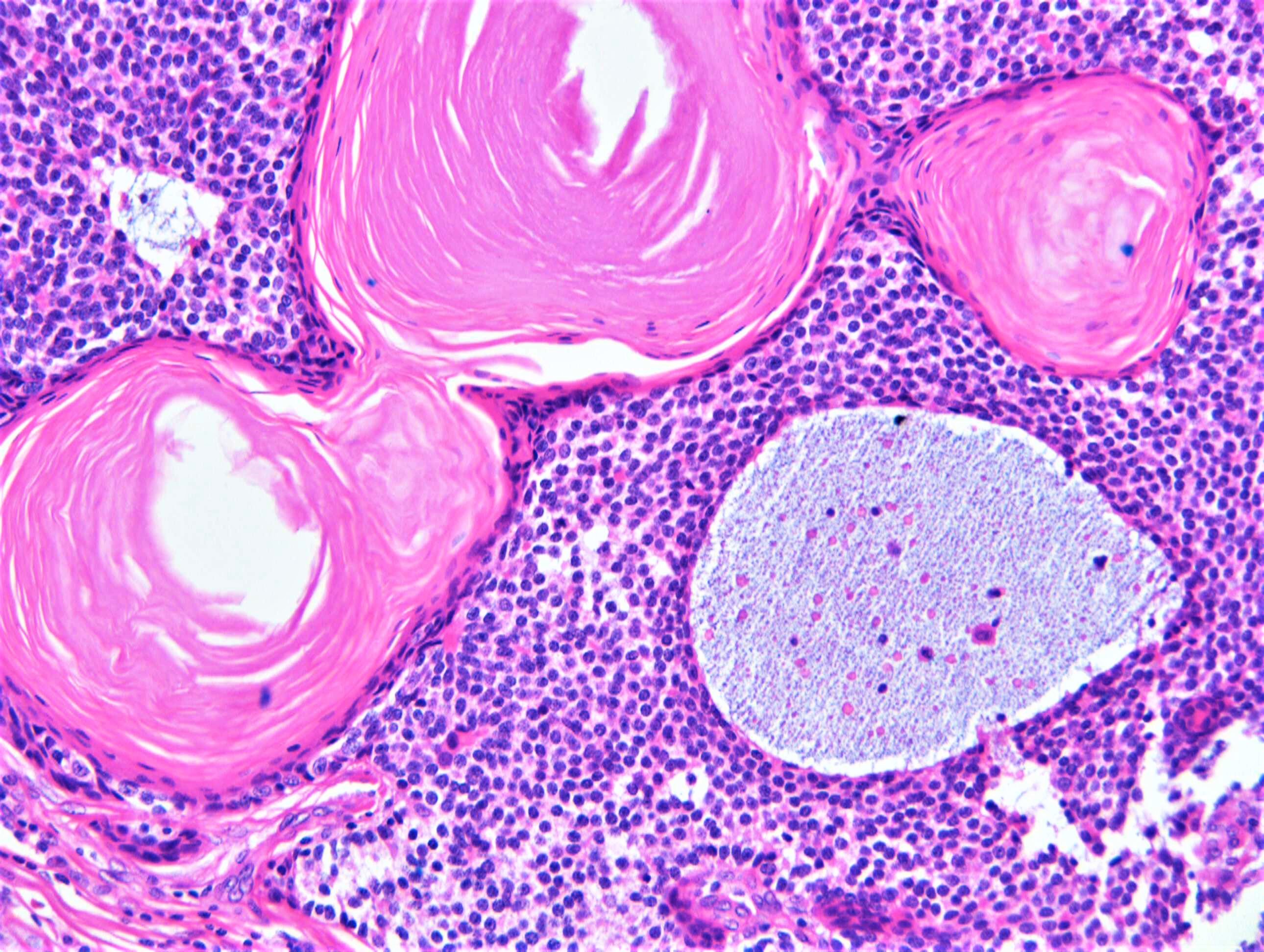
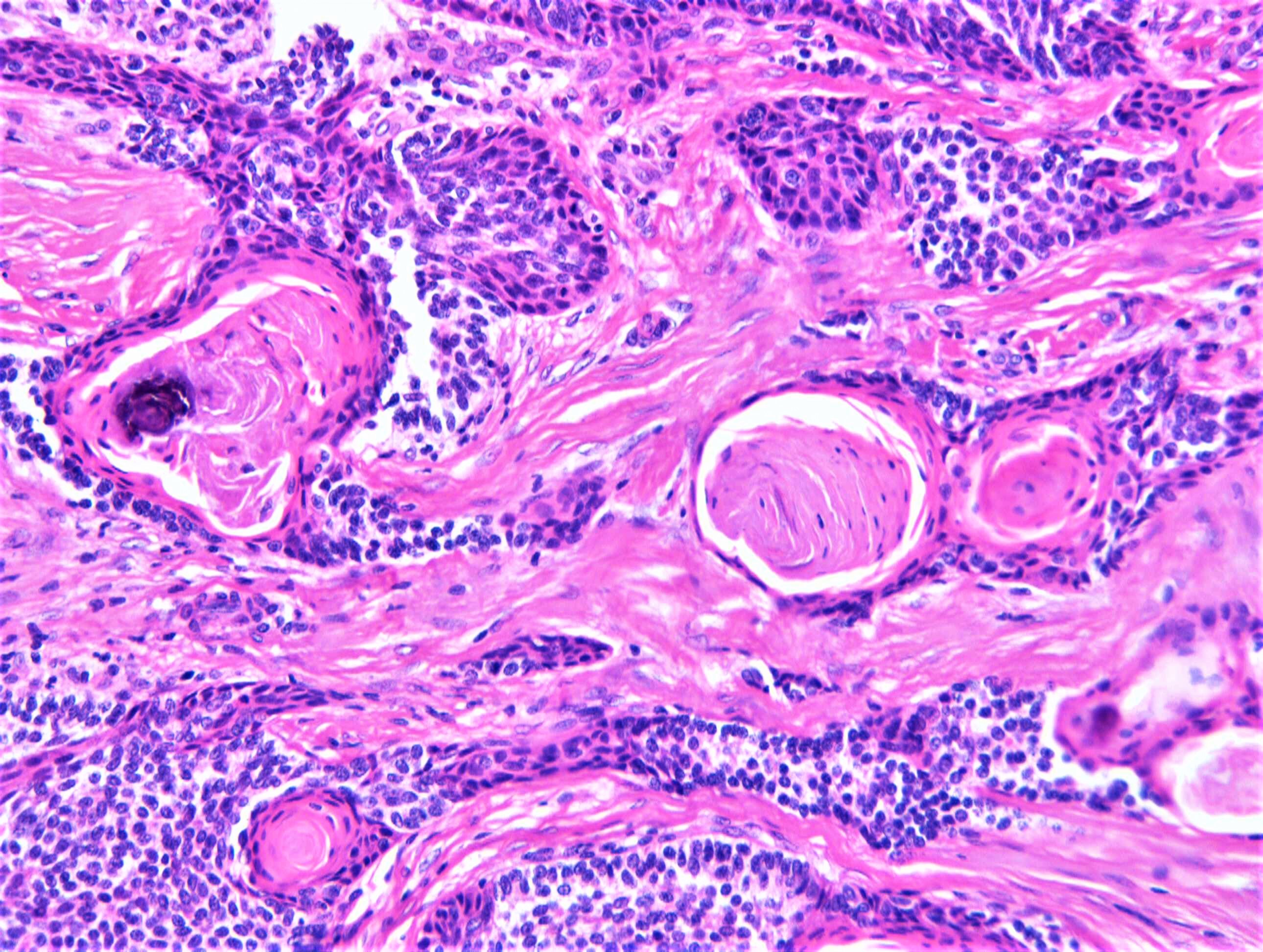
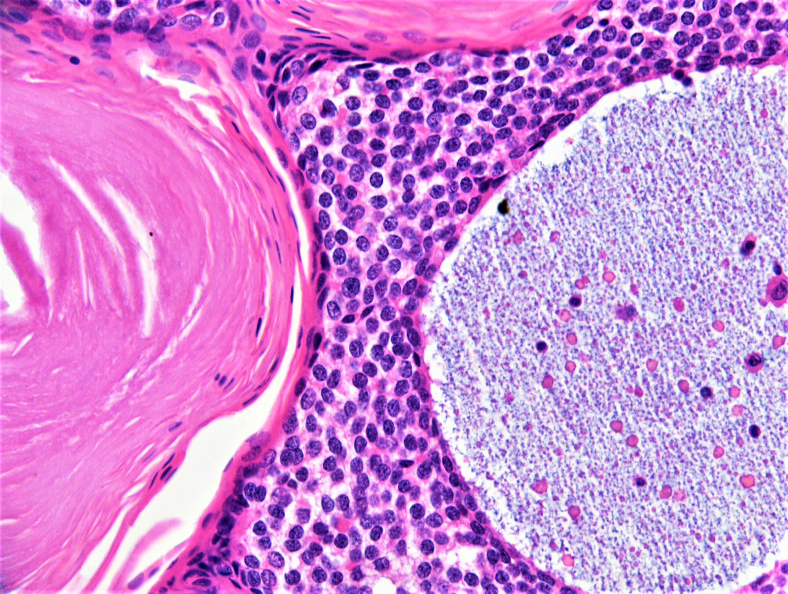
M45. Nodular lesion on the shin ?Dermatofibroma

Edited by Admin_Dermpath


  Report Record

User Feedback


vincenzo polizzi

Posted

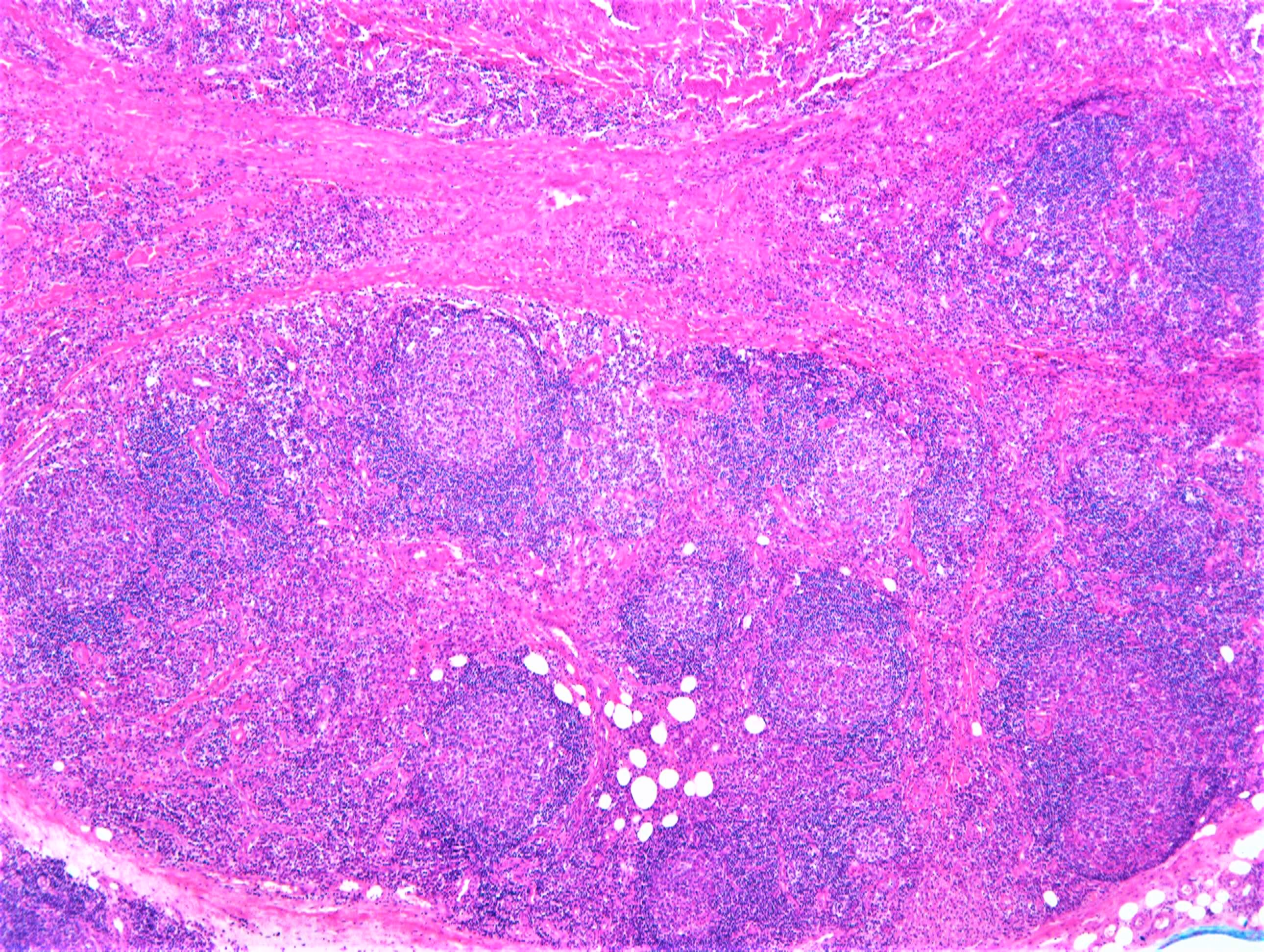
I find this case very confusing if thinking of a single pathology...but thinking of a pathological association I might venture the spot diagnosis of an adamantinoid trichoblastoma/Lymphadenoma + ALHE. 

Share this comment


Link to comment
share_externally

Anil Patki

Posted

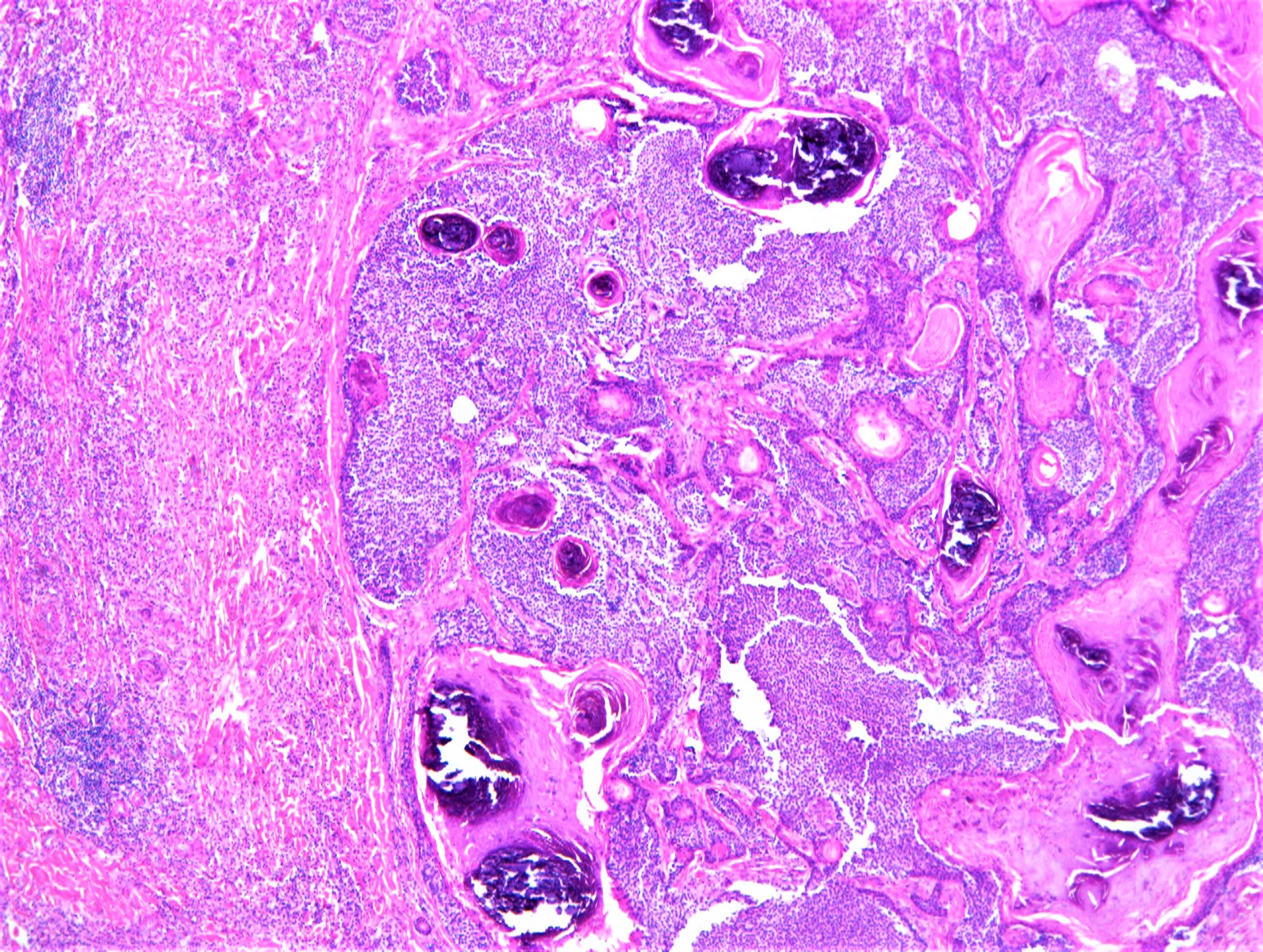
A poorly circumscribed dermal tumour with dark cells, hyalinized cells and foci of calcification. Some islands of dark cells show palisading. Eosinophils in the infiltrate create some confusion. Pilomatricoma is the thing I can think of.

Share this comment


Link to comment
share_externally

vincenzo polizzi

Posted

I want to correct ALHE with Kimura disease (is he an asian patient?).

Share this comment


Link to comment
share_externally

Raul Perret

Posted

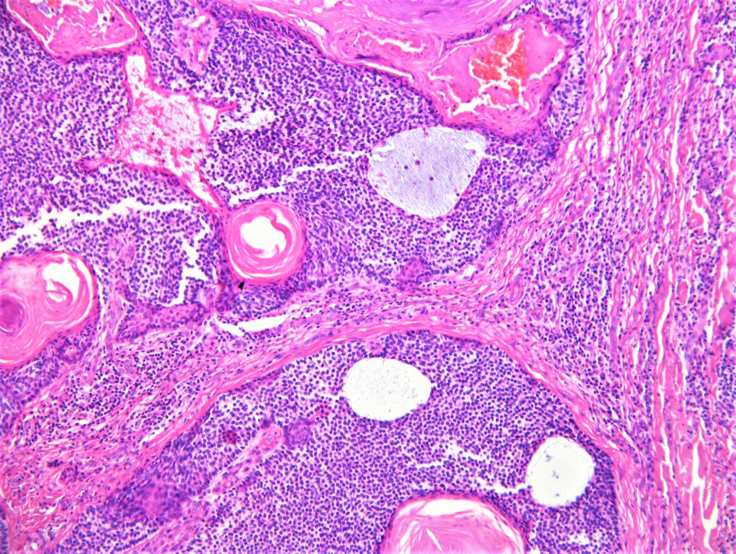
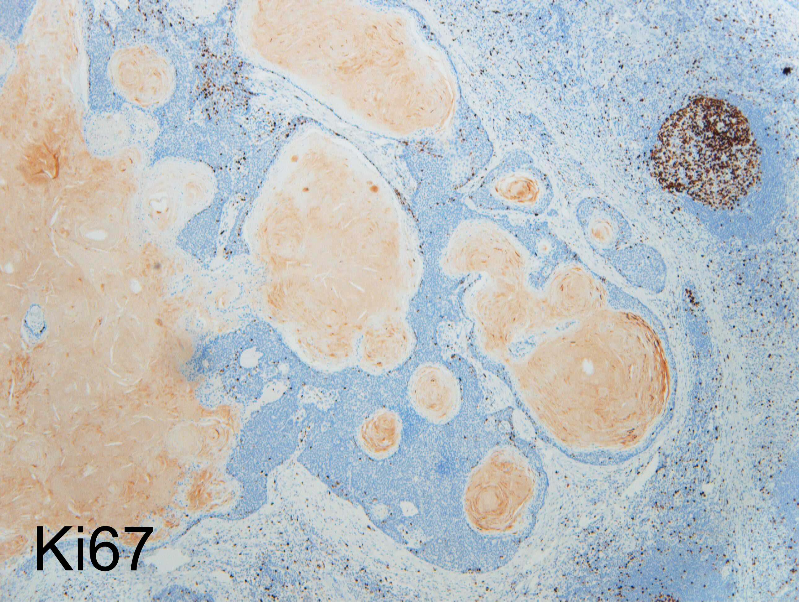
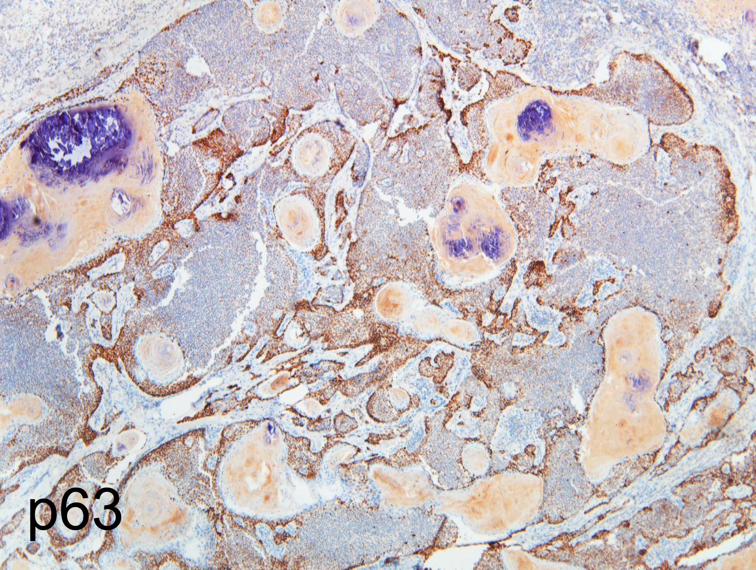
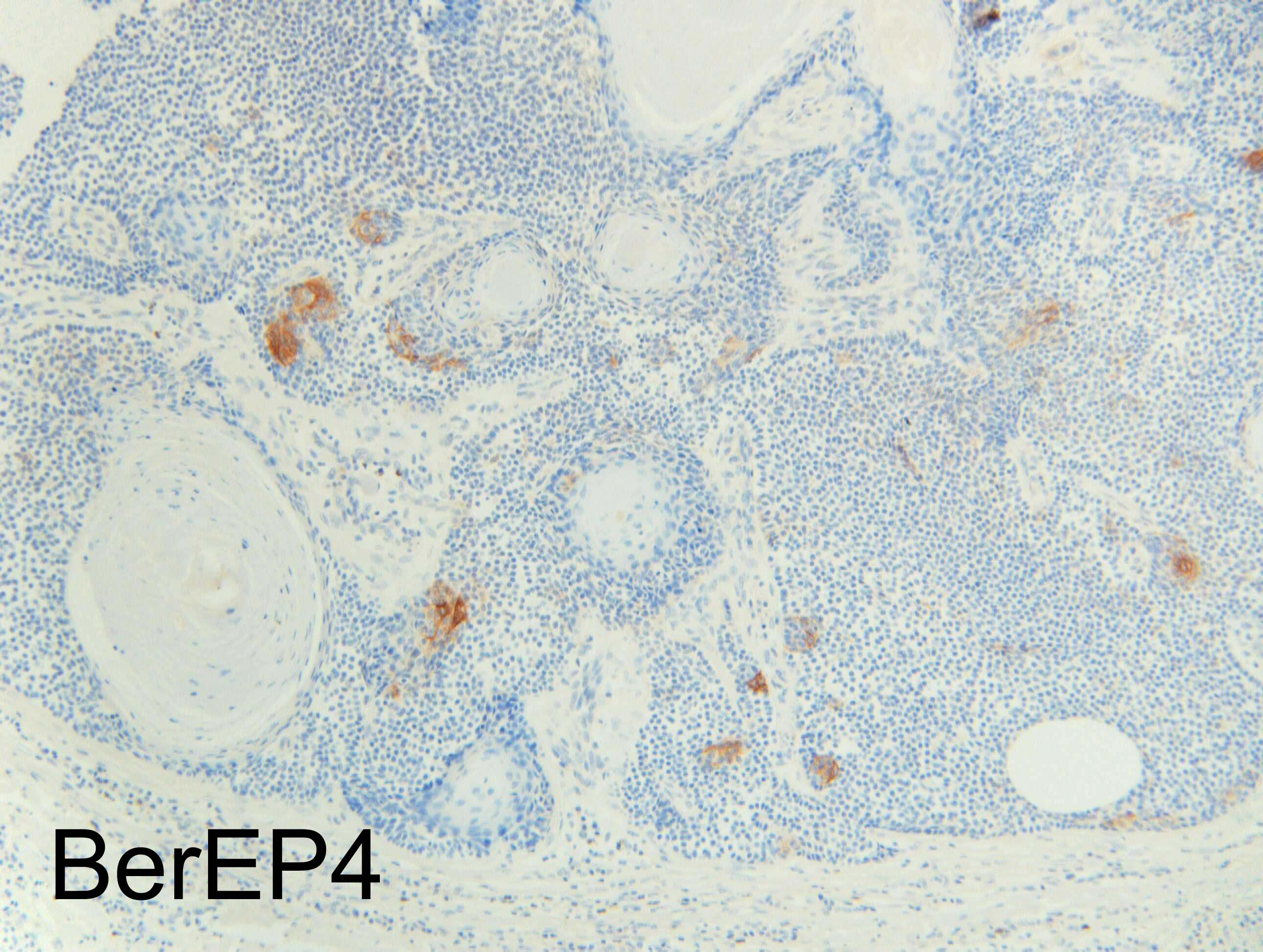
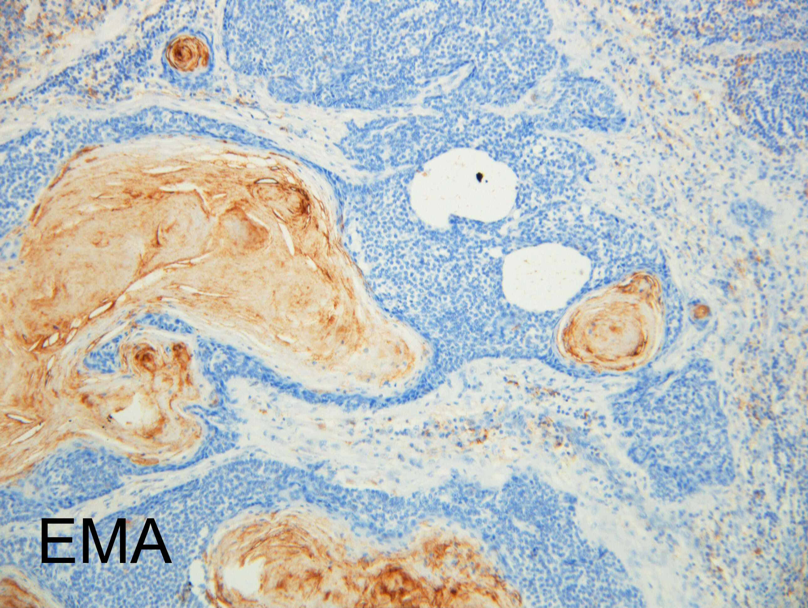
For me too this is a hard case. As Vincenzo I think there is a collision of pathologies, I would perform immunos for the lymphoid portion, could it be a B cell lymphoma (MALT) CD43, CD20, CD3, etc. The epithelial component is challenging too, on low power looks like pilomatricoma but I am less convinced at high power, I thought of BCC with prominent infundibular differentiation, follicular SCC? BerEP4, EMA, CEA, P53, ki67. Nice case

Share this comment


Link to comment
share_externally

Dr. Mona Abdel-Halim

Posted

Very dificult!

I am thinking of a pseudolymphomatous reaction surrounding an adnexal lesion. As for the nature of the adnexal lesion, it looks benign to me, not BCC and not fSCC. Definitly on high power, it is not pilomatricoma although it gives this impression on low power. There is epithelial mucin. I am thinking it could be inverted follicular keratosis??

Share this comment


Link to comment
share_externally

Alice Roberts

Posted

Possibly ALHE surrounding hidradenoma

Share this comment


Link to comment
share_externally

Sasi Attili

Posted

Difficult!!!! IFK with pseudolymphomatous reaction?

Share this comment


Link to comment
share_externally

Dr. Richard Carr

Posted

This is clearly a spot diagnosis except neither myself nor anyone else I shared the case with could give it a satisfactory name (I circulated it to about 20 friends and colleagues, many of national and some of international standing). My report favoured a low-grade follicular neoplasm (probably benign) with ALHE-like stromal reaction! If anyone sees another example we might publish a series!

Share this comment


Link to comment
share_externally


Join the conversation

You can post now and register later. If you have an account, sign in now to post with your account.

Guest
Add a comment...

×   Pasted as rich text.   Paste as plain text instead

  Only 75 emoji are allowed.

×   Your link has been automatically embedded.   Display as a link instead

×   Your previous content has been restored.   Clear editor

×   You cannot paste images directly. Upload or insert images from URL.

Loading...

×
×
  • Create New...