Jump to content
In this section we have spot diagnoses posted on a daily basis since June 2010, now over 4000! You can review the archived cases and read the suggested diagnoses by users and the final comment by the contributors.
Case are uploaded each week day by 10 am UK time with the correct diagnosis will generally be posted at 8 pm UK time. Why not view the most recent spot diagnosis and proffer a diagnosis?

Case Number : Case 1747 - 7 February - Dr Uma Sundram Posted By: Guest

Please read the clinical history and view the images by clicking on them before you proffer your diagnosis.
Submitted Date :
   (0 reviews)

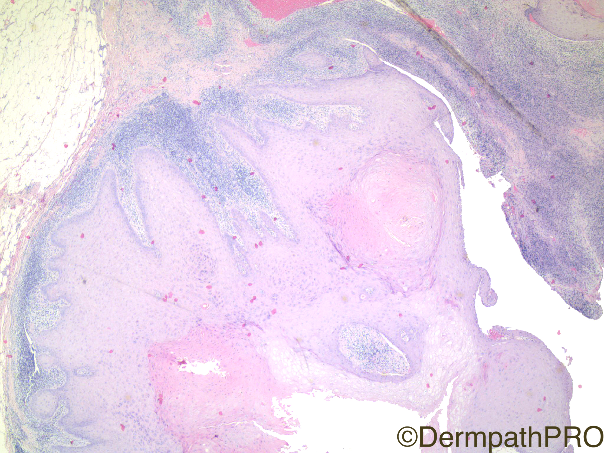
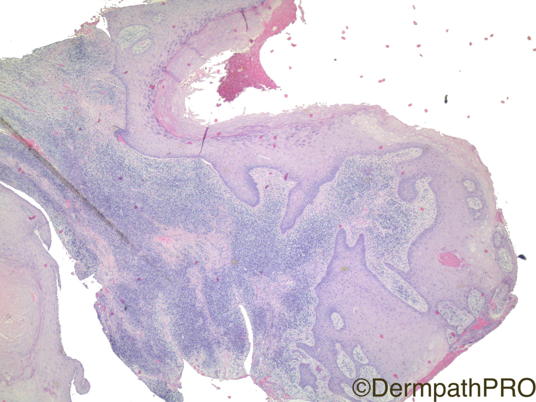
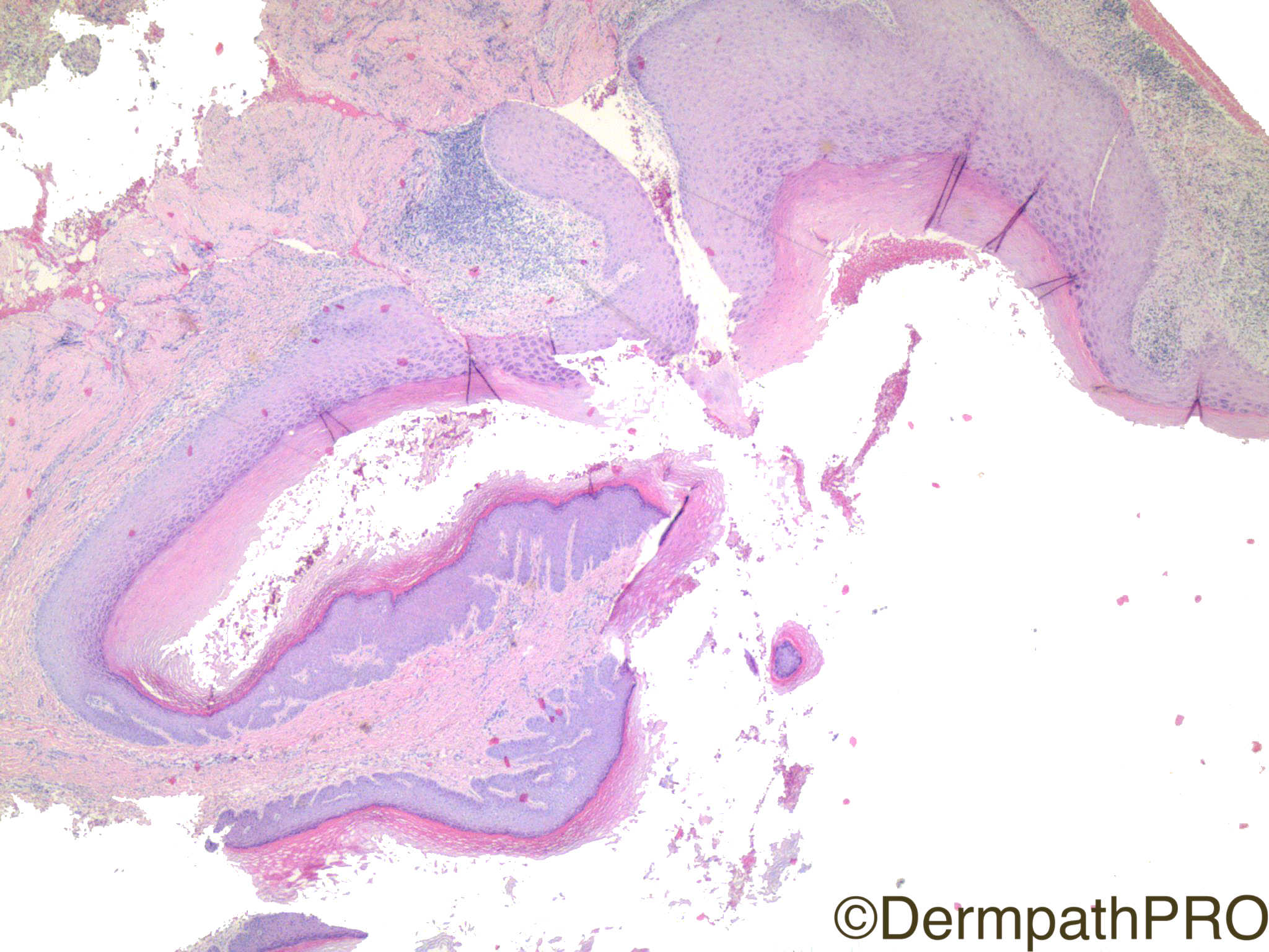
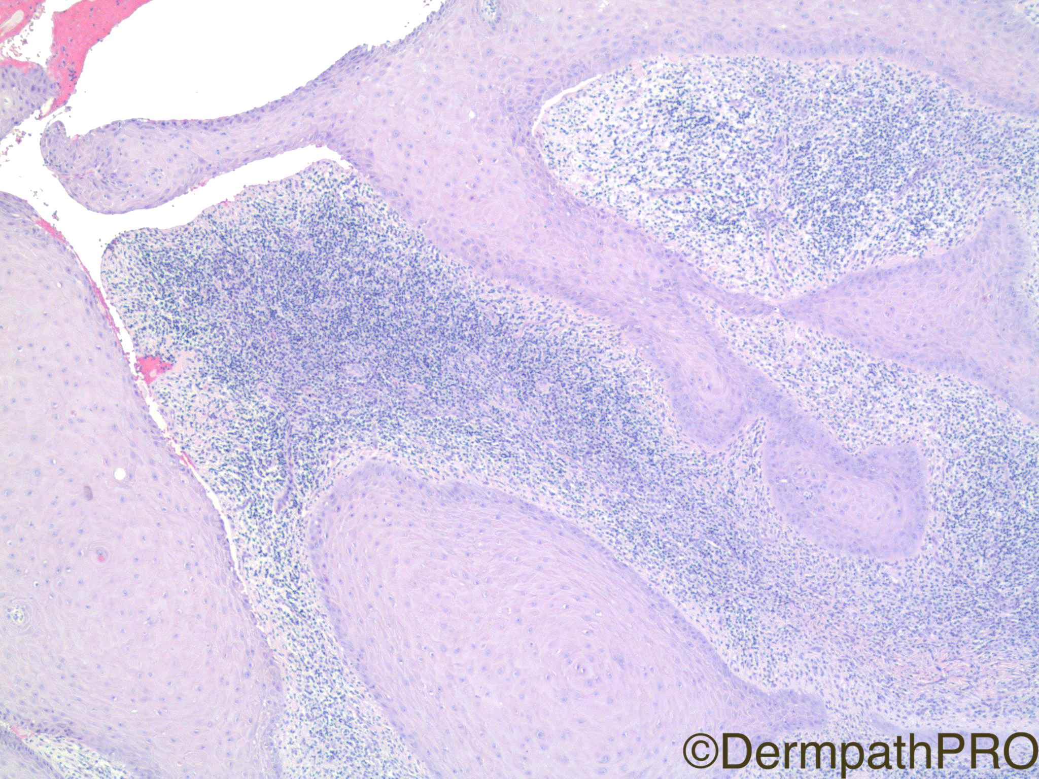
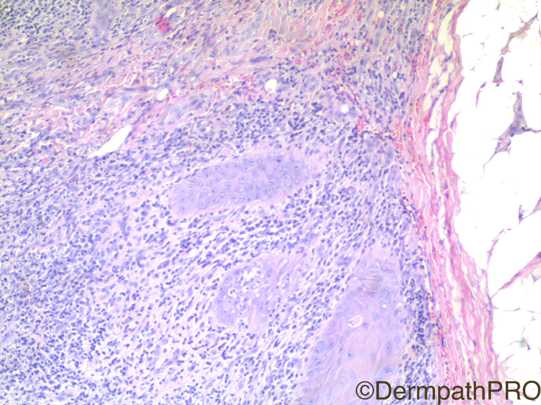
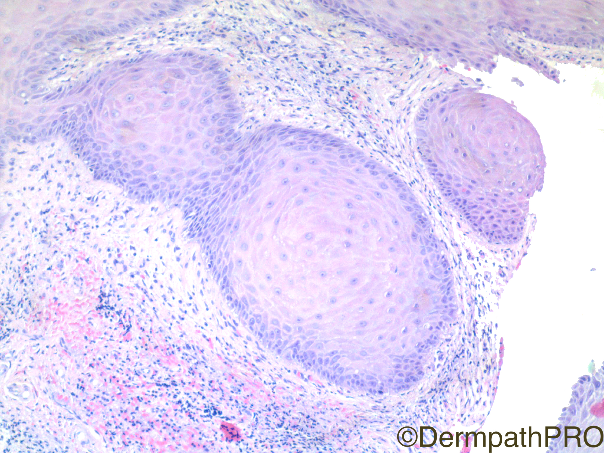
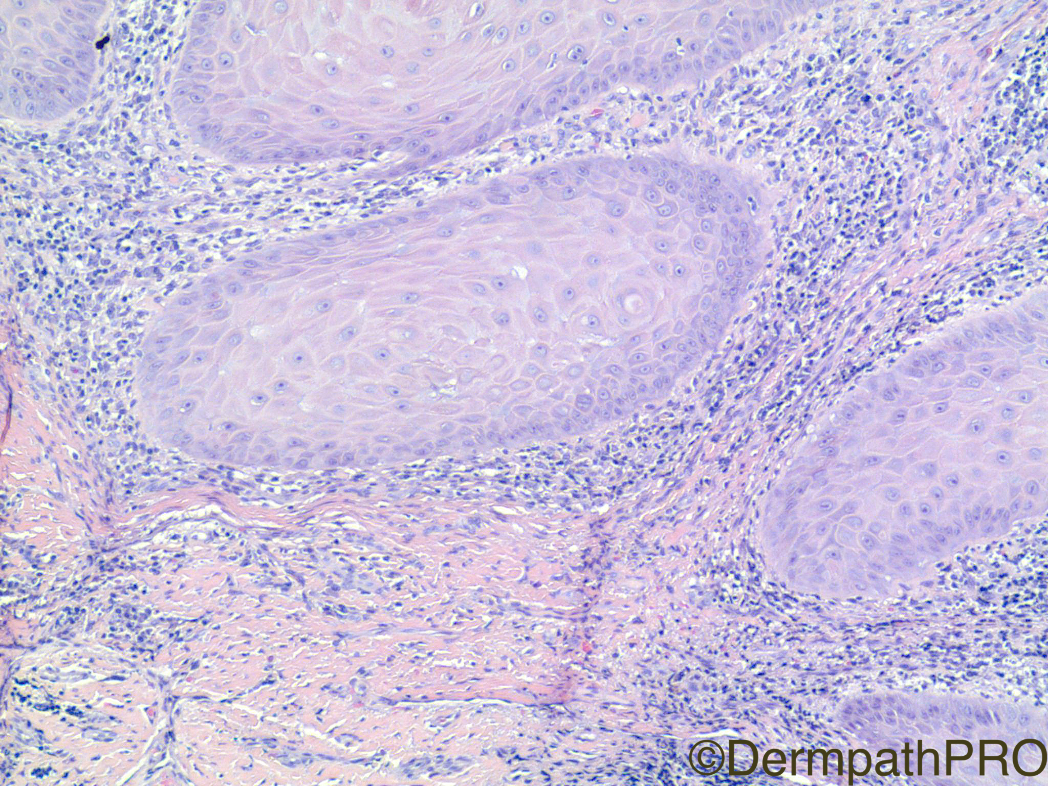
Clinical History: 45 year old man with lesion on back.

Case Posted by Dr Uma Sundram


  Report Record

User Feedback


Admin_Dermpath

Posted

Dr Uma Sundram has an intriguing Spot Diagnosis Case for you today, enjoy


Geoff Cross - DermpathPRO Projects

Share this comment


Link to comment
share_externally

vincenzo polizzi

Posted

Deep desmoplastic dermal reaction, pallor of the epithelium, and attempts of keratin pearls within the downward epithelial ramifications, make me think of Verrucous/(Cuniculatum)Carcinoma

But I know this is a tricky and  controversial diagnosis...

Share this comment


Link to comment
share_externally

urmilapandey

Posted

?scc arising in hypertrophic lichen planus

Share this comment


Link to comment
share_externally

I liked the above mentioned hypothesis of SCC arising in LP

Share this comment


Link to comment
share_externally

Robledo F. Rocha

Posted

Regressing keratoacanthoma. Neoplastic nests are embedded in a chronic inflamed and fibrotic stroma and they are composed by squamous cells whose nuclei are bland and whose cytoplasm become larger and more eosinophilic toward the center of the nests.

Share this comment


Link to comment
share_externally

Dr. Mona Abdel-Halim

Posted

As Robledo, I also thought of regressing KA

Share this comment


Link to comment
share_externally

Guest parasa gayatri devi

Posted

Agree with SCC in hypertrophic lichenplanus 

Share this comment


Link to comment
share_externally

msofopoulos

Posted

I cant see any atypia. I think more of a resolving (regressing) keratoacanthoma

Share this comment


Link to comment
share_externally

Raul Perret

Posted (edited)

I cannot decide based only on these images between regressing ka vs hypertrophic lichen planus. I do not think there is cancer here though. Favour regressing ka

Edited by Raul Perret

Share this comment


Link to comment
share_externally

Nitin Khirwadkar

Posted

Regressing squamoproliferative lesion, most suggestive of a regressing keratoacanthoma. Not convinced that this is invasive SCC.

Share this comment


Link to comment
share_externally

Alice Roberts

Posted

I'm considering hypertrophic LP +/- KA/ well diff SCC

Share this comment


Link to comment
share_externally

Alice Roberts

Posted

I don't think the lesion looks like it's regressing, but there is definitely lichenoid lymphocytic inflammation. Given the patients age and lack of atypia, I'm not sure I want to call this SCC. Tough.

Share this comment


Link to comment
share_externally

Uma Sundram

Posted

I tried to photograph the fact that the lesion was a cystic one, and hence the diagnosis is proliferating epidermoid cyst, with consideration for an invasive squamous cell carcinoma arising in a proliferating epidermoid cyst. I agree with many of you that the threshold for invasion has not been reached. I loved the discussion in this case because it’s a basic benign vs malignant one. We spend a lot of time writing literature about really rare entities for which you will ask for a consultant’s help anyway. We don’t spend a lot of time discussing cases where the decision making is somewhat basic but REALLY crucial, and has a huge impact on patient care. So, if this lesion were flat, the differential diagnosis would include regressing KA vs hypertrophic LP vs well differentiated invasive squamous cell carcinoma. All are reasonable ideas; the threshold between pseudoepitheliomatous hyperplasia and well differentiated invasive SCC is a well known moving target. I came down on the side of NOT being invasive but this is a difficult case! Well done!

Share this comment


Link to comment
share_externally

Dr. Richard Carr

Posted

Great case and agree with your comments Uma.  I think there is so much learning from the routine sign-out. Like you I was favouring infundibular (epidermoid) cyst with scarring/inflammation etc. giving rise to the proliferating borders - however like many bench cases tucked away in the pile it's not easy!

Share this comment


Link to comment
share_externally


Join the conversation

You can post now and register later. If you have an account, sign in now to post with your account.

Guest
Add a comment...

×   Pasted as rich text.   Paste as plain text instead

  Only 75 emoji are allowed.

×   Your link has been automatically embedded.   Display as a link instead

×   Your previous content has been restored.   Clear editor

×   You cannot paste images directly. Upload or insert images from URL.

Loading...

×
×
  • Create New...