Jump to content
In this section we have spot diagnoses posted on a daily basis since June 2010, now over 4000! You can review the archived cases and read the suggested diagnoses by users and the final comment by the contributors.
Case are uploaded each week day by 10 am UK time with the correct diagnosis will generally be posted at 8 pm UK time. Why not view the most recent spot diagnosis and proffer a diagnosis?

Case Number : Case 1730 - 13 January - Dr Richard A Carr Posted By: Guest

Please read the clinical history and view the images by clicking on them before you proffer your diagnosis.
Submitted Date :
   (0 reviews)

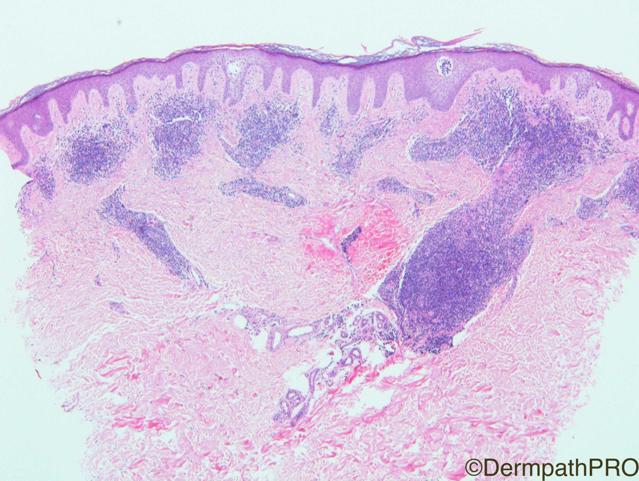
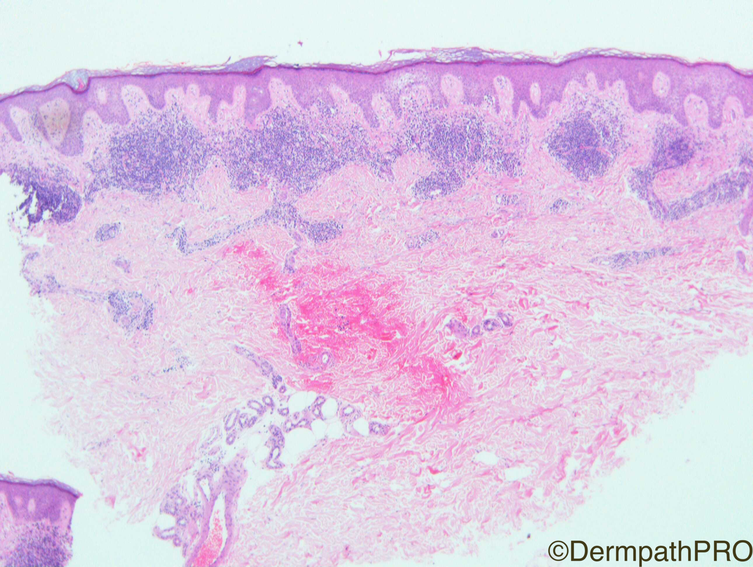
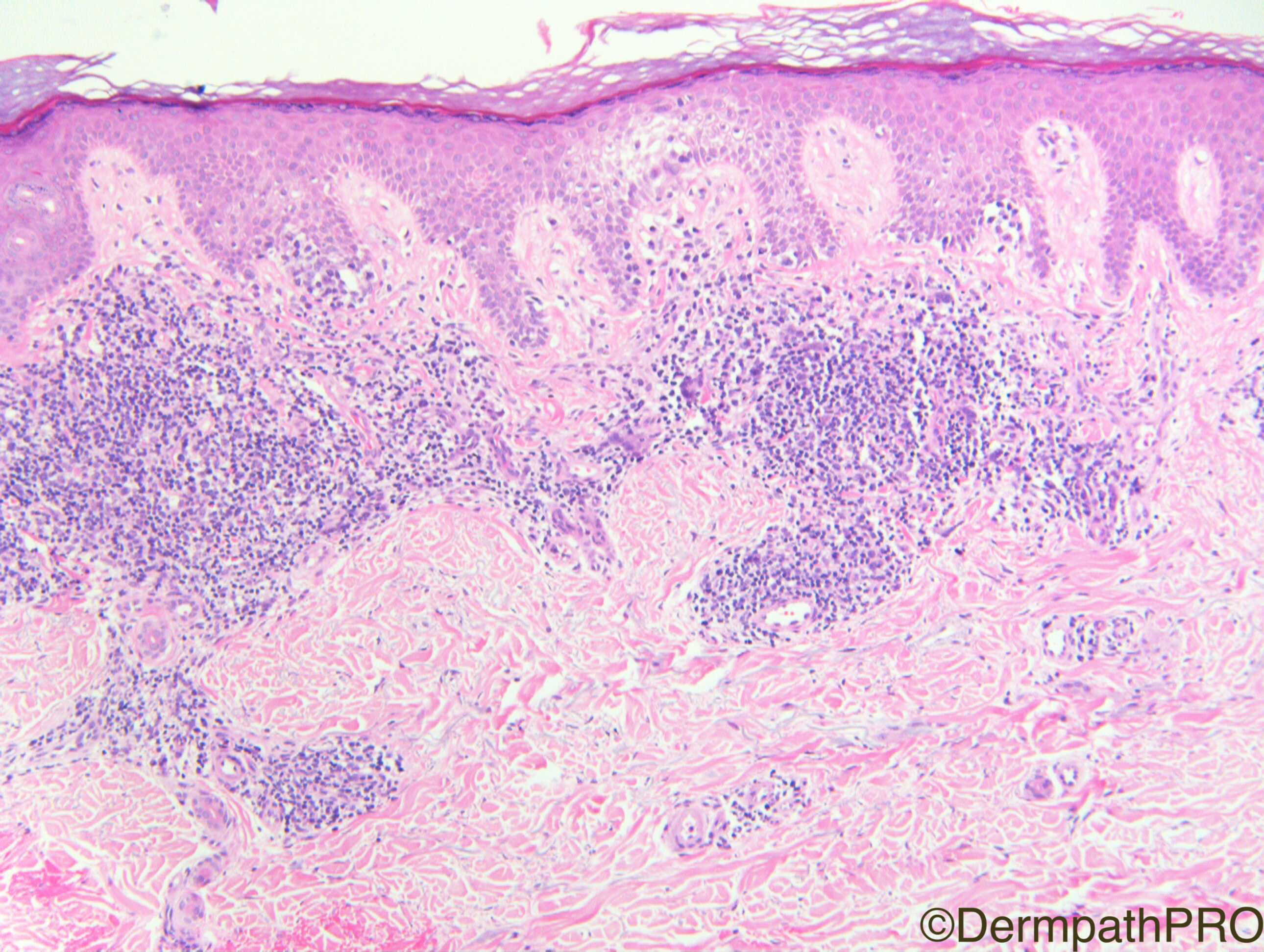
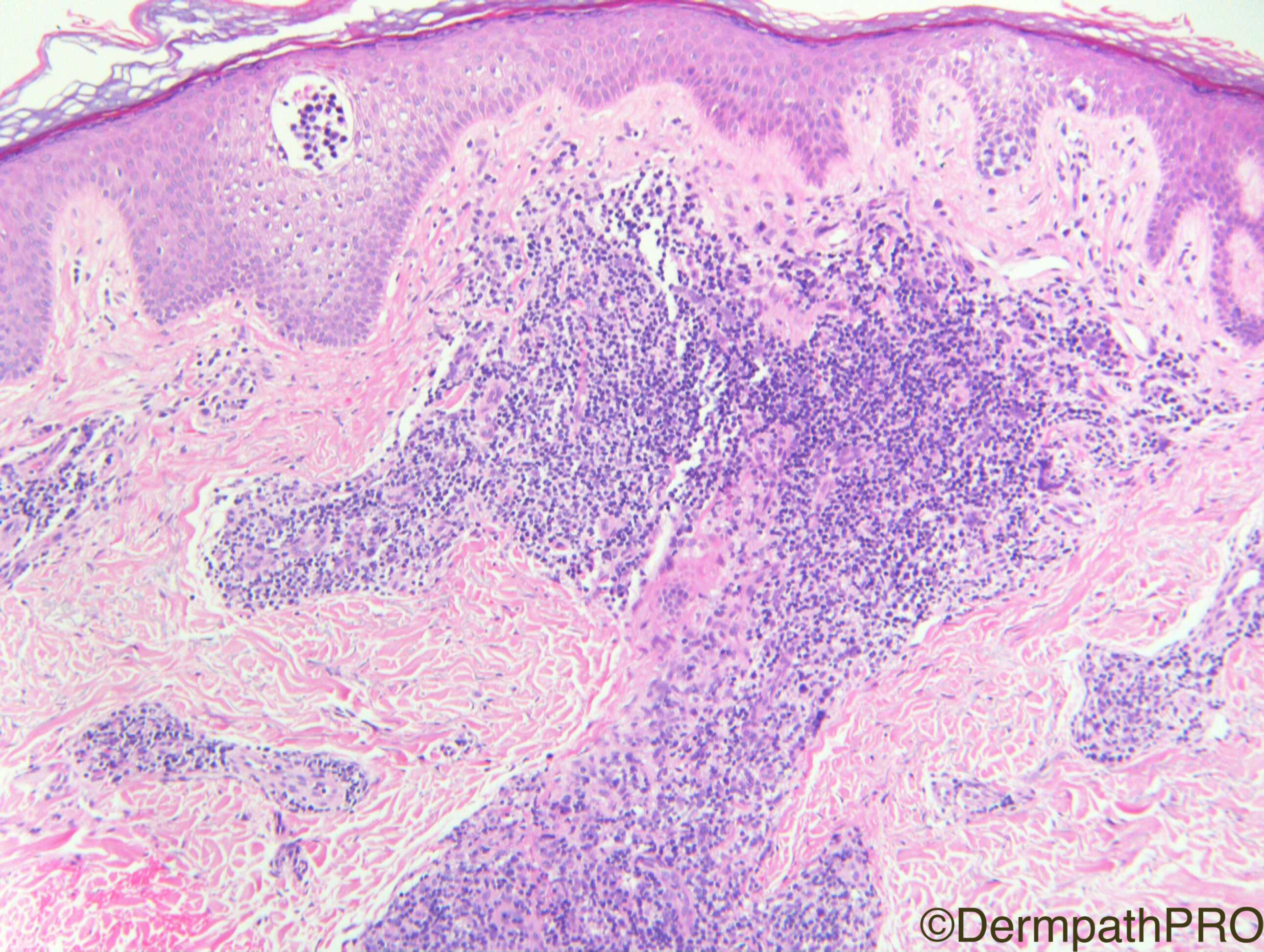
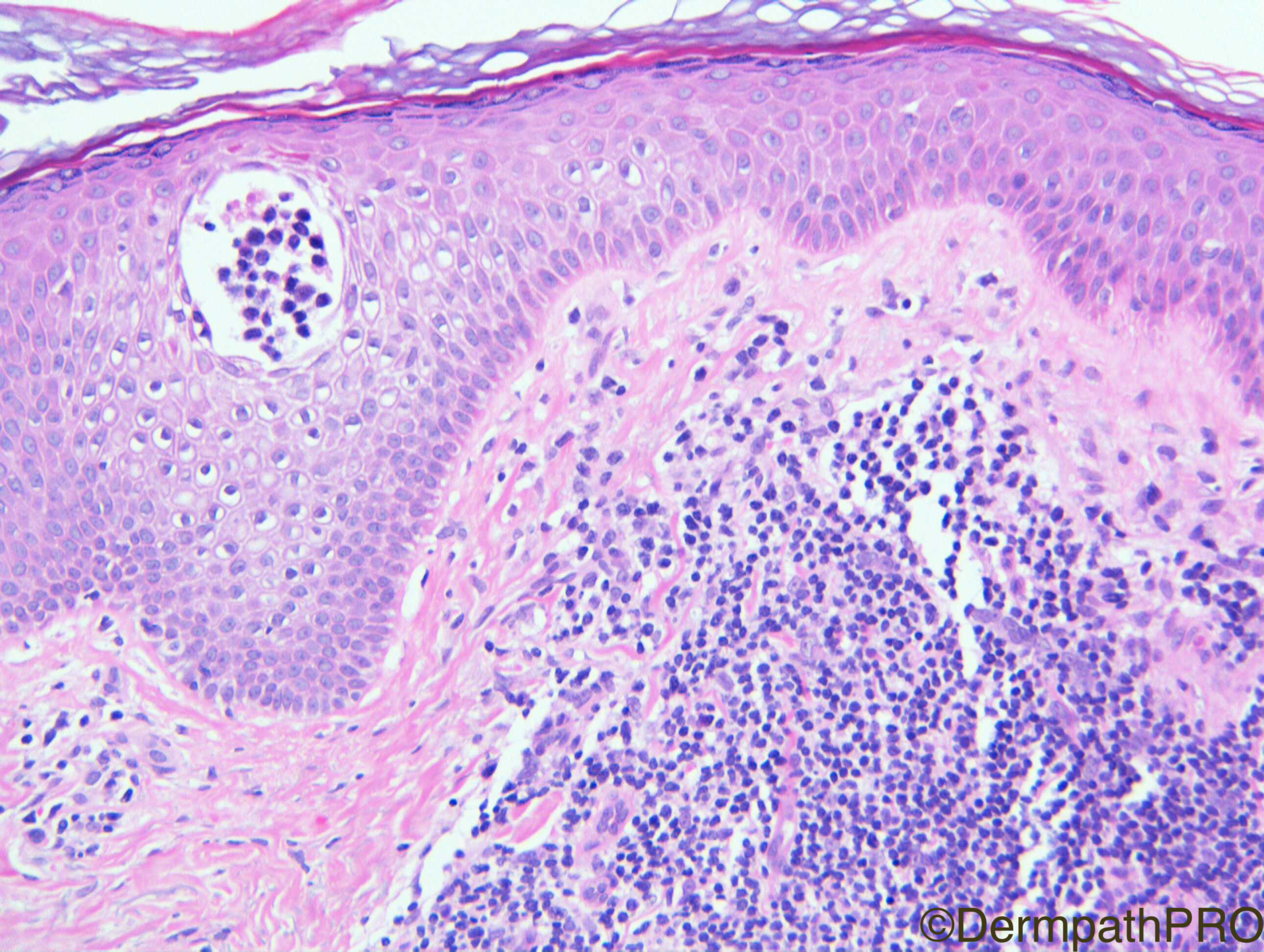
Clinical History: F60. Wrist. 2-3 years of erythematous papules + plaques, wrists, ankle/dorsum of hands. ?Granuloma annulare.

Case Posted by Dr Richard A Carr
Edited by Admin_Dermpath


  Report Record

User Feedback


Admin_Dermpath

Posted

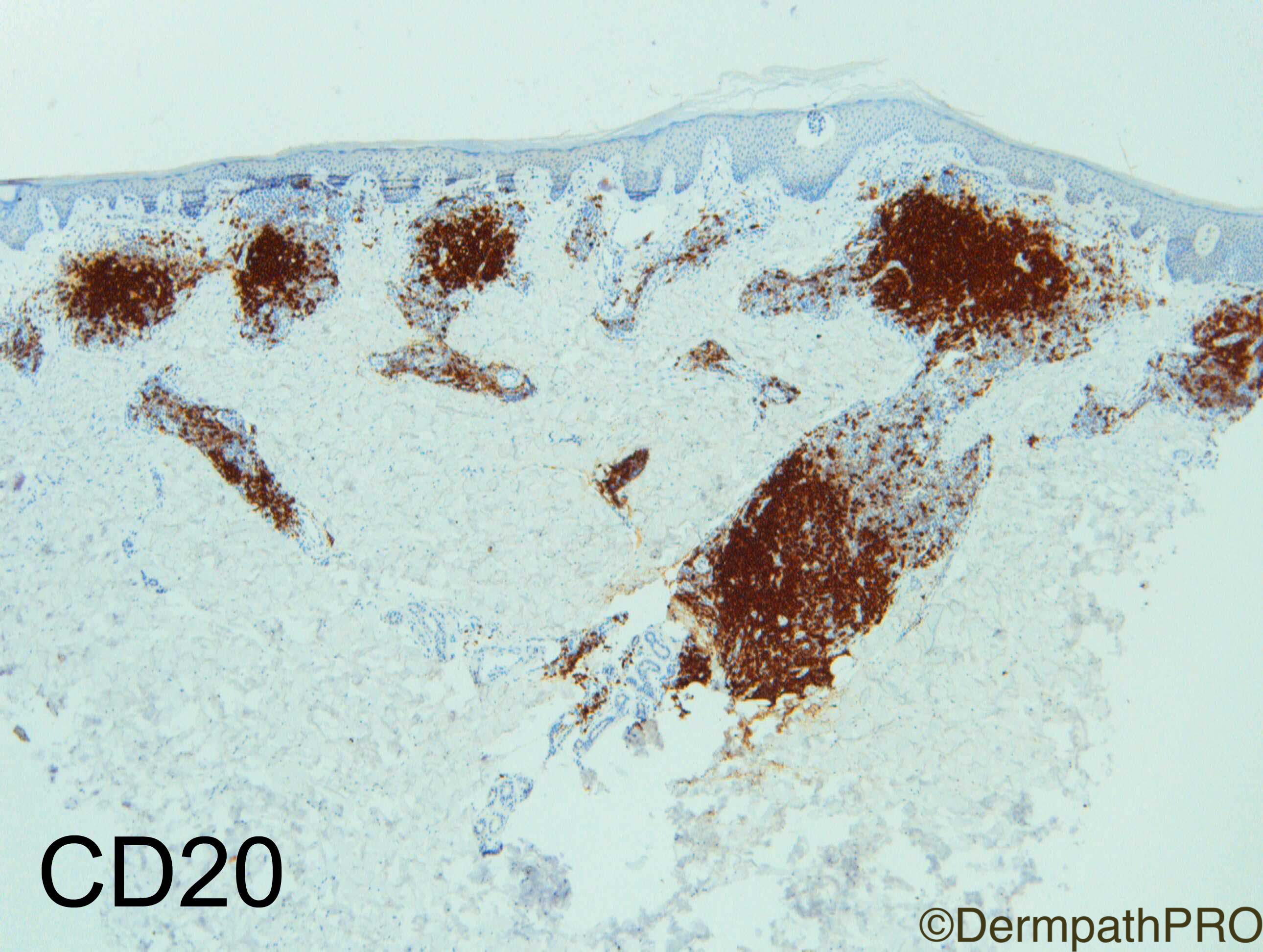
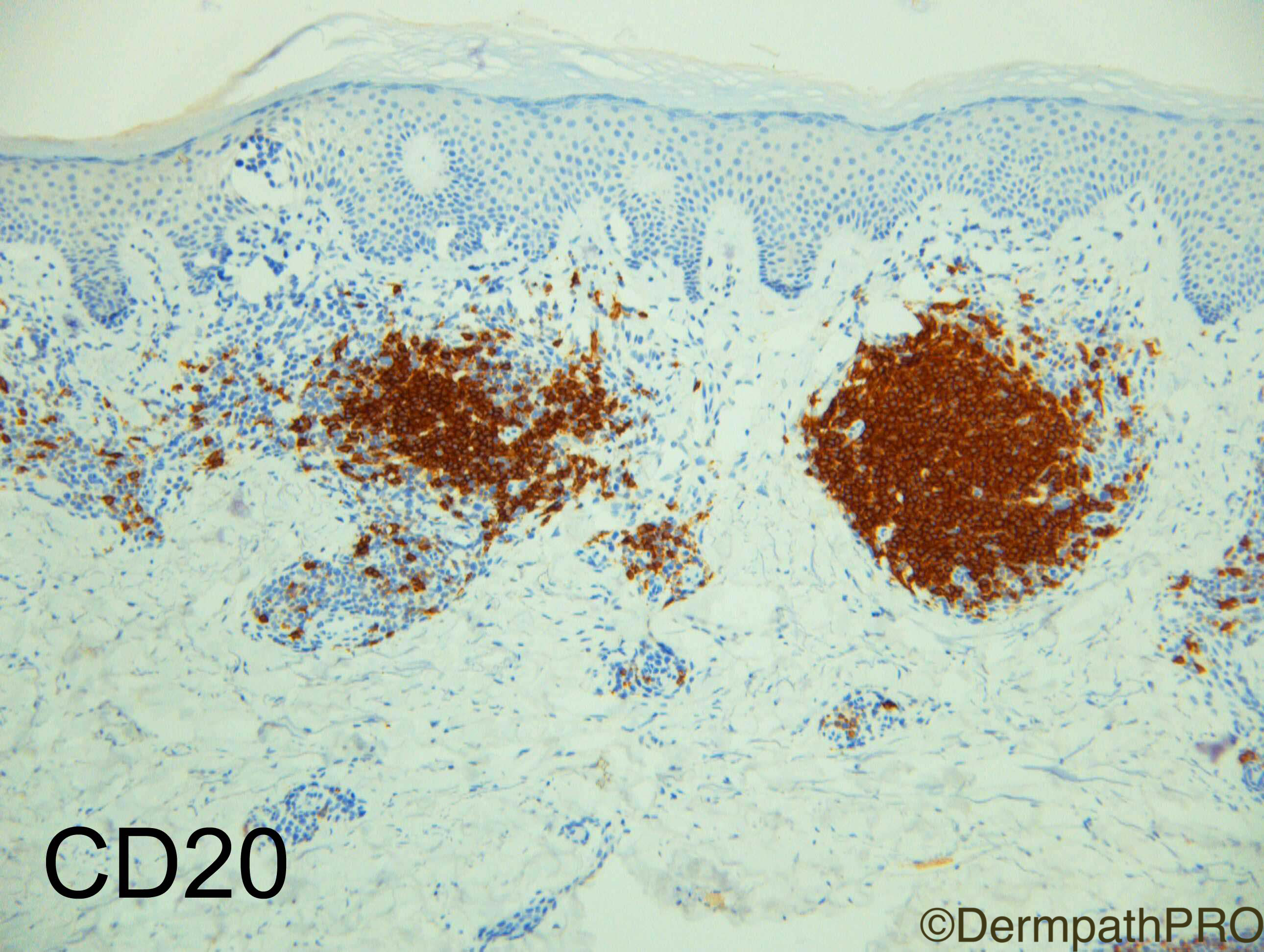
Here is your Friday the 13th Special from Dr Richard A Carr with IHC images to be added at 6pm-ish GMT, I hope this is not too scary a case  :-)


Geoff Cross - DermpathPRO Projects

Share this comment


Link to comment
share_externally

vincenzo polizzi

Posted

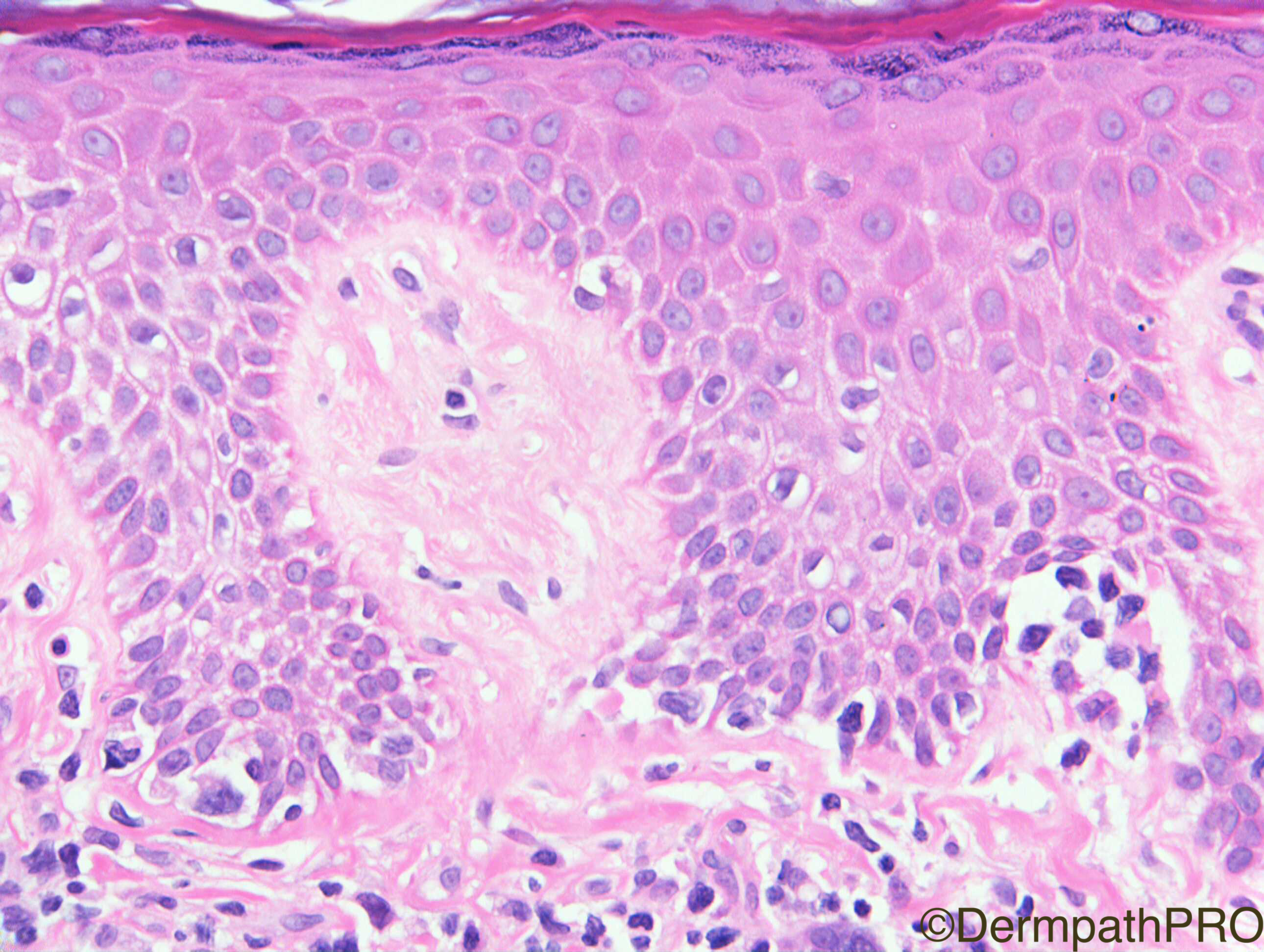
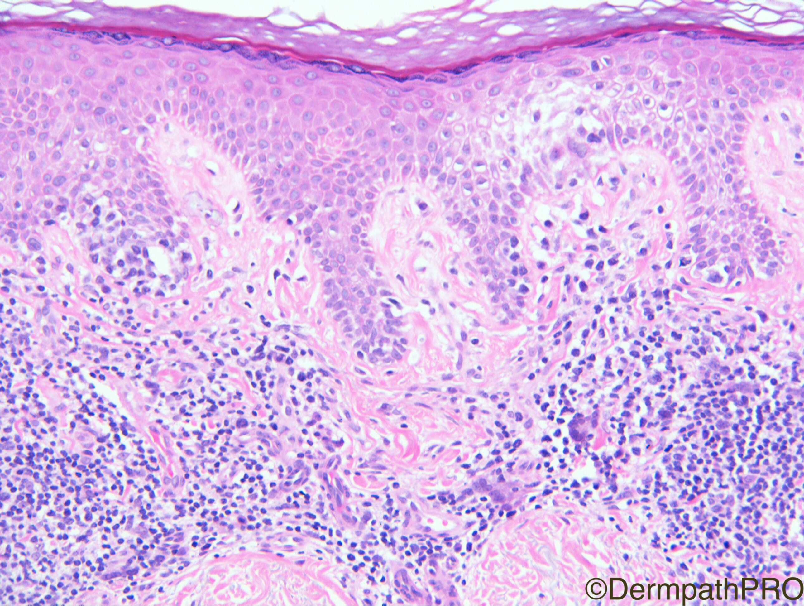
Clinical history and histological features ( lamellar fibrosis of papillary dermis, Pautrier abscess, linear lymphocytic infiltration of basal layer, atypical epidermal lymphocytes larger than intradermal ones ) make me favor Mycosis Fungoides. 

Share this comment


Link to comment
share_externally

Robledo F. Rocha

Posted

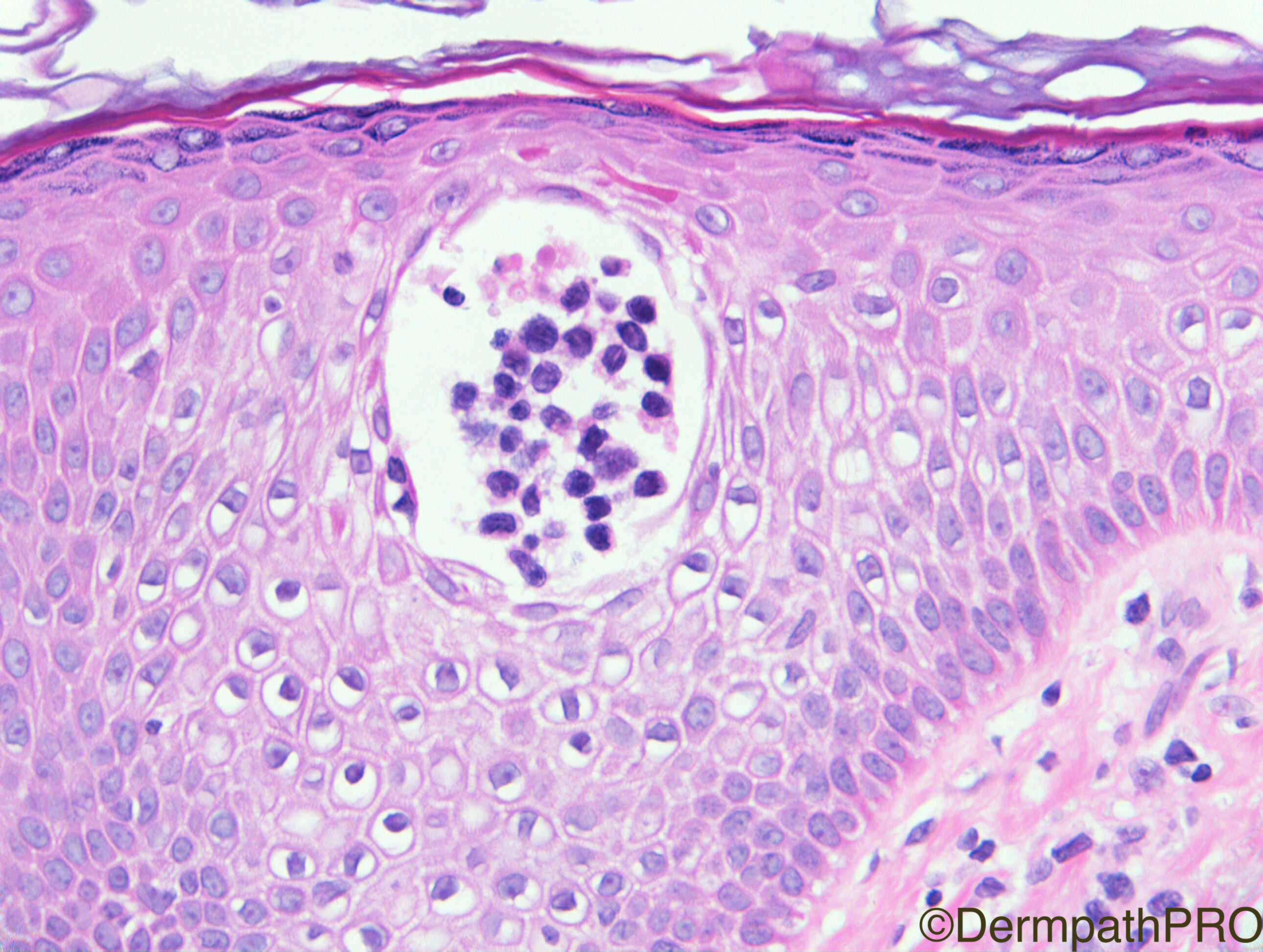
Sites of the lesions are not the expected ones for mycosis fungoides, so maybe it is a spongiotic dermatitis with a few epidermotropic lymphocytes lined up along basal layer and a Langerhans microabscess in the fashion of a Pautrier simulant.

Share this comment


Link to comment
share_externally

Nitin Khirwadkar

Posted

Agree with Robledo. Eczema, almost EAC like in image 1.

Share this comment


Link to comment
share_externally

vincenzo polizzi

Posted

Erythema annulare centrifugum-like mycosis fungoides.

Ceyhan AM, et al. Australas J Dermatol. 2011.

Share this comment


Link to comment
share_externally

Raul Perret

Posted

Also thought of spongiotic dermatitis, the dermal infiltrate is quite prominent though.

Share this comment


Link to comment
share_externally

Dr. Mona Abdel-Halim

Posted

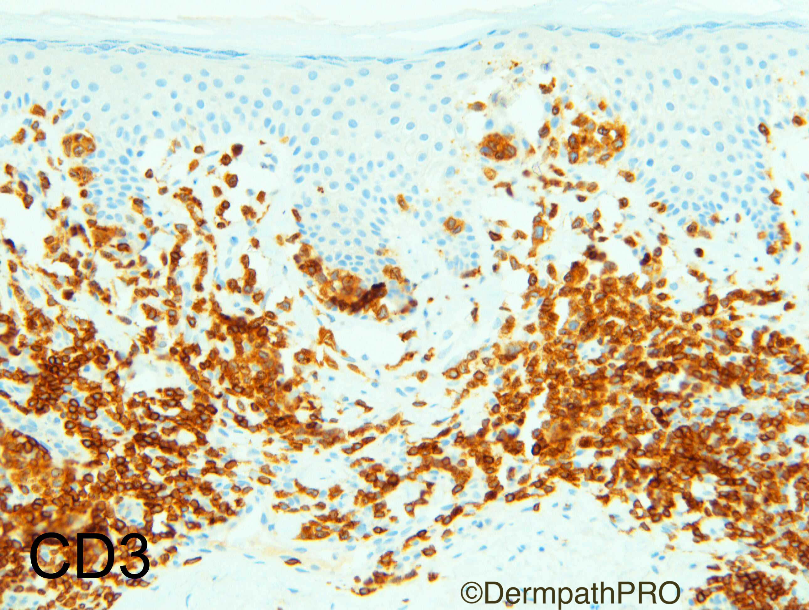
I would love to c a CD3 and CD1a first to verify what we r seeing in the last image. A true Pautrier's microabscess or a Pautrier simulant. 

But provisionally, I am favoring a spongiotic reaction. To me, the epidermis is too spongiotic for MF and overall looks inflammatory.

The dermal infiltrate is quite dense though. Some coat sleeve impression. 

Some basilar lymphocytes?? Seems annular in configuration (since the ddx is GA). So may be it is EAC with pautrier's simulant. Waiting for the immunos!!!


Share this comment


Link to comment
share_externally

I think this is a spongiotic dermatitis simulating MF with those Pautrier-like microabscesses. I would like to see immunostains for CD3 and CD1a as well, but at H&E it looks like langerhans cells. I agree with Mona´s observation that the clinical impression of GA may be a clue for EAC or contact dermatitis.

Share this comment


Link to comment
share_externally

Arash Daryakar

Posted

I like to rule out MF by IHC in this case.Although,spongiosis is too prominent to be MF.

Share this comment


Link to comment
share_externally

Arash Daryakar

Posted

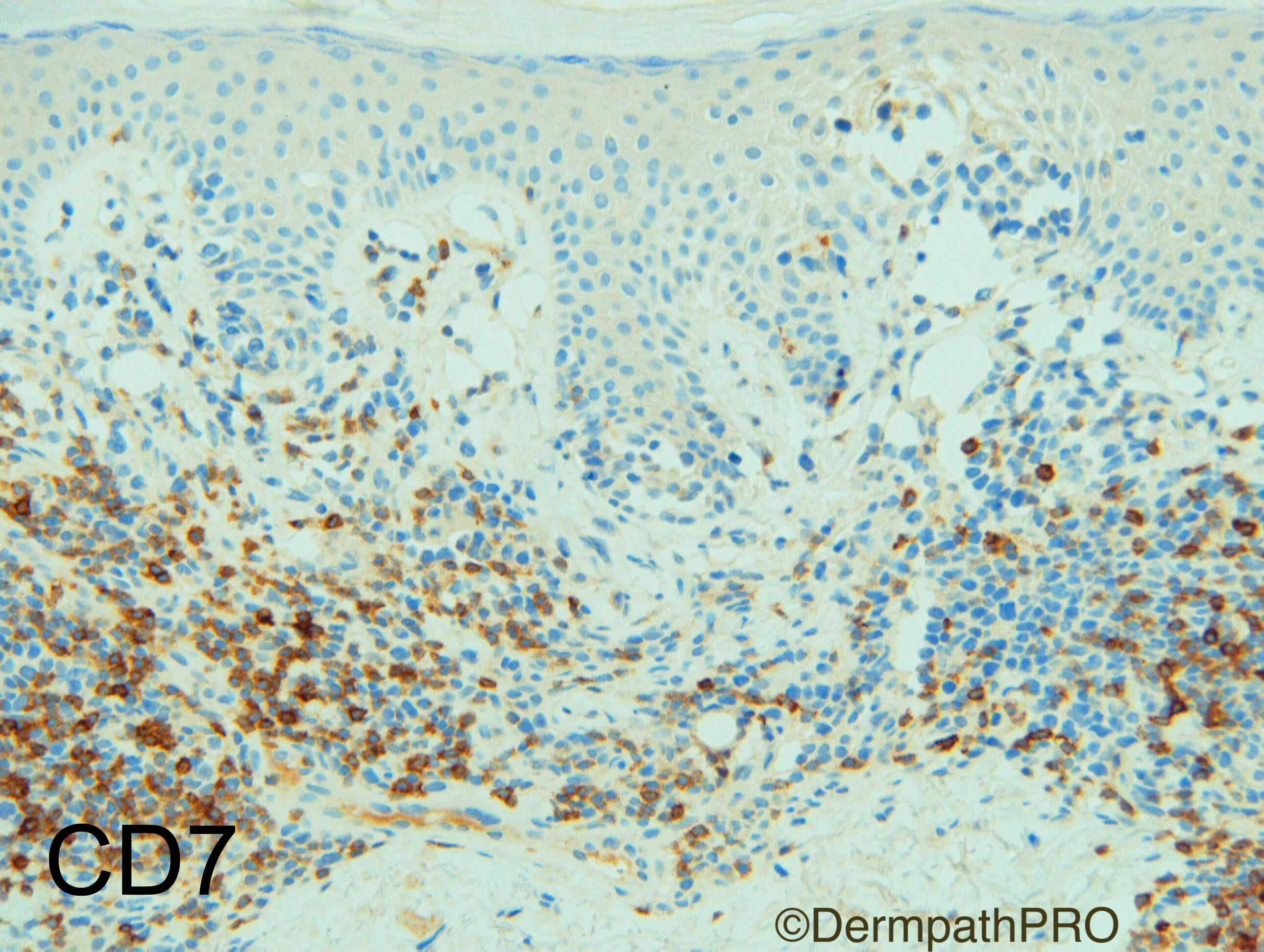
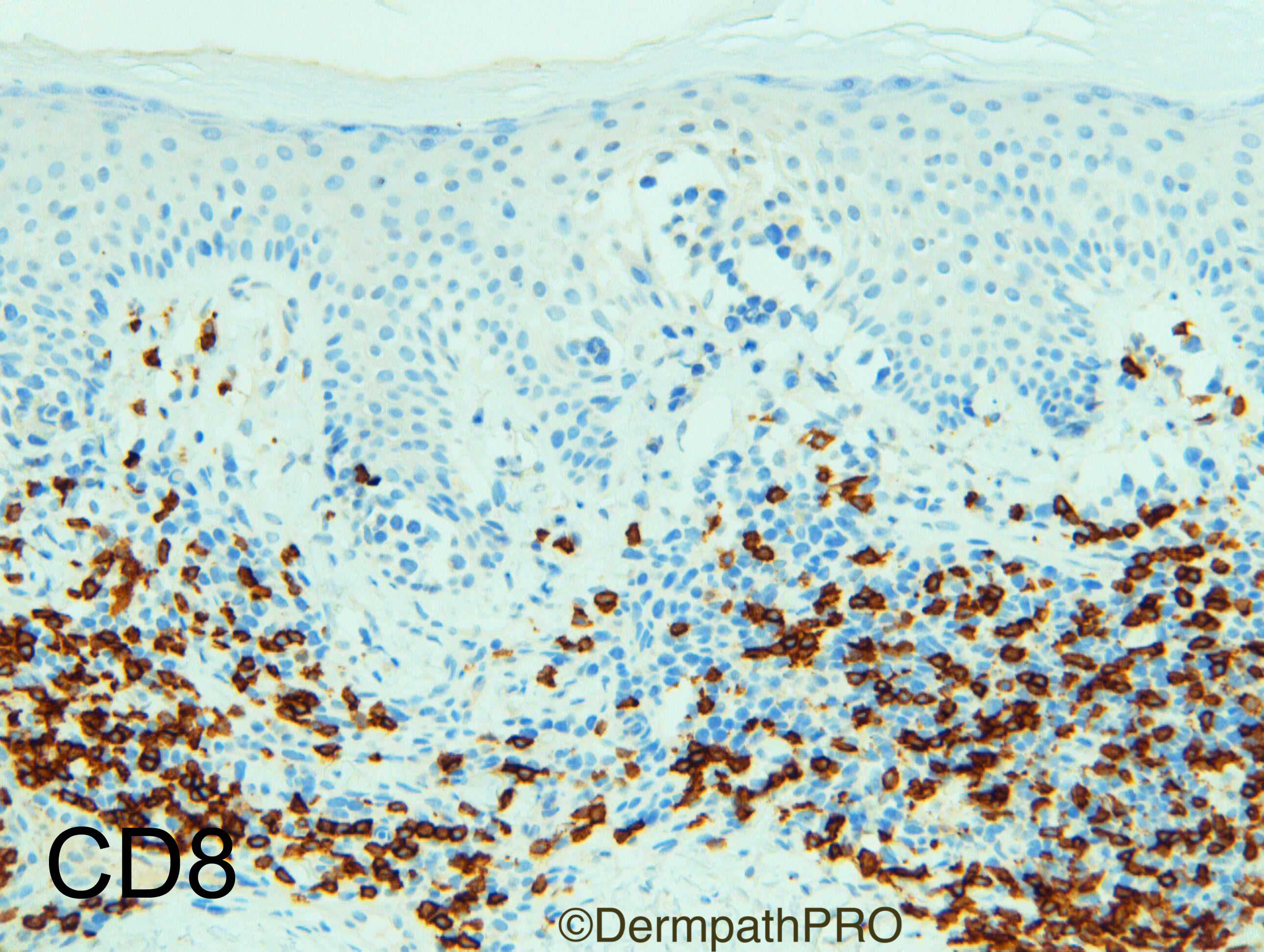
lt seems intraepidermal lymphocytes have loss of CD7 and CD8 neg . am i right?

so its more likely to be MF

Share this comment


Link to comment
share_externally

vincenzo polizzi

Posted

Let me explain in details why favor MF over the other mimics:

1) the epidermal abscess is a true pautrier for me, because nucleus are hyperchromatic, lymphocytic, in opposite to vescicular and reniform of LC.  And the epidermal lymphocytes are twice large than dermal ones  

2) it's too important for me the "lamellar fibrosis " of papillary dermis. It isn't diagnostic off course but it's an useful clue to MF. 

3) 2/3 years of clinical history fit in with chronic disease than acute/subacute one ( like the lamellar fibrosis fit in better with a long time junctional epidermal dermal process  ). 

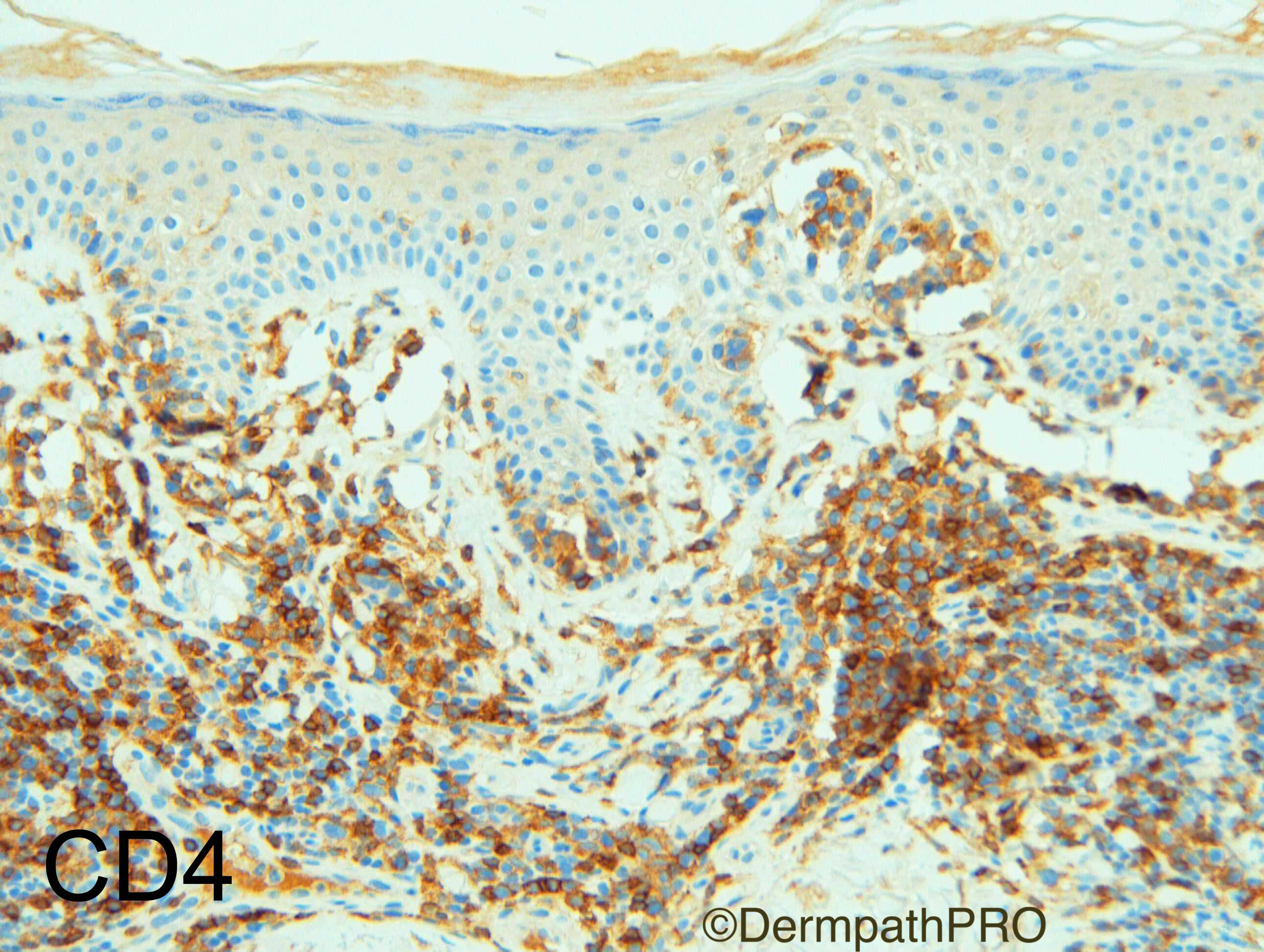
4) IHC isn't conclusive alone, but it fit very well in with the above, doesn't it?

Share this comment


Link to comment
share_externally

Dr. Richard Carr

Posted

Well done Vincenzo.  I agree with your diagnosis favouring mycosis fungoides (CD3+, CD4weak, CD7 loss, CD8-, clonal for TRG and TRB) currently being observed (pT1b skin limited disease). We can see spongiosis in mycosis fungoides although often the shear number of epidermotropic, hyperchromatic, lymphocytes remains disproportionate to the degree of spongiosis.  I agree with considering a Langerhan's cell pseudo-pautrier microabscess although Langerhan's cells typically have somewhat paler nuclei with more open chromatin and gentle folded (reniform) nuclei with more abundant pale eosinophilic cytoplasm (surprisingly could not find an example of such on dermpathpro - keep your eyes out though). I found the rather monotonous peri-vascular B-cells of some confusion, never having seen this pattern before so asked for B-cell clonality and this showed a weak polyclonal pattern). I'm guessing it was the latter pattern that led to some confusion with erythema annulare.

Share this comment


Link to comment
share_externally

Raul Perret

Posted

Well done Vincenzo, another thing learned today!

Share this comment


Link to comment
share_externally

Robledo F. Rocha

Posted

After immunohistochemical images, this is mycosis fungoides with odd clinical presentation and with odd microscopic features. Diseases definitively do not read Dermatopathology Textbooks!

Share this comment


Link to comment
share_externally


Join the conversation

You can post now and register later. If you have an account, sign in now to post with your account.

Guest
Add a comment...

×   Pasted as rich text.   Paste as plain text instead

  Only 75 emoji are allowed.

×   Your link has been automatically embedded.   Display as a link instead

×   Your previous content has been restored.   Clear editor

×   You cannot paste images directly. Upload or insert images from URL.

Loading...

×
×
  • Create New...