Jump to content
In this section we have spot diagnoses posted on a daily basis since June 2010, now over 4000! You can review the archived cases and read the suggested diagnoses by users and the final comment by the contributors.
Case are uploaded each week day by 10 am UK time with the correct diagnosis will generally be posted at 8 pm UK time. Why not view the most recent spot diagnosis and proffer a diagnosis?

Case Number : Case 846 - 13th September Posted By: Guest

Please read the clinical history and view the images by clicking on them before you proffer your diagnosis.
Submitted Date :
   (0 reviews)

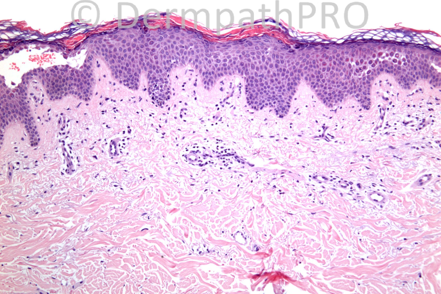
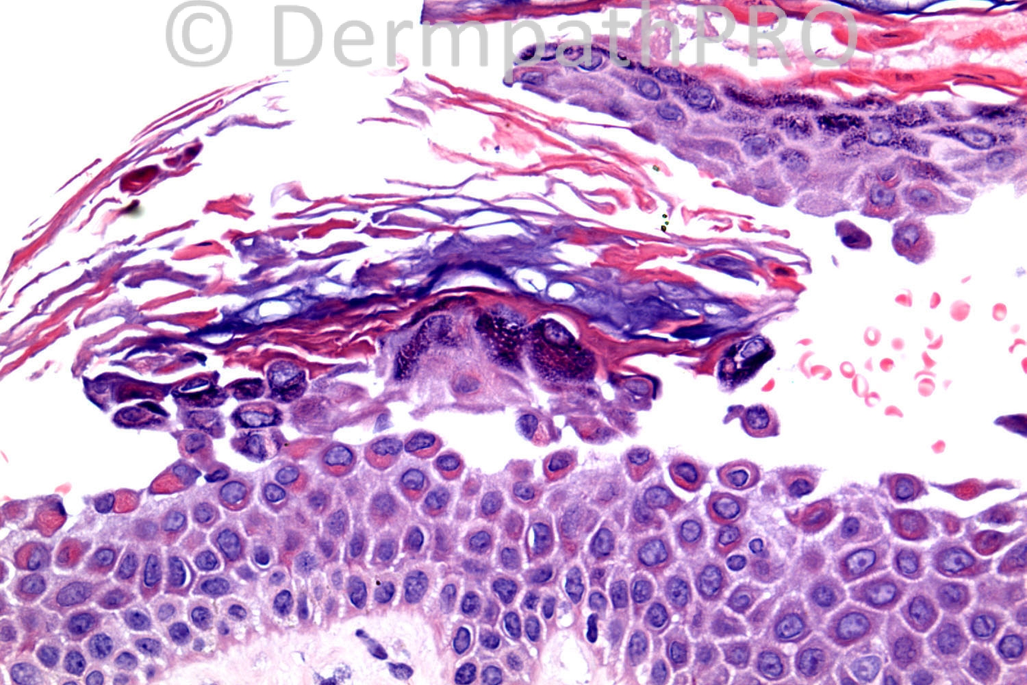
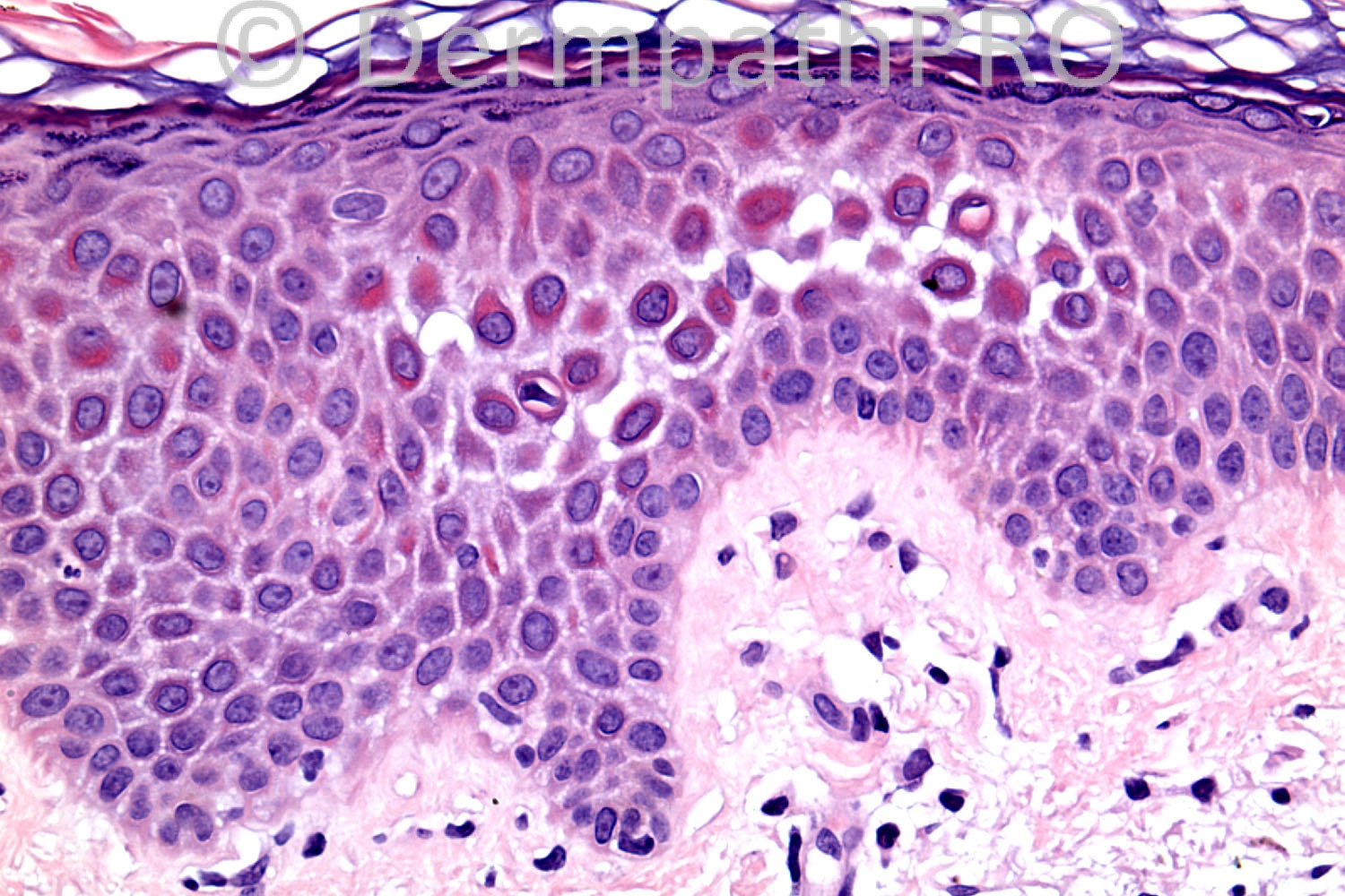
87 years old female. Eroded areas upper back and shoulder, one with collapsed blister.

Case posted by Dr. Richard Carr.

  Report Record

User Feedback


Darier's Disease or Grover's Disease ( Darier's type)

Blister formation is unysual in Darier's but could happen
The seperation in in mid epidermis and not in the granular layer or sub corneal which make superficial pemphigus less likely

I see too many dyskeratotic cells to call it superficial pemphigus
immunofloresence will tell the difference
clinically both Darie's and superficial pemphigus have some similar features
I think this case goes more with Darier's though the age is more typical for pemphigus
I will do immunofloresnece before giving a diagnosis

If immunofloresnce id positive I will call it pemphigus foliaceous ( although not happy from this diagnosis ) bit if negative I may think in drug induced pemphigus foliaceious or grovers'S Disease with Darier's features type.

Share this comment


Link to comment
share_externally

Dr. Mona Abdel-Halim

Posted

Superficial pemphigus (erythematosus or foliaceus)... Very beautifully demonstrated :-))

Share this comment


Link to comment
share_externally

Guest Graham Reilly

Posted

Pemphigus Foliaceus.High split with dyskeratotic keratinocytes.

Share this comment


Link to comment
share_externally

The split appears to be in upper reaches of the stratum spinosum, rather than at the subcorneal level. Also, there is (focally marked) spongiosis. This makes me slightly hesitant regarding the diagnosis of pemphigus foliaceus, and makes me wonder whether DIF might be negative and clinical features, although obviously not typical, might be compatible with Grover's disesae.

Share this comment


Link to comment
share_externally

Guest Marwa Fawzy

Posted

[color=#1C2837][font=arial, verdana, tahoma, sans-serif][size=4]Superficial pemphigus[/size][/font][/color]

Share this comment


Link to comment
share_externally

Pemphigus foliaceous, differential diagnosis in this case with Grover´s disease (that can simulate PF and also shows spongiosis). Need IFD to best differentiate. Upper back and shoulder are locations that favors PF.

Share this comment


Link to comment
share_externally

Arti Bakshi

Posted

[quote name='IgorSC' timestamp='1379073023']
Pemphigus foliaceous, differential diagnosis in this case with Grover´s disease (that can simulate PF and also shows spongiosis). Need IFD to best differentiate. Upper back and shoulder are locations that favors PF.
[/quote]
Agree with d/d of pemphigus foliaceous and Grover's.
The split does seem to be intrapidermal rather than subcorneal. IMF needed.

Share this comment


Link to comment
share_externally

Guest Pedro Aleixo

Posted

[color=#1C2837][font=arial, verdana, tahoma, sans-serif][size=4]Pemphigus foliaceous[/size][/font][/color]

Share this comment


Link to comment
share_externally

Robledo F. Rocha

Posted

Granular acantholytic blister with dyskeratotic keratinocytes and mild spongiosis. I think this is classic pemphigus foliaceus, but not the variant called pemphigus erythematosus, because lesions are not restricted to the face and there's no lupus findings, nor the variant called fogo selvagem, because I believe patient doesn't live in an endemic region.
I found acantholytic cleavage too diffuse to construe a diagnosis of Grover disease.
Case history is not compatible with bullous impetigo nor staphylococcal scalded skin syndrome.

Share this comment


Link to comment
share_externally

Guest Jim Davie MD

Posted

Pemphigus erythematosus (PE).

PE is often more limited in distribution than pemphigus vulgaris to upper back and face, so clinical makes sense. There is a combination of high level dyskeratosis and high-level acantholysis. There is episodic quality to this (given the checkerboard parakeratosis pattern).

A serologic survey showing positive indirect immunofluorescence (and often positive ANA) would help support a PE dx. Direct immunofluorescence is not too useful for differentiating pemphigus variants, but in PE there should be additional basement layer staining seen, in addition to common PV/PF pattern keratinocyte staining.

Share this comment


Link to comment
share_externally

[b]Subcorneal blisters[/b]
Dyskeratotic cells with hyperchromatic nuclei in granular layer (resembling the ‘grains’ found in Darier’s disease) - scattered acantholytic keratinocytes
Pemphigus foliaceus and erythematosus
Direct immunofluorescence is expected to show intercellular staining for IgG and C3 in both affected and normal skin. The lupus band test is sometimes positive in uninvolved skin in the pemphigus erythematosus (Senear–Usher syndrome) variant of pemphigus foliaceus

Share this comment


Link to comment
share_externally

PS The split is high in the granular layer or directly beneath the stratum corneum.

Share this comment


Link to comment
share_externally

In the rare mixed forms of the disease (pemphigus foliaceus/ Darier’s disease) both suprabasal acantholysis AND subcorneal clefting with acantholysis are present.

Share this comment


Link to comment
share_externally

Four histological patterns may be seen in Grover’s disease – the Darier like pattern, the Hailey–Hailey-like pattern, the pemphigus vulgaris like pattern, and the spongiotic pattern – all with negative immunofluorescence unless coexistence occurs.

Share this comment


Link to comment
share_externally

Guest Tiberiu Tebeica

Posted

In this clinical setting I favor pemphigus foliaceus. DIF needed to confirm this diagnosis. Grover's if DIF negative. Darier's is unlikely, unless patient with positive h/o Darier's.

Share this comment


Link to comment
share_externally

Coexistence of pemphigus foliaceus and Grover’s disease has been reported.

Share this comment


Link to comment
share_externally

Dr. Richard Carr

Posted

Thank you for the excellent responses (very educational) and Mona's kind comments.

I must thank Dr Vivek Mudaliar again for sharing this lovely case of Pemphigus foliaceus with me. IMF studies showed intercellular IgG and C3 in the epidermis. As you know target antigen is desmoglein 3 that is mainly expressed in the upper epidermis (relatively less in oral epithelium - hence the absence of oral disease in Pf.)

Regarding dyskeratosis Weedon makes the following comments: In late lesions of pemphigus foliaceus the epidermis may be hyperplastic, with overlying focal parakeratosis and some orthokeratosis. Dyskeratotic cells with hyperchromatic nuclei and somewhat resembling the ‘grains’ found in Darier's disease are a distinctive feature of the granular layer (Fig. 6.2). In the rare mixed forms of the disease both suprabasal acantholysis and subcorneal clefting with acantholysis are present.

Enjoy the rest of your weekends.

Share this comment


Link to comment
share_externally

The target antigen is desmoglein 1 expressed in desmosomes of upper epidermis and is much less expressed than desmoglein 3 in oral mucosa. Desmoglein 3 is expressed in desmosomes of lower epidermis.

Share this comment


Link to comment
share_externally

i'm sure Richard ment Desmoglein 1 and not 3
thank you Gohar for your contribution

Share this comment


Link to comment
share_externally


Join the conversation

You can post now and register later. If you have an account, sign in now to post with your account.

Guest
Add a comment...

×   Pasted as rich text.   Paste as plain text instead

  Only 75 emoji are allowed.

×   Your link has been automatically embedded.   Display as a link instead

×   Your previous content has been restored.   Clear editor

×   You cannot paste images directly. Upload or insert images from URL.

Loading...

×
×
  • Create New...