Jump to content
In this section we have spot diagnoses posted on a daily basis since June 2010, now over 4000! You can review the archived cases and read the suggested diagnoses by users and the final comment by the contributors.
Case are uploaded each week day by 10 am UK time with the correct diagnosis will generally be posted at 8 pm UK time. Why not view the most recent spot diagnosis and proffer a diagnosis?

Case Number : Case 1934 - 27 Oct - Dr Richard Carr Posted By: Guest

Please read the clinical history and view the images by clicking on them before you proffer your diagnosis.
Submitted Date :
   (0 reviews)

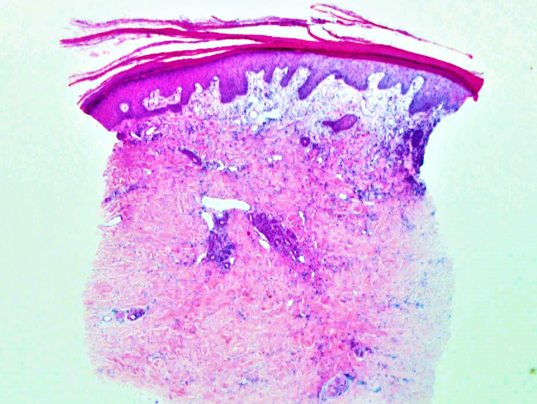
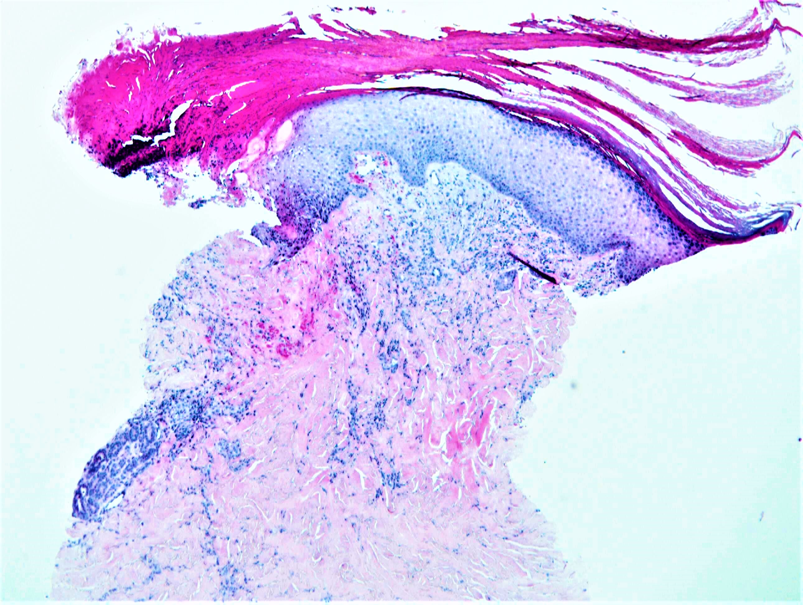
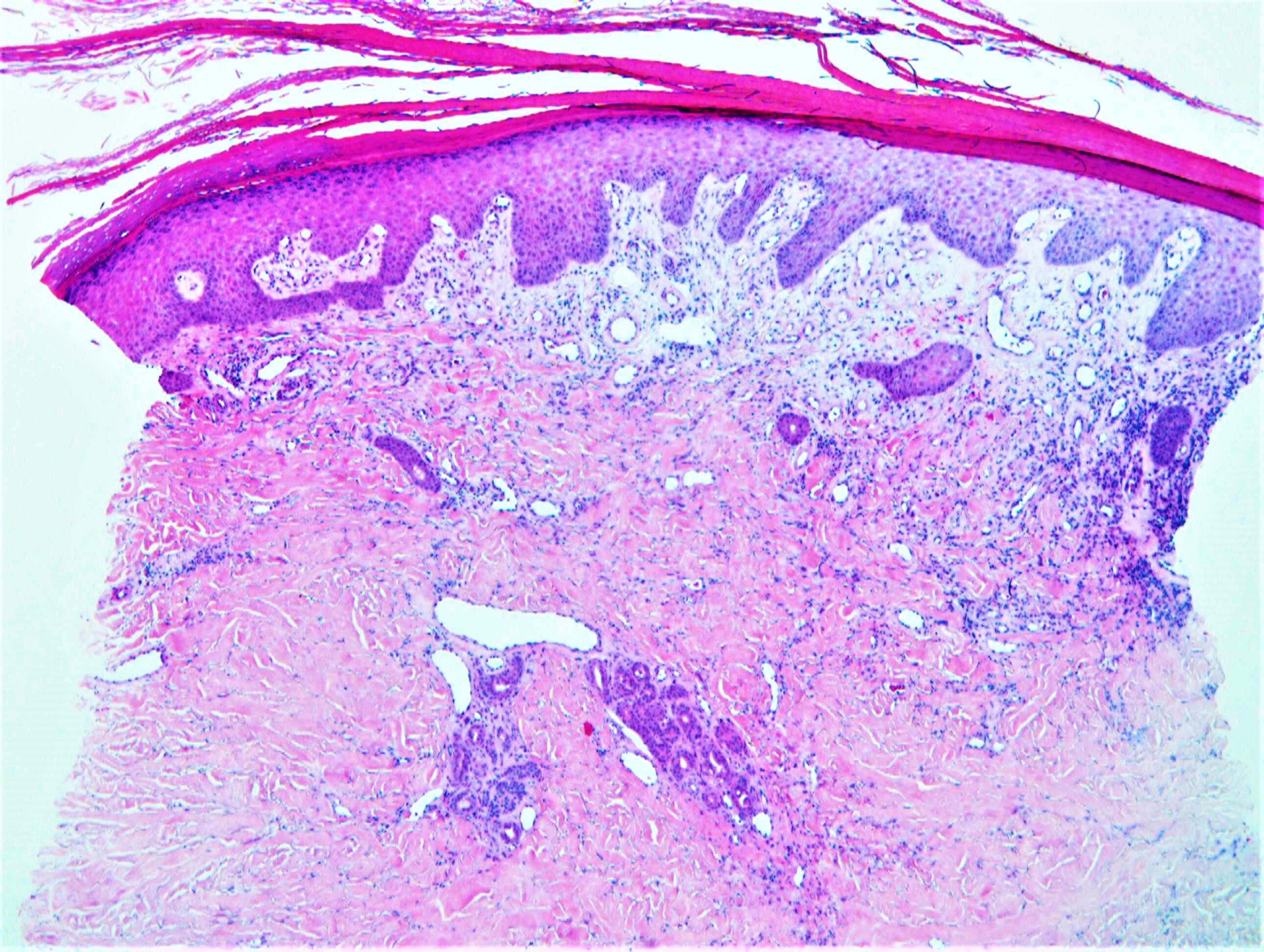
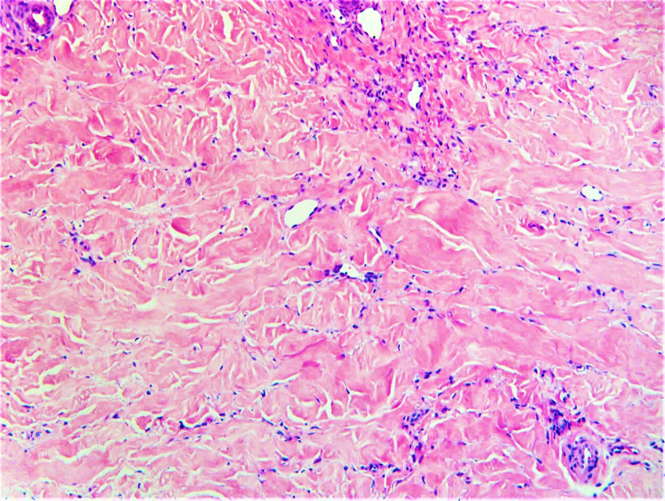
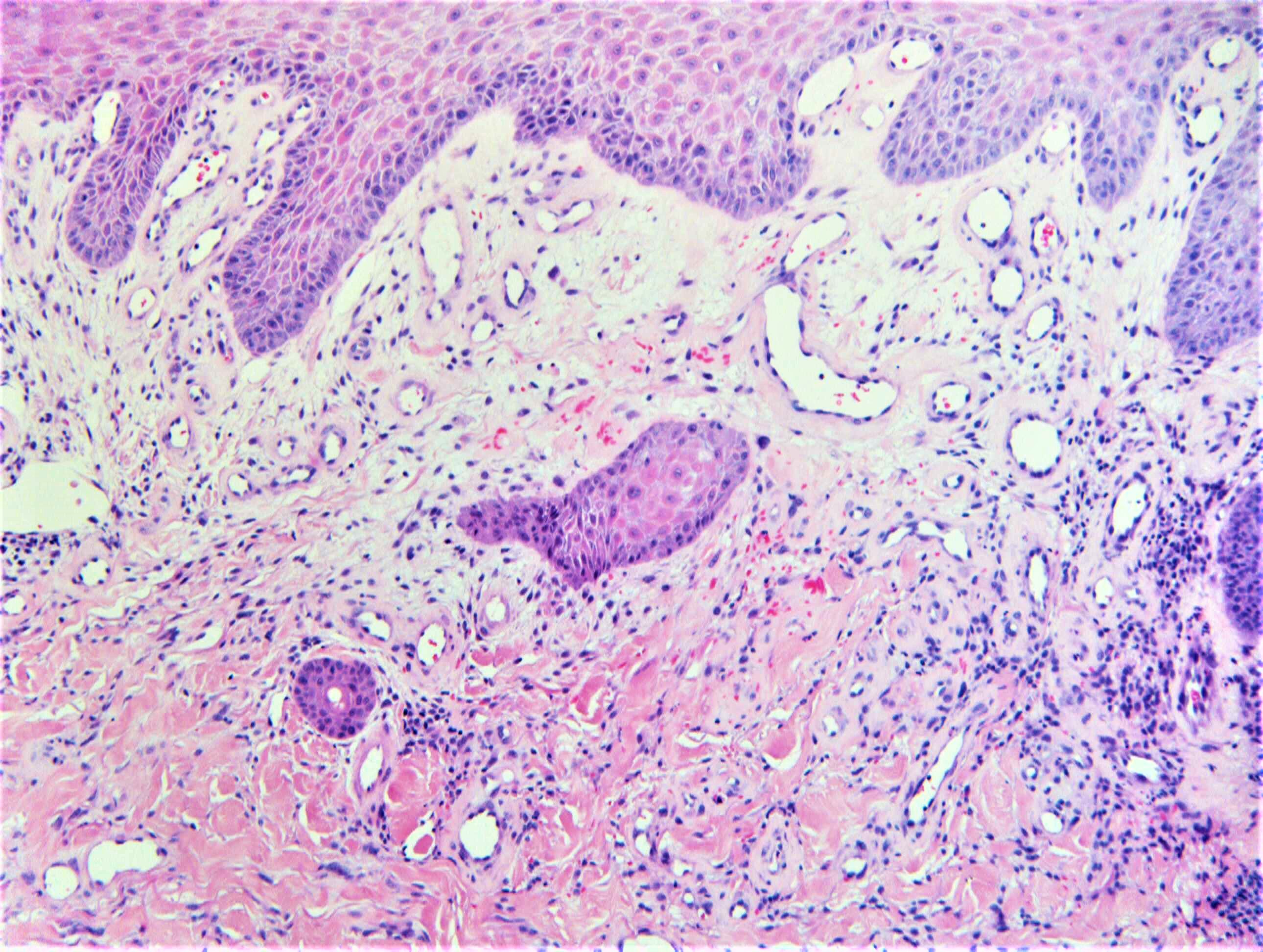
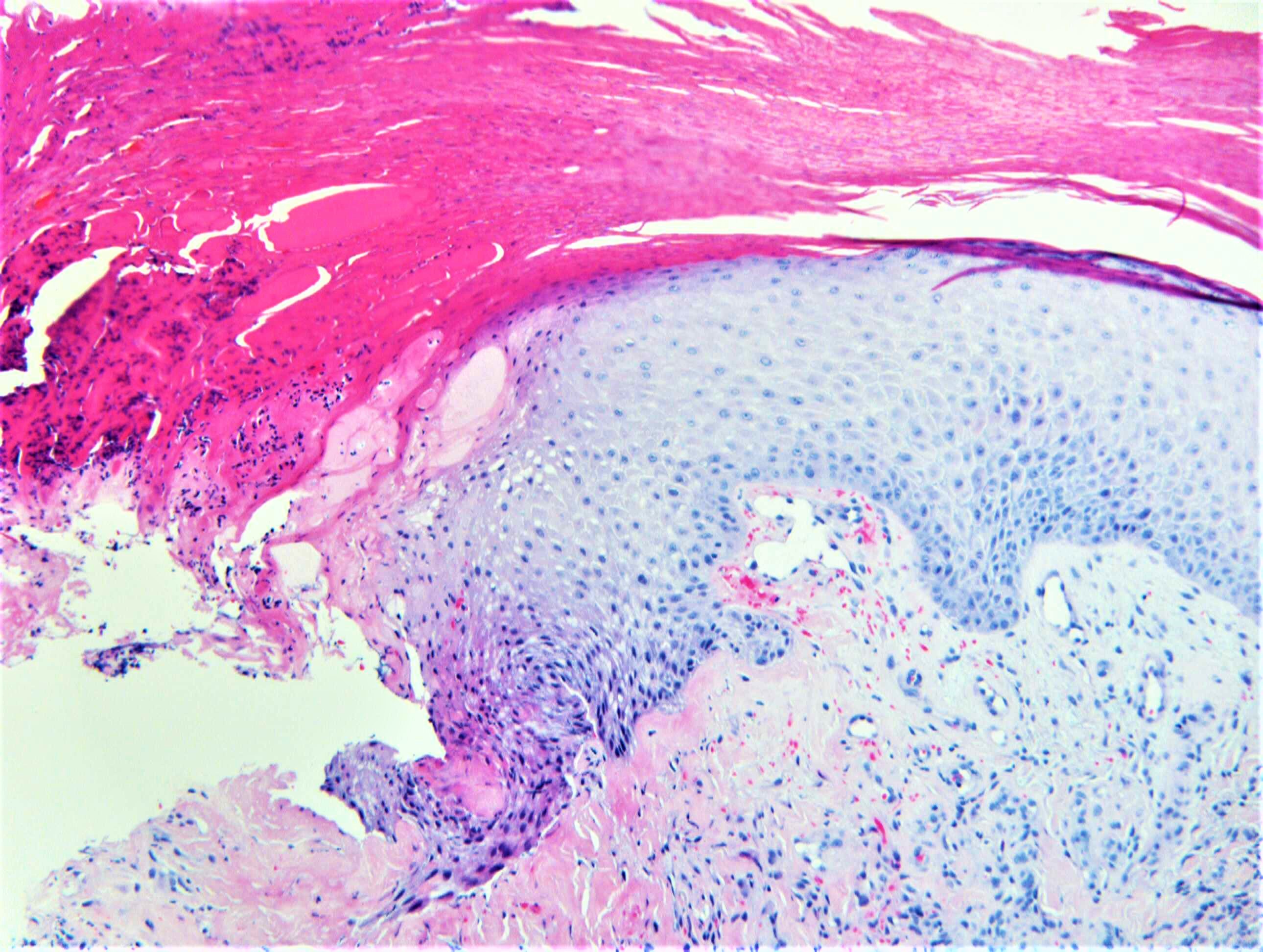
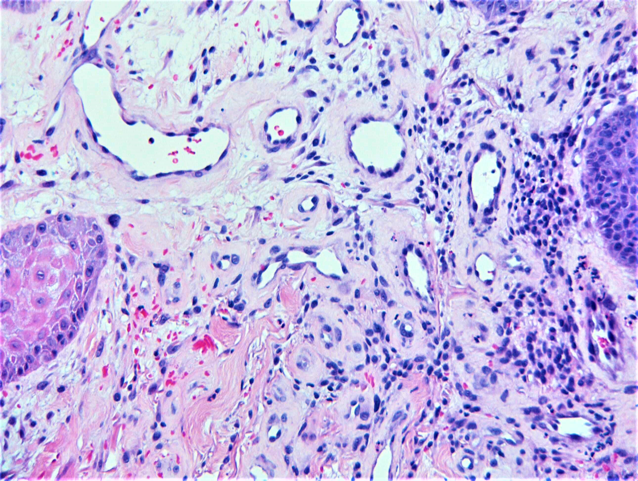
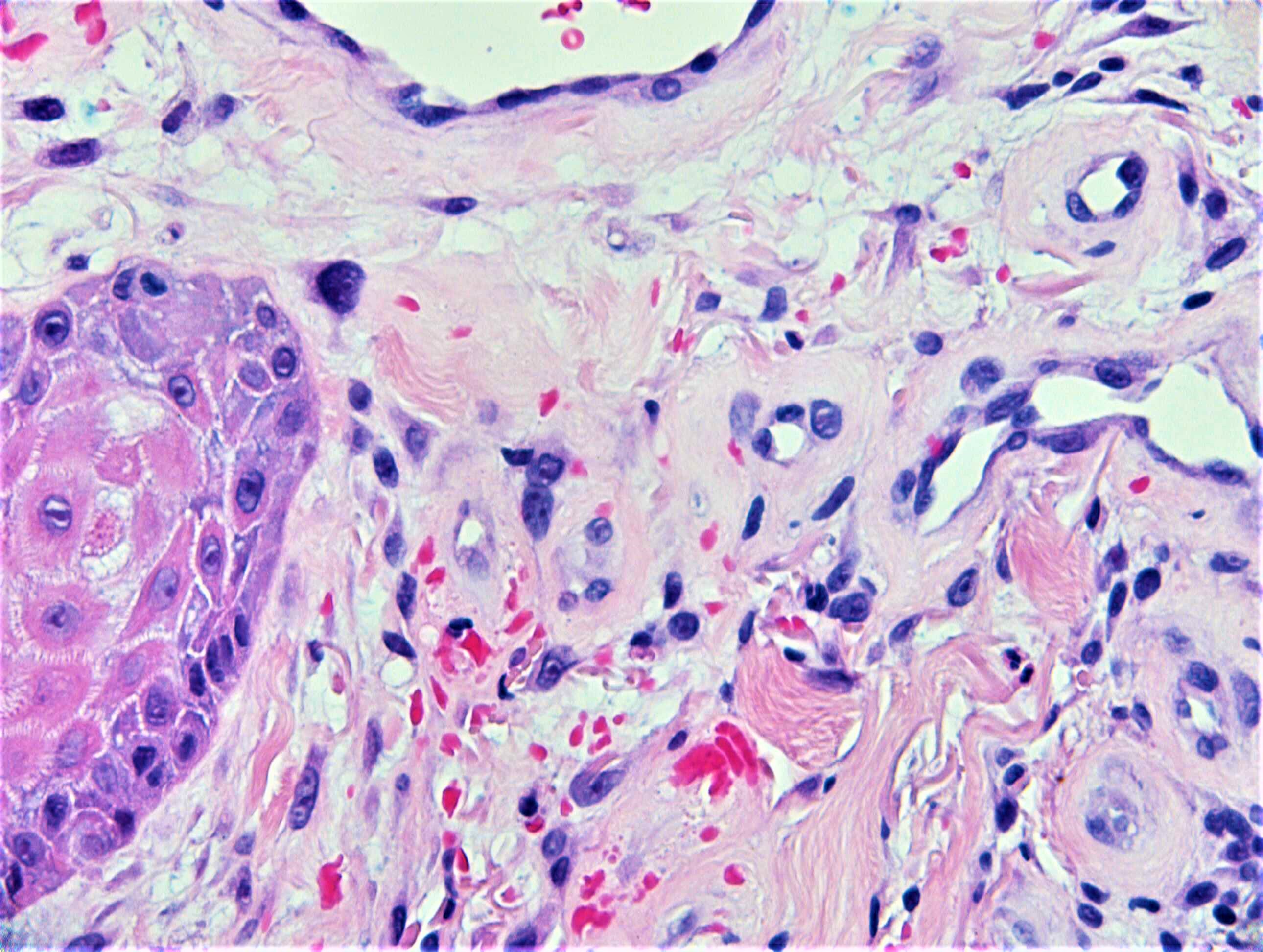
M85. Posterior thigh. Scaly. ?Bowen’s/BCC ?acanthosis

Edited by Admin_Dermpath


  Report Record

User Feedback


Hobnail hemangioma, possibly post- traumatic

Share this comment


Link to comment
share_externally

Sasi Attili

Posted

Not sure I can arrive at a specific diagnosis. Dermis appears scarred with a few inflammatory cells and dilated capillaries (site related/ post-inflammatory)

Share this comment


Link to comment
share_externally

Sasi Attili

Posted

Ulceration? History says- lesion?

7 hours ago, Admin_Dermpath said:

On behalf of Dr Richard Carr:


Diagnosis: ?Decubital / pressure ulceration


Share this comment


Link to comment
share_externally

Dr. Richard Carr

Posted

Well this is the sort of case you have to do some lateral thinking. This is how my brain worked it through. Reactive appearing epidermal hyperplasia with focal ulceration. Prominent vascularity in the upper dermis with a few extravasated RBC. Slight endothelial swelling and a prominent stromal fibroblast or two. Rather sclerotic appearing mid dermis. Elderly patient on the back of the thigh - I wonder if this is near the hip joint?  Could all this be secondary to local pressure akin to chondrodermatitis or so-called atypical decubital fibroplasia.

My Report: "Reactive/benign ulceration with vascular proliferation and sclerotic deeper collagen resembles morphoea or radiodermatitis but in view of anatomic location, could this be pressure related (atypical decubital fibroplasia/ischaemic fasciitis)?"

I often phrase my report conclusion in the form of a question as in this case and offer some indication as to my train of thoughts even if they don't quite fit. In this case clinical feed-back indicated it could be secondary to the effects of local pressure.

Share this comment


Link to comment
share_externally


Join the conversation

You can post now and register later. If you have an account, sign in now to post with your account.

Guest
Add a comment...

×   Pasted as rich text.   Paste as plain text instead

  Only 75 emoji are allowed.

×   Your link has been automatically embedded.   Display as a link instead

×   Your previous content has been restored.   Clear editor

×   You cannot paste images directly. Upload or insert images from URL.

Loading...

×
×
  • Create New...