Jump to content
In this section we have spot diagnoses posted on a daily basis since June 2010, now over 4000! You can review the archived cases and read the suggested diagnoses by users and the final comment by the contributors.
Case are uploaded each week day by 10 am UK time with the correct diagnosis will generally be posted at 8 pm UK time. Why not view the most recent spot diagnosis and proffer a diagnosis?

Case Number : Case 1913 - 28 Sept - Dr Arti Bakshi Posted By: Guest

Please read the clinical history and view the images by clicking on them before you proffer your diagnosis.
Submitted Date :
   (0 reviews)

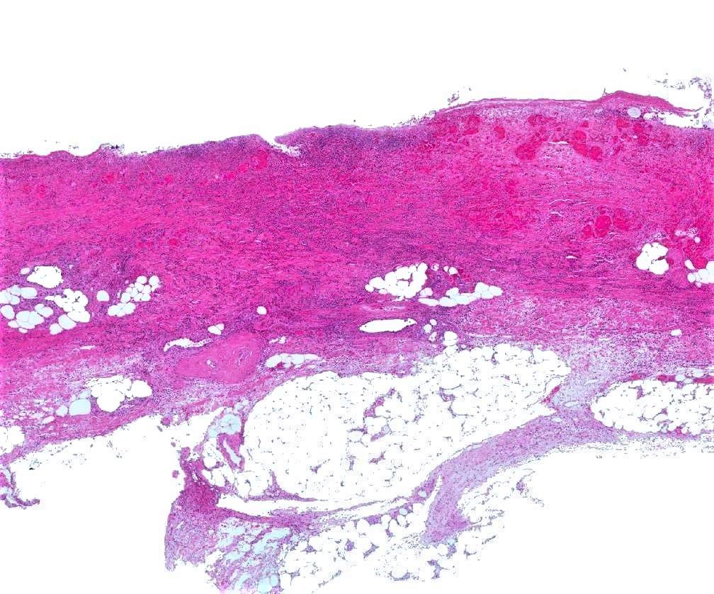
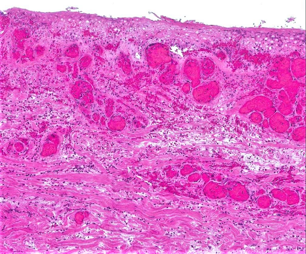
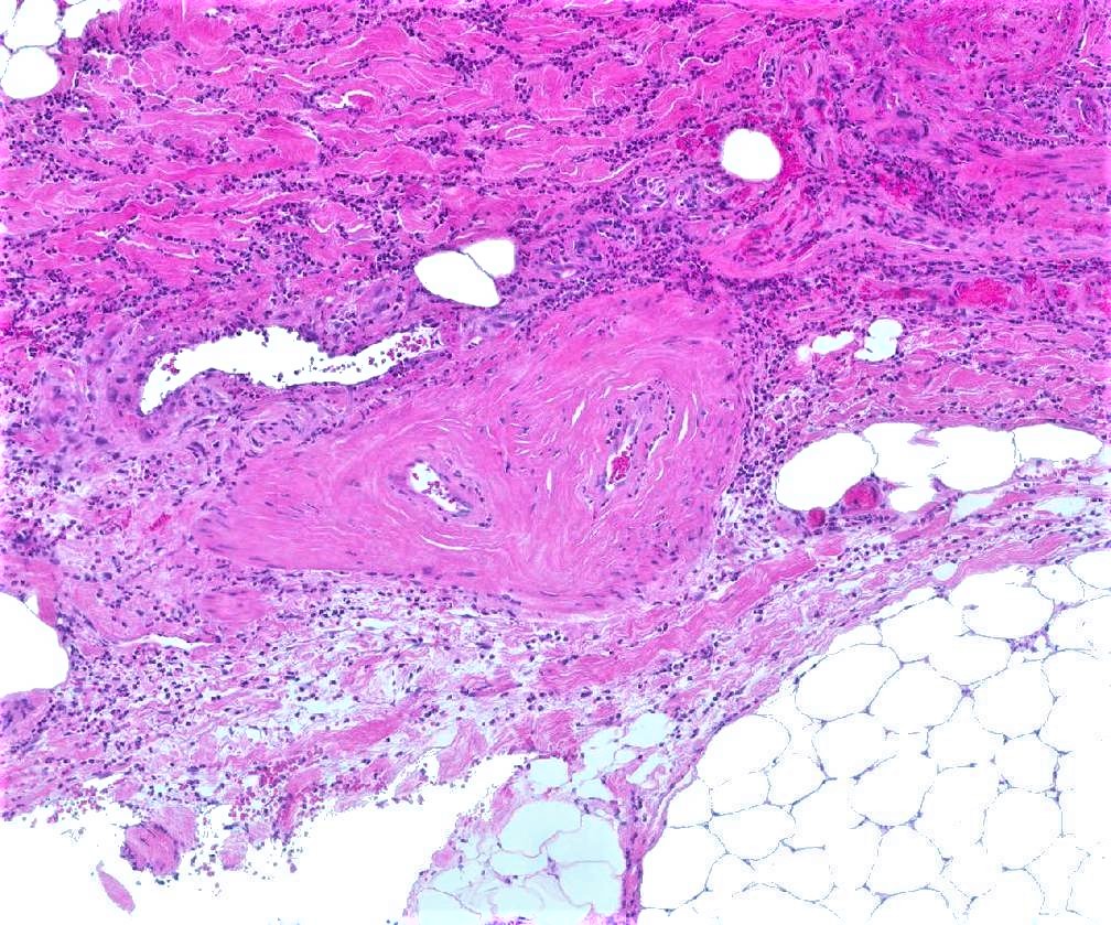
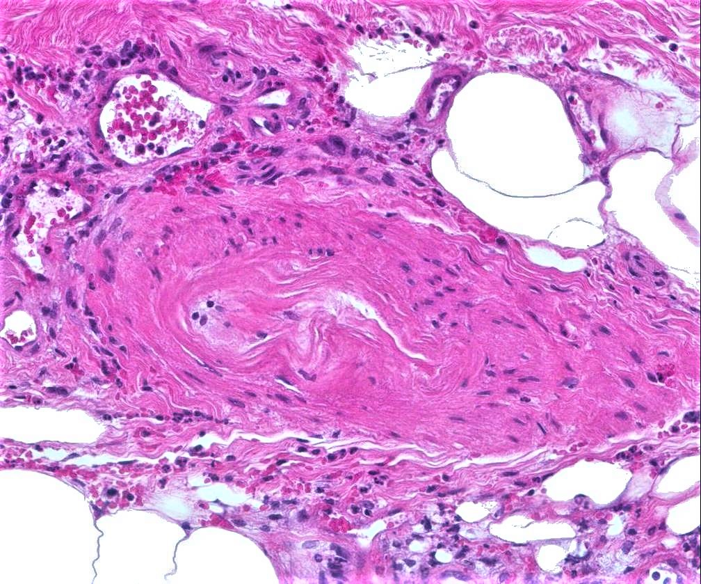
Middle aged male, 52 yrs, enlarging ulcer on leg, painful

Edited by Admin_Dermpath


  Report Record

User Feedback


urmilapandey

Posted

haven't seen this before, ?ischaemic ulcer secondary to arteriosclerosis

Share this comment


Link to comment
share_externally

vincenzo polizzi

Posted

Agree with Urmilapandey. There is some scleredema-like feature...?Diabetic microangiopathy ?Ulcerated Scleredema Diabeticorum

Share this comment


Link to comment
share_externally

This is livedoid vasculopathy according to me. Hyalinized thickened vessel walls, fibrin like deposits in lumina. Late stage is atrophie blanche

Share this comment


Link to comment
share_externally

Raul Perret

Posted

Made me think of Martorell's ulcer, Hypertension related or diabetes

Share this comment


Link to comment
share_externally

Saman Fatah

Posted

Agree with Raul, I thought of Martorell's hypertensive ischaemic leg ulcers too especially with the degree of changes of the subcutaneous arteriole. The occlusive superficial dermal vessels are probably secondary phenomenon. 

The ulcer is excruciating painful to patient, usually with many cardiovascular risk factors including hypertension (in almost all).

Share this comment


Link to comment
share_externally

Arti Bakshi

Posted

Yes this is Martorell's ulcer (or hypertensive ischaemic ulcer).

Typically, this occurs on lower legs of patients with uncontrolled hypertension and is excruciatingly painful. The patient had a duplex arterial and venous scan which was negative.

As noted in this case, the vessel type involved is an arteriole, hence the changes are typically seen in the vessels at the dermo-subcutaneous junction. There is arteriolar hyalinosis with intimal hyperplasia and almost complete narrowing of the lumen.

Here is a link to a rather similar case put by Dr Carr on dermpathpro a while ago. https://dermpathpro.com/spot-diagnosis-1/2015-spot-diagnoses/december-2015/case-1432-18-december-r1497/ . I did not get the diagnosis then, but having seen this case on dermpathpro, was able to make the correct diagnosis in my case.


Share this comment


Link to comment
share_externally


Join the conversation

You can post now and register later. If you have an account, sign in now to post with your account.

Guest
Add a comment...

×   Pasted as rich text.   Paste as plain text instead

  Only 75 emoji are allowed.

×   Your link has been automatically embedded.   Display as a link instead

×   Your previous content has been restored.   Clear editor

×   You cannot paste images directly. Upload or insert images from URL.

Loading...

×
×
  • Create New...