Jump to content
In this section we have spot diagnoses posted on a daily basis since June 2010, now over 4000! You can review the archived cases and read the suggested diagnoses by users and the final comment by the contributors.
Case are uploaded each week day by 10 am UK time with the correct diagnosis will generally be posted at 8 pm UK time. Why not view the most recent spot diagnosis and proffer a diagnosis?

Case Number : Case 1759 - 23 February - Dr Arti Bakshi Posted By: Guest

Please read the clinical history and view the images by clicking on them before you proffer your diagnosis.
Submitted Date :
   (0 reviews)

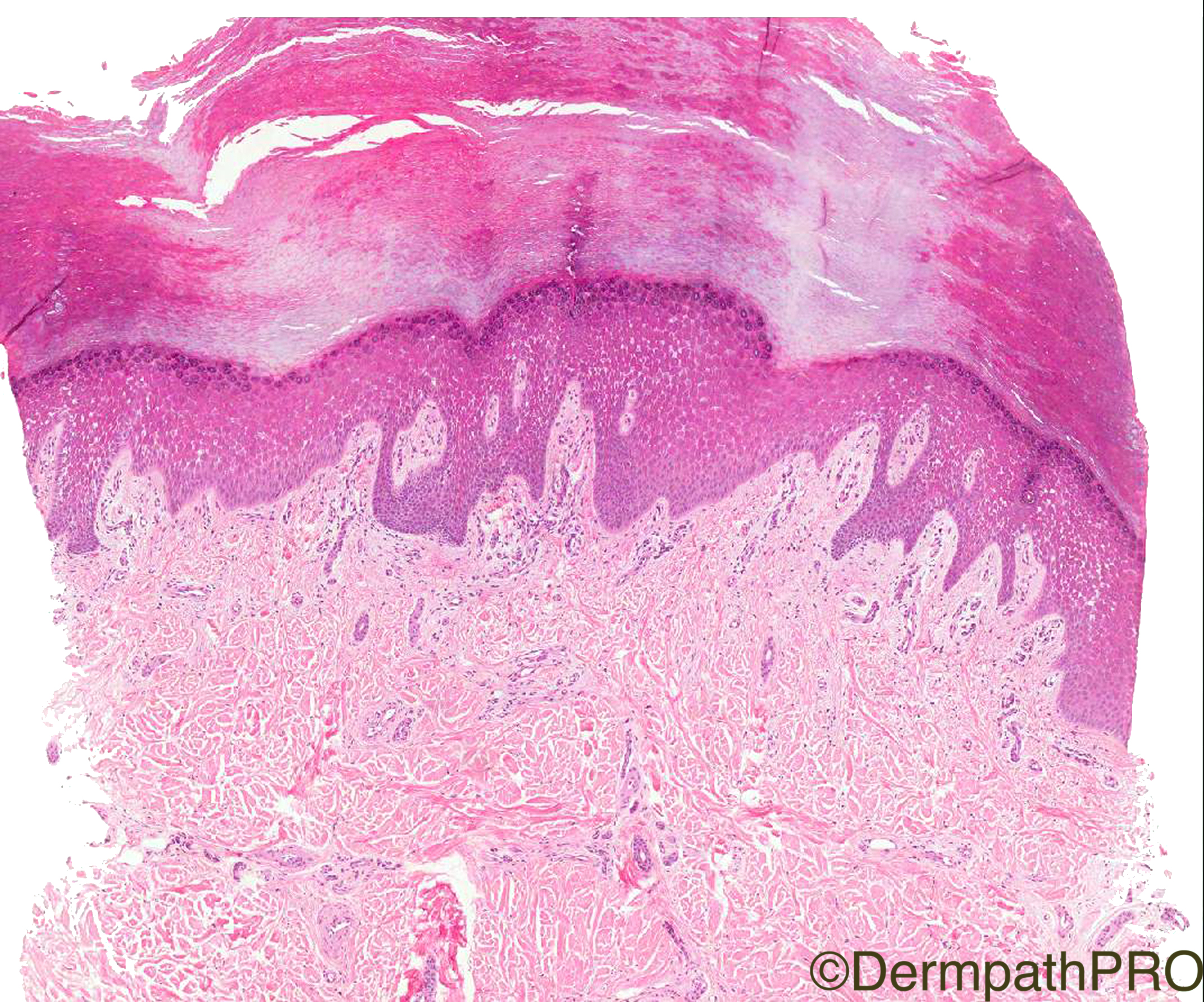
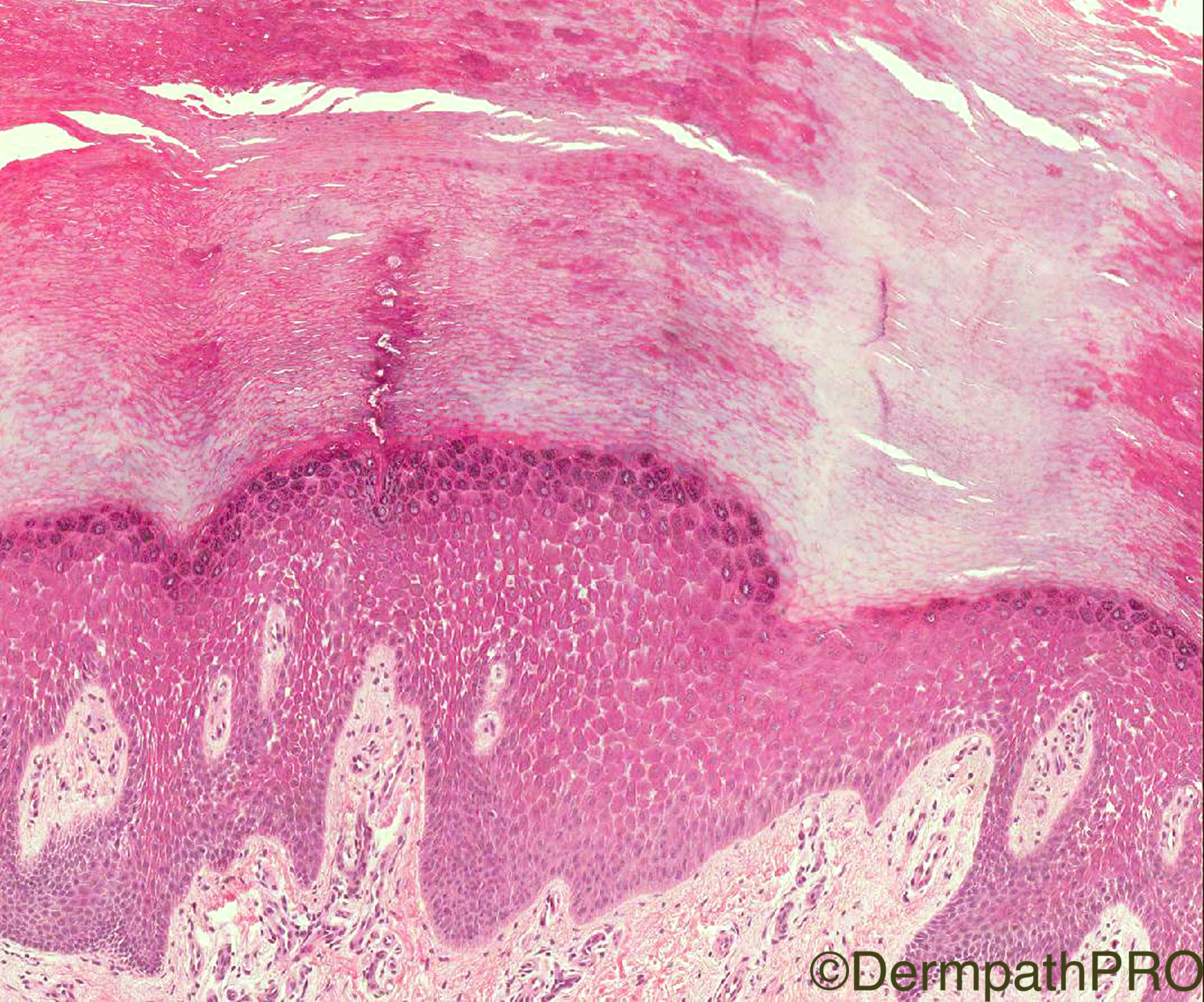
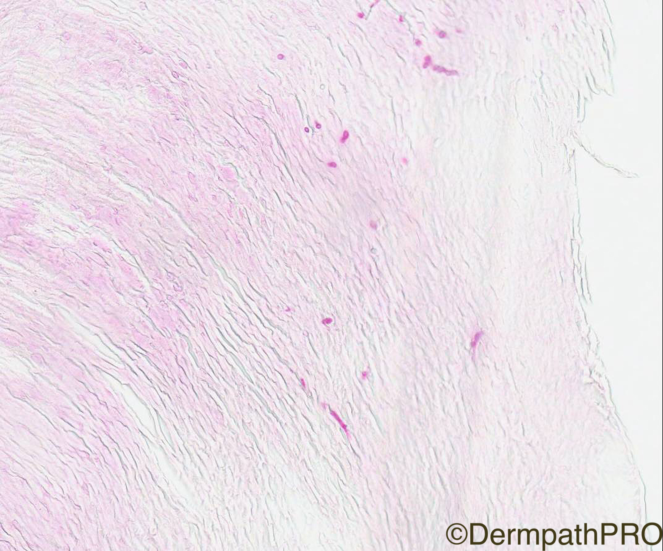
Clinical History: 40/M, diffuse thickening of palms and feet. H/o cardiac disease in family.

Case Posted by Dr Arti Bakshi

Edited by Admin_Dermpath


  Report Record

User Feedback


Admin_Dermpath

Posted

Storm Doris is assaulting the UK today, not good dog walking weather, so take a break and give Dr Arti Bakshi's Spot Case an examination and see what diagnosis you arrive at.


Geoff Cross - DermpathPRO Projects

Share this comment


Link to comment
share_externally

Raul Perret

Posted (edited)

Looks llike a diffuse palmoplantar keratoderma with acantholytic features. Here a nice article that could be related to the congenital heart disease mentioned. Maybe Carvajal or Naxos syndrome depending on the heart disease.

Edited by Raul Perret

Share this comment


Link to comment
share_externally

vincenzo polizzi

Posted

This is a tricky case for me: on the one hand the histology is consistent with a striated PPK ( acantholisis associated with hype rkeratosis and acanthosis ) on the other hand the clinical description is of a diffuse thickening of the palmoplantar skin...

Share this comment


Link to comment
share_externally

vincenzo polizzi

Posted

Very nice article, Raul. Thanks!

Now the tricky case has become a beautiful case for me,and I've learnt another pathological chapter...

Share this comment


Link to comment
share_externally

Arti Bakshi

Posted

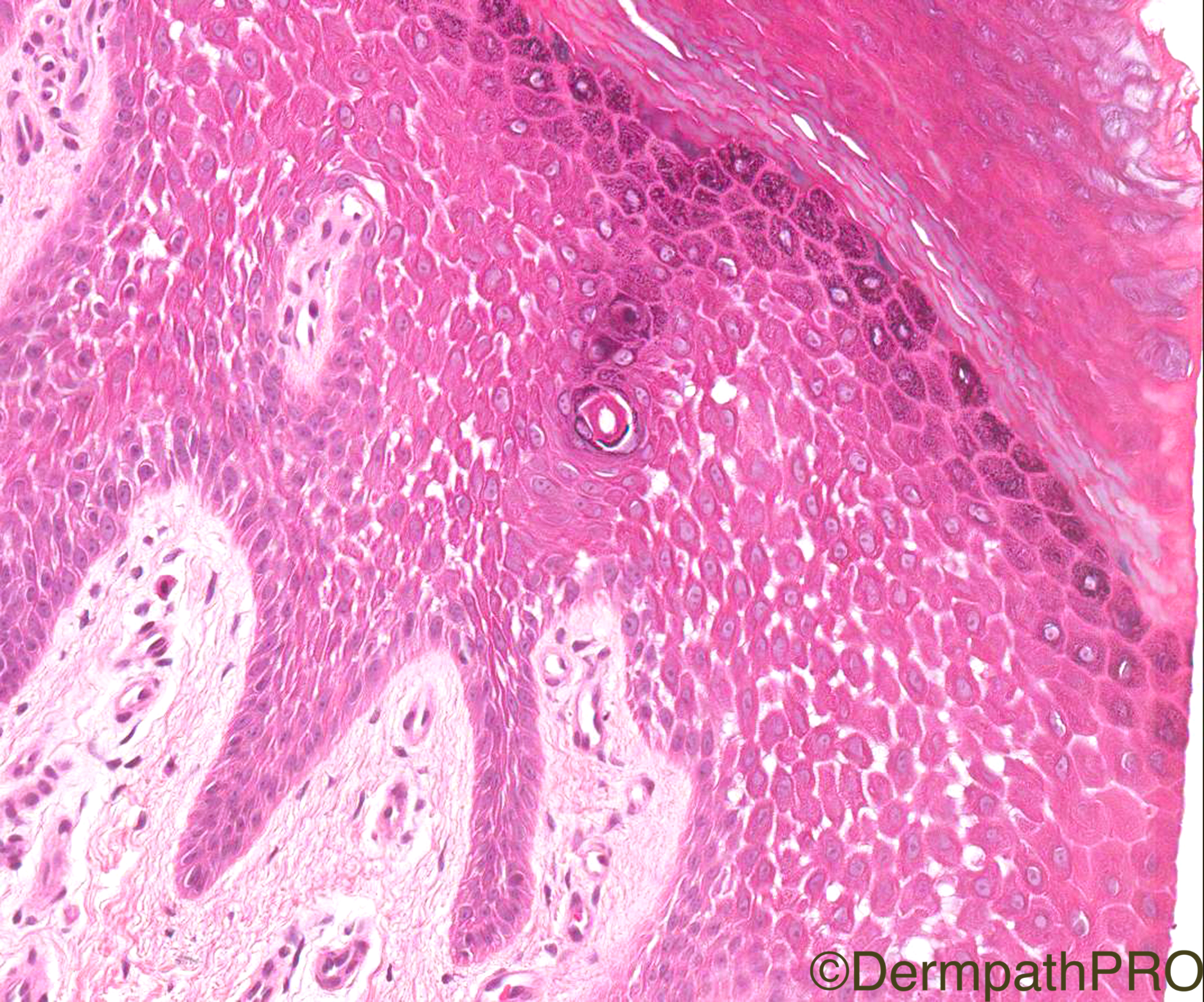
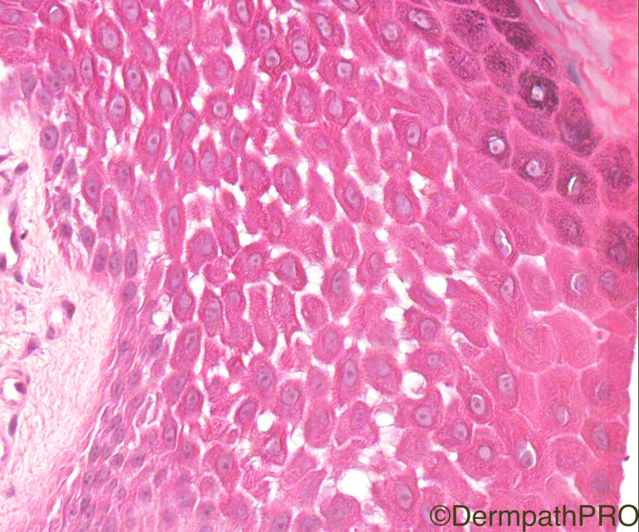
Both Raul and Vicenzo doing very well! Could I, at this point, ask a very basic morphological question from everyone. Would you consider the changes in the last 2 images as representing acantholysis or spongiosis??, its a dilemma that I went thru looking at this case......please feel free to answer this, even if you dont want to comment on the actual diagnosis!

Share this comment


Link to comment
share_externally

vincenzo polizzi

Posted

I'm sharing the same Arti's dilemma now ( before it was acantholysis for me, after the Arti's question I changed to spongiosis, and now...many doubts. The partially separated cells show a different cytoplasmic stain and some are lightly rounded and condensed...but it's an impression. May be a spongiosis instead, because there isn't any cells single isolated and intercellular connections are evenly obvious...

Share this comment


Link to comment
share_externally

Dr. Mona Abdel-Halim

Posted

Agree with Raul that this is mostly syndromal PPK with cardiac abnormalities...

As for Arti's question, I think most of the cells have more or less rounded countours favoring acantholysis. There is minimal infiltrate and no exocytosis. I think it is acantholoysis (hereditary non inflamatory keratinocyte dyscohesion)

Share this comment


Link to comment
share_externally

Raul Perret

Posted

I have the same opinion as Mona acantholysis for me. The presence of intercelullar bridges can be tricky as we are taught that they are evident mainly in spongiotic patterns. However if you check ancient cases of Hailey Hailey disease here and here you will see that some of the cells can still show intercellular bridges. Another point is that spongiosis is rarely well delimited within the epidermis as we see here (maybe in some cases of miliaria cristalina?)

Share this comment


Link to comment
share_externally

Robledo F. Rocha

Posted

I didn’t find any sign of inflammation, so agree with palmoplantar keratoderma (hyperkeratosis palmaris et plantaris).

About Arti’s query, spongiosis means intraepidermal intercellular edema, recognized microscopically by widened spaces between keratinocytes, chiefly in the spinous layer. Acantholysis results from dissolution of intercellular connections of the epidermal cells with loss of cohesion between them. In the images above, the intercellular bridges of the keratinocytes do not appear elongated and stretched as would be expected in spongiotic reactions. Actually, the epidermal cells have round shapes and are separated from one another, evincing their disadhesion due to destruction of the intercellular attachment, namely, an acantholytic process. 

Share this comment


Link to comment
share_externally

Arti Bakshi

Posted

Great discussion!

This is palmoplantar keratoderma, ofcourse. The patient was found to have Desmoplakin (DSP) gene mutation. His brothers and father also have PPK and one of his brothers has ARVD. None have woolly hair though.

With regards to acantholysis vs spongiosis, I have to admit I found it quite tricky in this case. To compound matters, there were also fungal hypahe in the stratum corneum, indicative of fungal superinfection. (will ask Geoff to add an image). One of the dictums with PPK is that presence of spongiosis should prompt a PAS stain to look for fungi! But I agree with the comments above that there is complete lack of inflammation which would be odd for a spongiotic process. To my eye, there was stretching of intercellular bridges...but Raul's comment is pertinent in this regard that one has to be wary of this feature in early stages of acantholysis. From a pathogenetic point of view this makes sense (particularly in hereditary cases)  as the keratinocytes start separating off from each other due to weakened junctions even before they are completely lost. I also agree that there is some early rounding up of keratinocytes, again favouring acantholysis. In this case, it doesn't matter as the mutation is already known, but diagnosing an  acantholytic form of PPK can help point towards the underlying mutation. 

Well done all and thanks for all your comments! 

Share this comment


Link to comment
share_externally

Mariantonieta Tirado

Posted

I can't see very well but there might be also a secondary dermatophyte infection.

Share this comment


Link to comment
share_externally

Dr. Richard Carr

Posted

I think there is clear cut spongolysis (you heard it here first folks!).

Share this comment


Link to comment
share_externally


Join the conversation

You can post now and register later. If you have an account, sign in now to post with your account.

Guest
Add a comment...

×   Pasted as rich text.   Paste as plain text instead

  Only 75 emoji are allowed.

×   Your link has been automatically embedded.   Display as a link instead

×   Your previous content has been restored.   Clear editor

×   You cannot paste images directly. Upload or insert images from URL.

Loading...

×
×
  • Create New...